. 2021 Oct;8(10):2040-2051.
doi: 10.1002/acn3.51453. Epub 2021 Sep 15.

Role of cellular prion protein in splenic CD4+ T cell differentiation in cerebral ischaemic/reperfusion

Affiliations

Role of cellular prion protein in splenic CD4+ T cell differentiation in cerebral ischaemic/reperfusion

Baizhuo Zhang et al. Ann Clin Transl Neurol. 2021 Oct.

Abstract

Objective: Cellular prion protein (PrPC ), the primary form of prion diseases pathogen, has received increasing attention for its protective effect against ischaemic stroke. Little is known about its role in peripheral immune responses after cerebral ischaemia/reperfusion (I/R) injury. This study is to detect the variation of splenic CD4+ T lymphocytes differentiation and the concentration of inflammatory cytokines after murine cerebral I/R injury in the context of PRNP expression as well as its influence on the ischaemic neuronal apoptosis.

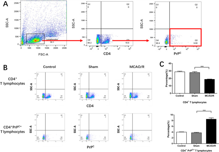
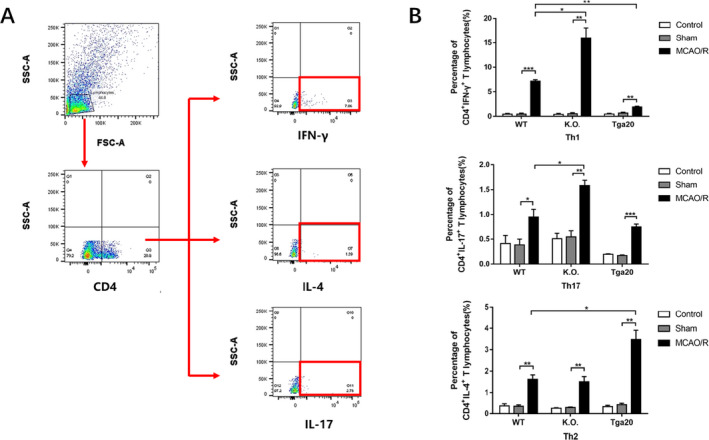
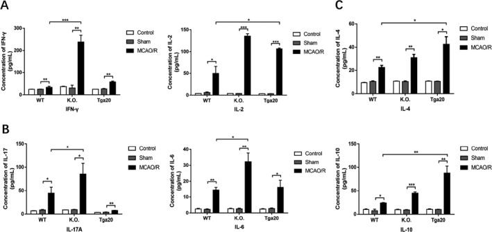
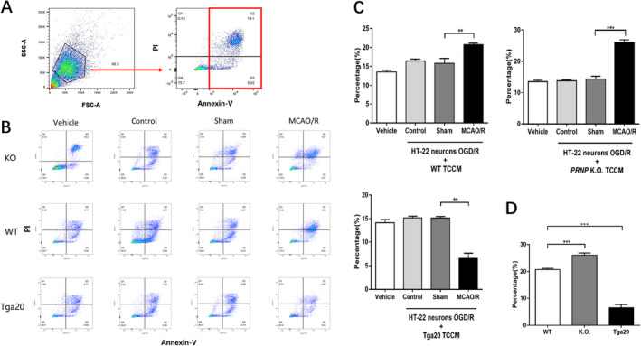
Methods: We established the cerebral ischaemic murine model of different PRNP genotypes. We detected the percentage of splenic CD4+ PrPC+ T cells of PRNP wild-type mice and the ratio of splenic Th1/2/17 lymphocytes of mice of different PRNP expression. The relevant inflammatory cytokines were then measured. Oxygen glucose deprivation/reperfusion (OGD/R) HT22 mouse hippocampal neurons were co-cultured with the T-cell-conditioned medium harvested from the spleen of modelled mice and then the neuronal apoptosis was detected.

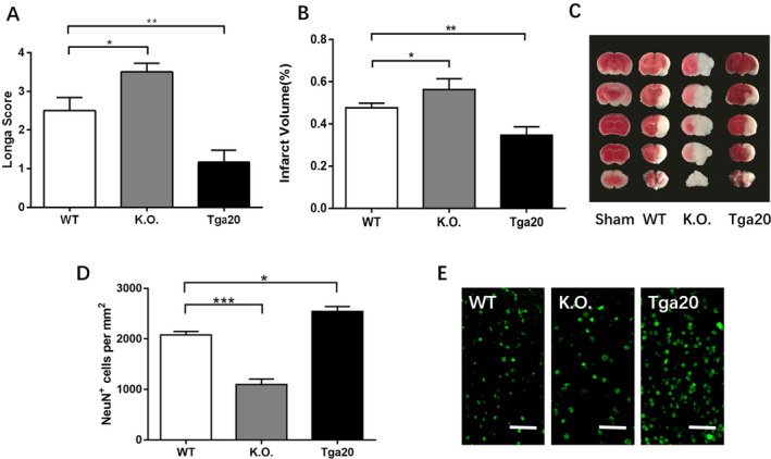
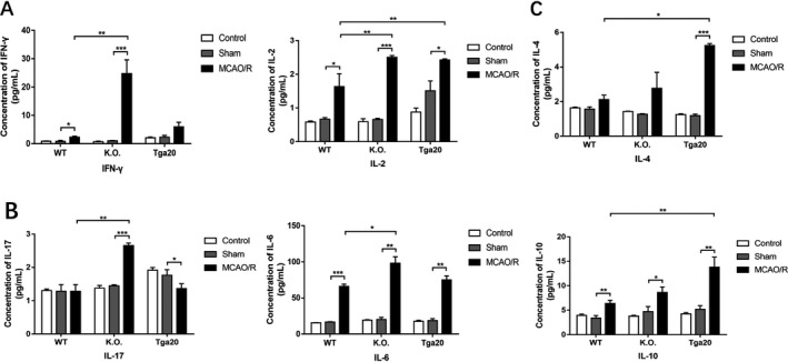
Results: CD4+ PrPC+ T lymphocytes in wild-type mice elevated after MCAO/R. PRNP expression deficiency led to an elevation of Th1/17 phenotypes and the promotion of pro-inflammatory cytokines, while PRNP overexpression led to the elevation of Th2 phenotype and upregulation of anti-inflammatory cytokines. In addition, PrPC -overexpressed CD4+ T cells weakened the apoptosis of OGD/R HT-22 murine hippocampal neurons caused by MCAO/R CD4+ T-cell-conditioned medium, while PrPC deficiency enhanced apoptosis.

Interpretation: PrPC works as a neuron protector in the CNS when I/R injury occurs and affects the peripheral immune responses and defends against stroke-induced neuronal apoptosis.


Conflict of interest statement

None.

Figures

Figure 1
Figure 1
The comparison of lesion severity of experimental animals (neurological score, infarction volume and NeuN+ cell density) of different genotypes after 90 min of ischaemia and 24h of recovering. The percentage of infarction volume was calculated by TTC staining (WT control n = 6, sham n = 6, tMCAO/R n = 6). (A). The Longa score of each genotype after MCAO/R injury. (B). The infarction volume of each genotype. (C) The example of the TTC staining of each group. (D) The neuronal density of each genotype determined by NeuN immunohistochemistry in the striatum. (E) The representative photographs of neuronal density of each genotype. Scale bars: 50 μm. Analysis of variance (ANOVA), and post hoc Duncan’s test was used to assess differences among multiple groups and data are displayed as mean ± standard deviation (SD), *P < 0.05, **P < 0.01, ***P < 0.001.
Figure 2
Figure 2
The post‐stroke expression of CD4+ and CD4+PrPC+ T cells in WT group (control n = 6, sham n = 6, tMCAO/R n = 6). (A) An example showing the gating strategy used to identify CD4+ T cells and CD4+PrPC+ T cells after MCAO/R injury of each genotype in the presence of the anti‐ CD4 anti‐ CD230 (PrP) mAbs. The first gate was set on physical parameters, then on CD4+ events and then on CD230+ events. (B) The example of the analysis of the CD4+ and CD4+PrPC+ T cell percentage through flow cytometry. The Q3 quadrant represented the CD4+ T cells (upper line) and the CD4+PrPC+ T cells (lower line). (C) The proportion of CD4+ T cells and CD4+PrPC+ T cells Experiments were carried out in biologically triplicate. Analysis of variance (ANOVA), and post hoc Duncan’s test was used to assess differences among multiple groups and data are displayed as mean ± standard deviation (SD), *** P < 0.001.
Figure 3
Figure 3
The percentage of splenic Th1/2/17 phenotypes of each genotype. (WT n = 6, KO n = 6, Tga20 n = 6) after stimulated by PMA/iono and protein transport inhibitor for 5h. (A) An example showing the gating strategy used to identify Th1/2/17 phenotypes respectively. The first gate was set on physical parameters and then on CD4+ events. Then IFN‐γ+, IL‐4+ and IL‐17+ events were separately detected and showed in the Q3 quadrant. (B) The ratio of Th1/2/17 lymphocytes respectively. Experiments were carried out in biologically triplicate. Analysis of variance (ANOVA), and post hoc Duncan’s test and Dunnett’s test were used to assess differences among multiple groups and data are displayed as mean ± standard deviation (SD), *P < 0.05, **P < 0.01, ***P < 0.001.
Figure 4
Figure 4
Flow cytometry‐based evaluation of concentrations of Th1/17‐related pro‐inflammatory and Th2‐related anti‐inflammatory cytokines in cultured T lymphocyte supernatants stimulated by PMA/iono for 5h. (WT n = 6, KO n = 6, Tga20 n = 6). (A) The expression of Th1‐related pro‐inflammatory cytokines. (B) The expression of Th17‐related pro‐inflammatory cytokines. (C) The expression of Th2‐related anti‐inflammatory cytokines. Experiments were carried out in biologically triplicate. Analysis of variance (ANOVA), and post hoc Duncan’s test and Dunnett’s test were used to assess differences among multiple groups and data are displayed as mean ± standard deviation (SD), *P < 0.05, **P < 0.01, ***P < 0.001.
Figure 5
Figure 5
Flow cytometry‐based evaluation of concentrations of Th1/17‐related pro‐inflammatory and Th2‐related anti‐inflammatory cytokines in serum (WT n = 6, KO n = 6, Tga20 n = 6). (A) The expression of Th1‐related pro‐inflammatory cytokines. (B) The expression of Th17‐related pro‐inflammatory cytokines. (C) The expression of Th2‐related anti‐inflammatory cytokines. Experiments were carried out in biologically triplicate and data are displayed as mean ± standard deviation (SD), *P < 0.05, **P < 0.01, ***P < 0.001.
Figure 6
Figure 6
Effects of PrPC‐affected TCCM of each genotype on HT‐22 neuronal cell lines subjected to OGD/R injury. The vehicle group (n = 6), OGD/R+ control TCCM group (n = 6), OGD/R+ sham TCCM group (n = 6) and OGD/R+ MCAO/R TCCM group (n = 6). (A) An example showing the gating strategy to identify the apoptosis of HT‐22 murine hippocampal after co‐culture with TCCM in the presence of annexin V. The apoptotic neurons were showed in Q2 and Q3 quadrant. (B) Examples of cell apoptosis rates in HT‐22 murine hippocampal cells in each group of different genotypes. (C) The percentage of apoptotic OGD/R HT‐22 neurons after co‐cultured with TCCM of different genotype. (D) The comparison of OGD/R HT‐22 neurons apoptosis with MCAO/R TCCM of different genotype. Experiments were carried out in biologically triplicate and data are displayed as mean ± standard deviation (SD), **P < 0.01, ***P < 0.001.

References

    1. Liu T, Clark RK, McDonnell PC, et al. Tumor necrosis factor‐alpha expression in ischemic neurons. Stroke. 1994;25(7):1481‐1488. doi:10.1161/01.STR.25.7.1481 - DOI - PubMed
    1. Wang X, Yue T‐L, Young PR, et al. Expression of interleukin‐6, c‐Fos, and zif268 mRNAs in rat ischemic cortex. J Cereb Blood Flow Metab. 1995;15(1):166‐171. doi:10.1038/jcbfm.1995.18 - DOI - PubMed
    1. Wang X, Barone FC, Aiyar NV, Feuerstein GZ. Interleukin‐1 receptor and receptor antagonist gene expression after focal stroke in rats. Stroke. 1997;28(1):155‐162. doi:10.1161/01.STR.28.1.155 - DOI - PubMed
    1. Yoshimura A. Post‐ischemic inflammation regulates neural damage and protection. Front Cell. 2014;8:1‐8. doi:10.3389/fncel.2014.00319 - DOI - PMC - PubMed
    1. Offner H, Subramanian S, Parker SM, et al. Experimental stroke induces massive, rapid activation of the peripheral immune system. J. Cereb Blood Flow Metab. 2006;26:654‐665. doi:10.1038/sj.jcbfm.9600217 - DOI - PubMed

Publication types