. 2021 Sep 30;125(38):10748-10758.
doi: 10.1021/acs.jpcb.1c06054. Epub 2021 Sep 15.

Shedding Light on Thermally Induced Optocapacitance at the Organic Biointerface

Affiliations

Shedding Light on Thermally Induced Optocapacitance at the Organic Biointerface

Gaia Bondelli et al. J Phys Chem B. .

Abstract

Photothermal perturbation of the cell membrane is typically achieved using transducers that convert light into thermal energy, eventually heating the cell membrane. In turn, this leads to the modulation of the membrane electrical capacitance that is assigned to a geometrical modification of the membrane structure. However, the nature of such a change is not understood. In this work, we employ an all-optical spectroscopic approach, based on the use of fluorescent probes, to monitor the membrane polarity, viscosity, and order directly in living cells under thermal excitation transduced by a photoexcited polymer film. We report two major results. First, we show that rising temperature does not just change the geometry of the membrane but indeed it affects the membrane dielectric characteristics by water penetration. Second, we find an additional effect, which is peculiar for the photoexcited semiconducting polymer film, that contributes to the system perturbation and that we tentatively assigned to the photoinduced polarization of the polymer interface.


Conflict of interest statement

The authors declare no competing financial interest.

Figures

Figure 1
Figure 1
Laurdan as a polarity-sensitive probe. (a) Laurdan chemical structure. (b) Laurdan Jablonski diagram, illustrating absorption (exc. 370 nm, red line), solvent relaxation to, and emission from two different excited states: LE state, with emission near 460 nm, and ICT state in more polar environments, with emission at longer λ. A schematic representation of the corresponding emission maxima of Laurdan in ordered and disordered phases is shown.
Figure 2
Figure 2
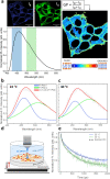
Laurdan probe monitors membrane fluidity in cells through a shift in the emission spectrum. (a) Laurdan fluorescence images of HEK-293 cells recorded simultaneously in the blue channel (425–475 nm, IB) and green channel (500–550 nm, IG) and relative Laurdan emission spectrum. On the right, fluidity map obtained applying the GP formula for each pixel, pseudocolored as indicated by the scale with GP ranging from −1 to 1. The scale bar is 20 μm. (b–e) TRPL measurements on HEK-293 cells grown on glass substrates. Deconvolution of the Laurdan emission spectra in HEK-293 cells recorded at 22 °C (b) and 40 °C (c), upon excitation at 370 nm, fitted with two Gaussian curves centered at 460 and 510 nm, used in the GP evaluation. Obtained values go toward more negative values (disordered state). (d) Schematic representation of the sample used in this study, heated by a Peltier plate. (e) Laurdan PL kinetics, integrated in the range of 440–650 nm, fitted to biexponential decay functions: dynamics become faster when temperature is increased.
Figure 3
Figure 3
TRPL measurements on HEK-293 cells grown on P3HT:PCBM films. Deconvolution of the Laurdan emission spectra, recorded with excitation at 370 nm only (a) and at 370 nm + CW 561 nm (46 mW/mm2) (b), fitted with two Gaussian curves centered at 460 and 510 nm, used in the GP evaluation. Measurements were performed on the same cell spot. PL contribution from the polymeric film was avoided integrating only the range of 100–2000 ps. (c) Schematic representation of the sample used in this study. The laser beam illuminates the back of the sample, where the P3HT:PCBM film is located, while the PL excitation 370 nm beam impinges the sample from the opposite side, where the cells stained with Laurdan are plated. (d) Laurdan PL kinetics, integrated in the range of 440–550 nm, fitted to double exponential decay curves: dynamics become faster upon illumination with a 561 nm CW laser.
Figure 4
Figure 4
Laurdan PL as a function of 561 nm CW laser power. (a) Average (n = 6) ΔGP for TRPL measurement with 370 nm excitation only and TRPL measurement with 370 nm + CW 561 nm excitation; error bars represent the standard deviation. (b) Laurdan TRPL decays at increasing 561 nm CW laser power: dynamics become faster, going from 0 mW/mm2 (exc. 370 nm only) to 46 mW/mm2 and were fitted to double exponential decay curves.

References

    1. Potter S. M. Distributed Processing in Cultured Neuronal Networks. Prog. Brain Res. 2001, 130, 49–62. 10.1016/S0079-6123(01)30005-5. - DOI - PubMed
    1. Manfredi G.; Lodola F.; Paternó G. M.; Vurro V.; Baldelli P.; Benfenati F.; Lanzani G. The Physics of Plasma Membrane Photostimulation. APL Mater. 2021, 9, 03090110.1063/5.0037109. - DOI
    1. DiFrancesco M. L.; Lodola F.; Colombo E.; Maragliano L.; Bramini M.; Paternò G. M.; Baldelli P.; Serra M. D.; Lunelli L.; Marchioretto M.; et al. Neuronal Firing Modulation by a Membrane-Targeted Photoswitch. Nat. Nanotechnol. 2020, 15, 296–306. 10.1038/s41565-019-0632-6. - DOI - PubMed
    1. Paternò G. M.; Colombo E.; Vurro V.; Lodola F.; Cimò S.; Sesti V.; Molotokaite E.; Bramini M.; Ganzer L.; Fazzi D.; D’Andrea C.; Benfenati F.; Bertarelli C.; Lanzani G. Membrane Environment Enables Ultrafast Isomerization of Amphiphilic Azobenzene. Adv. Sci. 2020, 7, 190324110.1002/advs.201903241. - DOI - PMC - PubMed
    1. Paternò G. M.; Bondelli G.; Sakai V. G.; Sesti V.; Bertarelli C.; Lanzani G. The Effect of an Intramembrane Light-Actuator on the Dynamics of Phospholipids in Model Membranes and Intact Cells. Langmuir 2020, 36, 11517–11527. 10.1021/acs.langmuir.0c01846. - DOI - PubMed

Publication types