. 2021 Sep 15;13(611):eaba7791.
doi: 10.1126/scitranslmed.aba7791. Epub 2021 Sep 15.

A small-molecule SUMOylation inhibitor activates antitumor immune responses and potentiates immune therapies in preclinical models

Affiliations

A small-molecule SUMOylation inhibitor activates antitumor immune responses and potentiates immune therapies in preclinical models

Eric S Lightcap et al. Sci Transl Med. .

Abstract

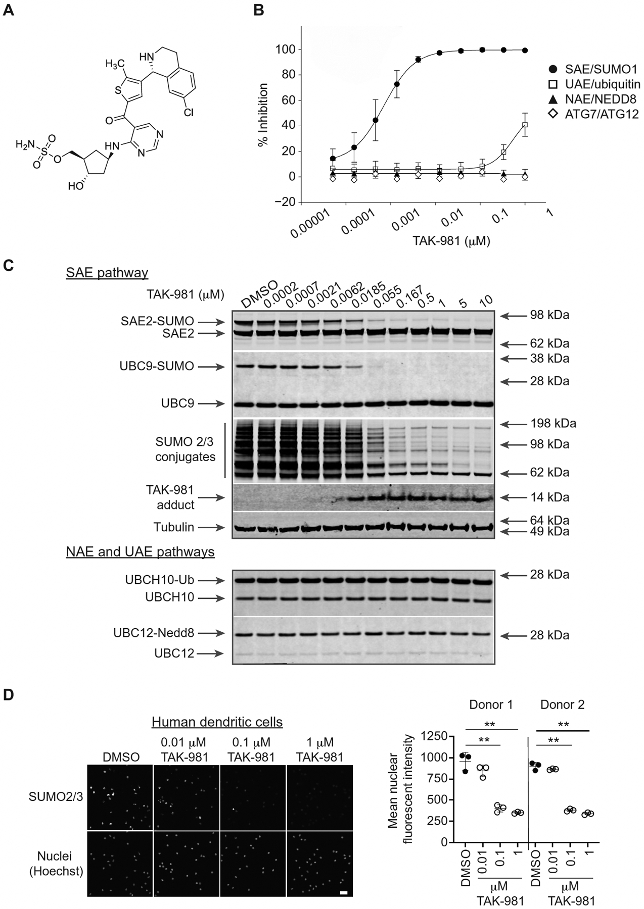
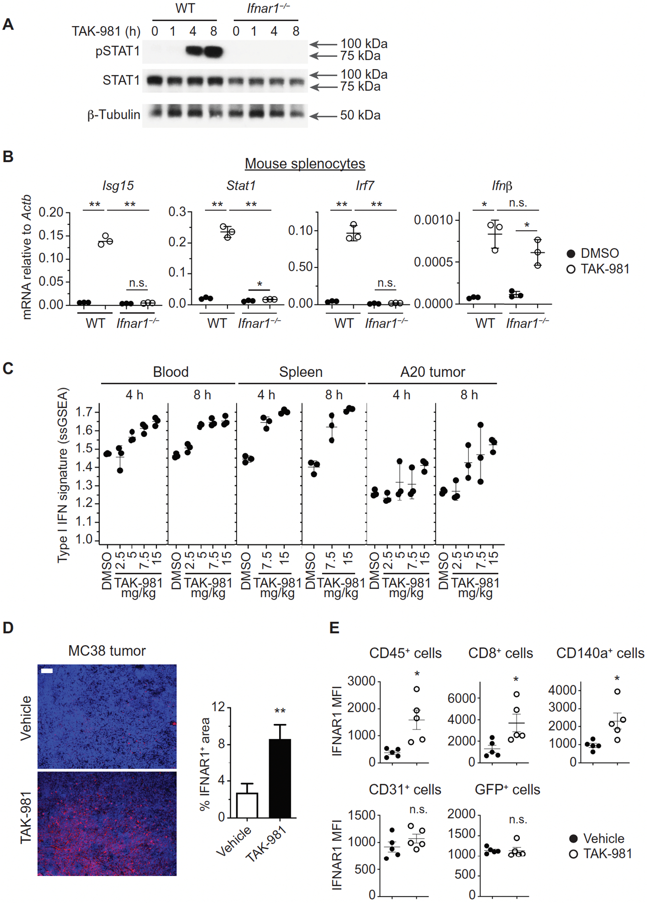
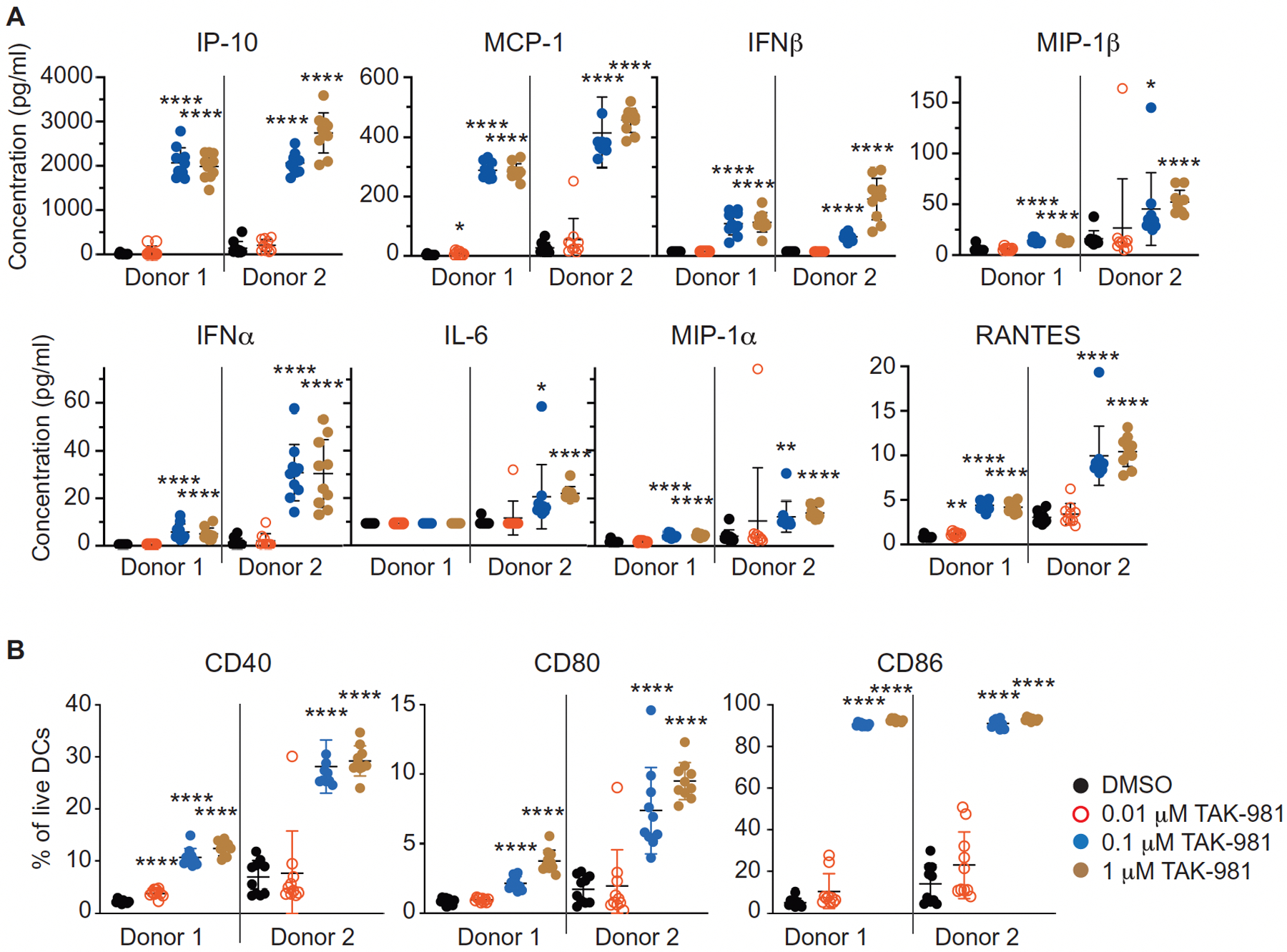
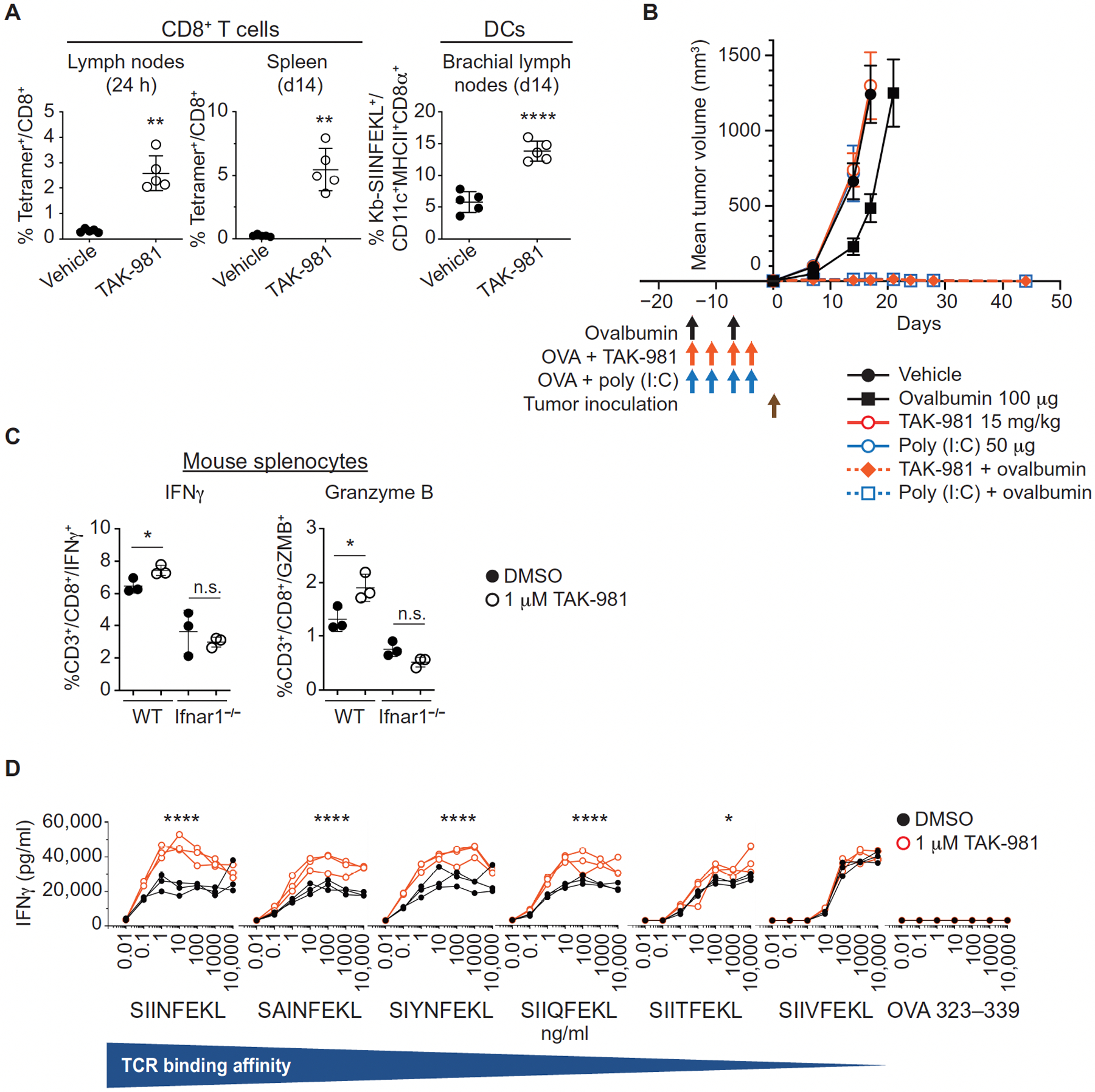
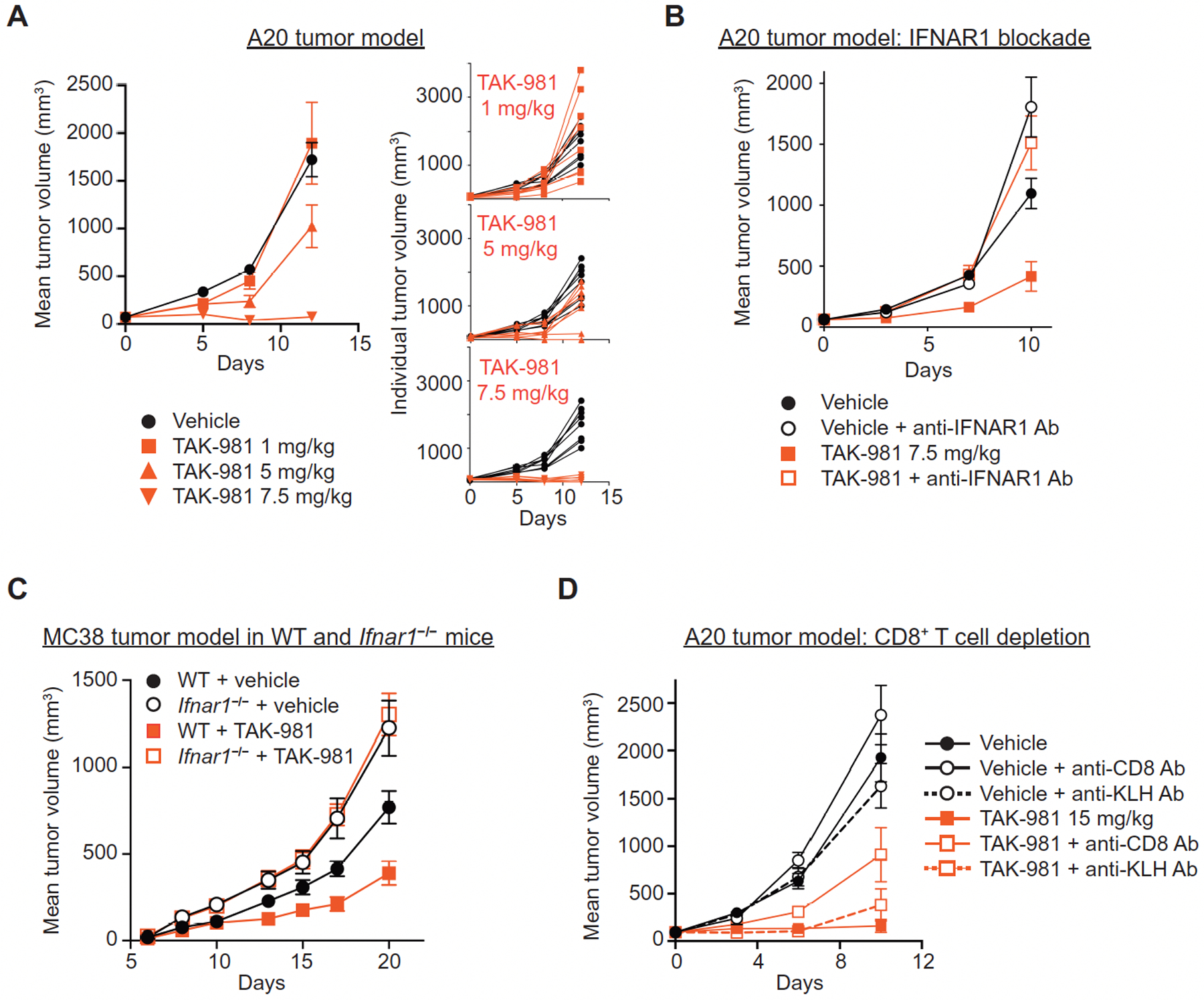
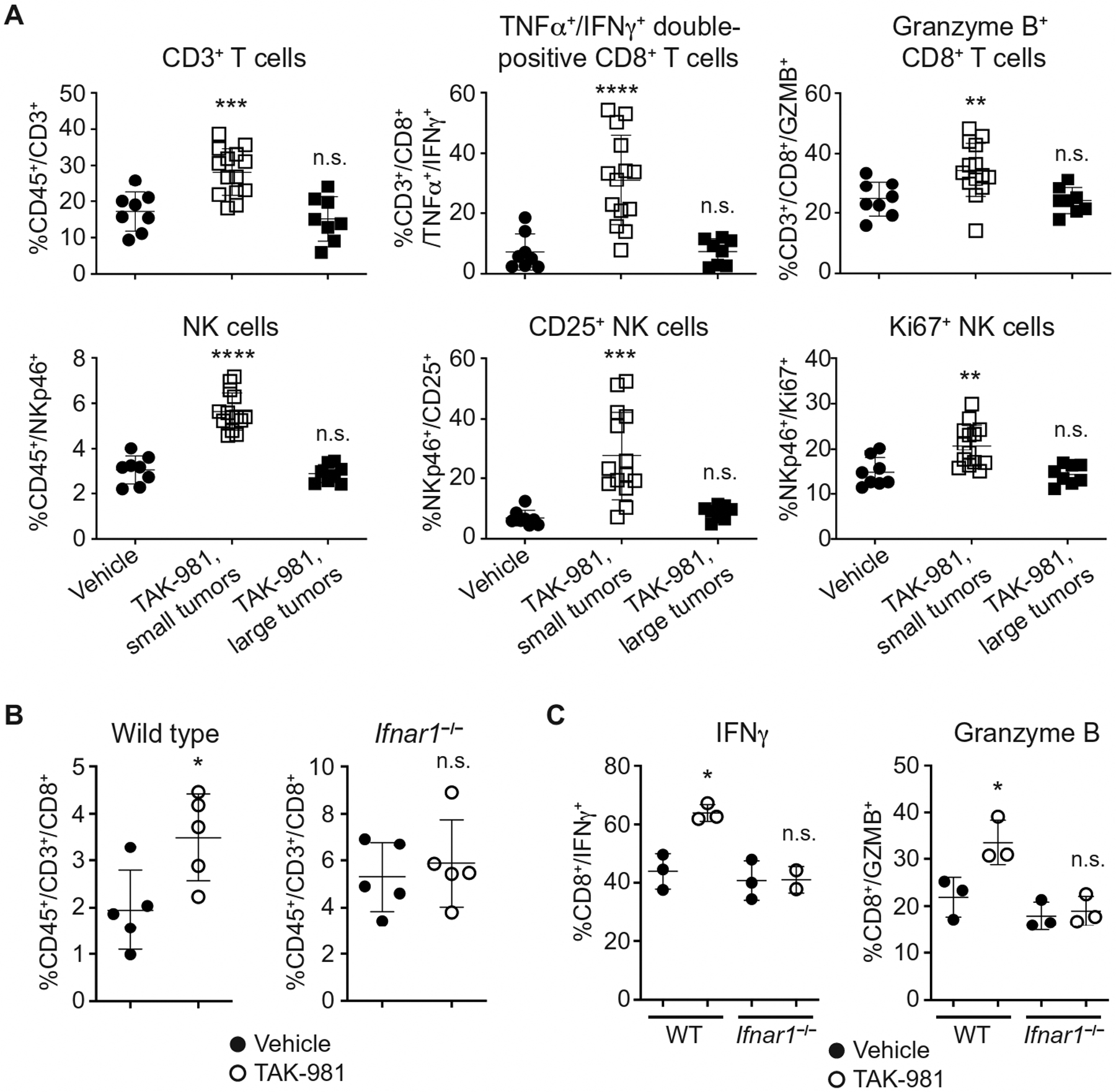
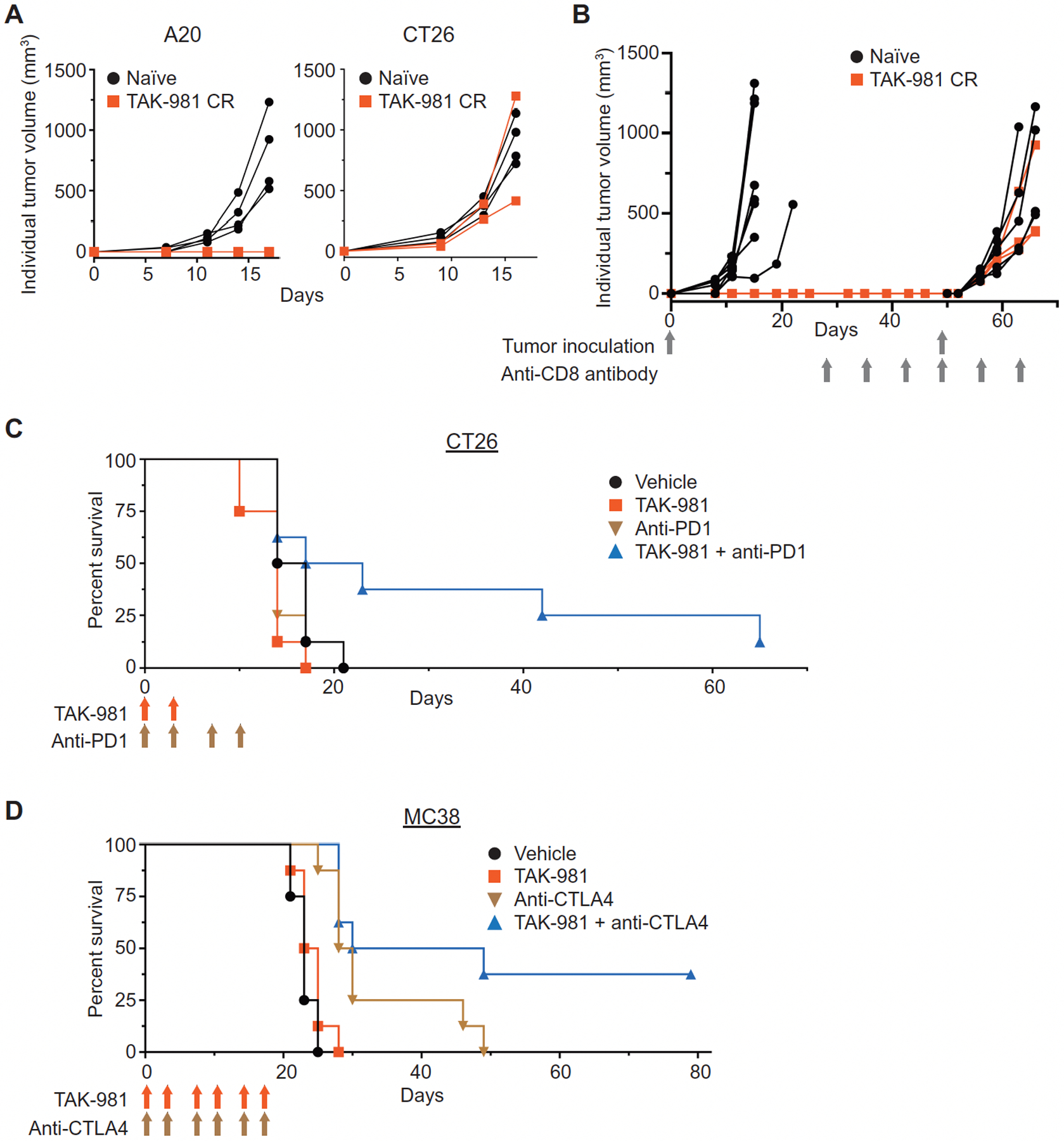
SUMOylation, the covalent conjugation of small ubiquitin-like modifier (SUMO) proteins to protein substrates, has been reported to suppress type I interferon (IFN1) responses. TAK-981, a selective small-molecule inhibitor of SUMOylation, pharmacologically reactivates IFN1 signaling and immune responses against cancers. In vivo treatment of wild-type mice with TAK-981 up-regulated IFN1 gene expression in blood cells and splenocytes. Ex vivo treatment of mouse and human dendritic cells promoted their IFN1-dependent activation, and vaccination studies in mice demonstrated stimulation of antigen cross-presentation and T cell priming in vivo. TAK-981 also directly stimulated T cell activation, driving enhanced T cell sensitivity and response to antigen ex vivo. Consistent with these observations, TAK-981 inhibited growth of syngeneic A20 and MC38 tumors in mice, dependent upon IFN1 signaling and CD8+ T cells, and associated with increased intratumoral T and natural killer cell number and activation. Combination of TAK-981 with anti-PD1 or anti-CTLA4 antibodies improved the survival of mice bearing syngeneic CT26 and MC38 tumors. In conclusion, TAK-981 is a first-in-class SUMOylation inhibitor that promotes antitumor immune responses through activation of IFN1 signaling. TAK-981 is currently being studied in phase 1 clinical trials (NCT03648372, NCT04074330, NCT04776018, and NCT04381650) for the treatment of patients with solid tumors and lymphomas.


Figures

Fig. 1.
Fig. 1.. TAK-981 is a potent and selective inhibitor of SUMOylation.
(A) Chemical structure of TAK-981, ((1R,2S,4R)-4-((5-(4-((1R)-7-chloro-1,2,3,4-tetrahydroisoquinolin-1-yl)-5-methylthiophene-2-carbonyl)pyrimidin-4-yl)amino)-2-hydroxycyclopentyl)methyl sulfamate. (B) E1–E2 homogeneous time-resolved fluorescence (HTRF) transthiolation assay to assess the activity of TAK-981 against recombinantly purified SAE, UAE, NAE and ATG7. Each of the E1 assays was tested at an ATP concentration near the Km for each enzyme. The points represent averages of nine replicate individual biochemical assays, except for ATG7 which represents a single assay. Data are shown as mean +/− SD. (C) Representative Western blot analysis (of n = 2 independent experiments) of HCT116 cells treated for 4h with increasing concentrations of TAK-981. The status of SUMO 2/3 protein conjugation to the E1 (SAE) and E2 (UBC9) enzymes, as well as the presence of SUMO 2/3 conjugates and formation of the TAK-981-SUMO adduct, was assessed. (D) Dose-dependent suppression of SUMO 2/3 conjugates in human DCs. DCs were isolated from the PBMCs of two donors, treated with TAK-981 or dimethylsulfoxide (DMSO) for 3h in triplicate, fixed and stained with anti-SUMO 2/3 antibody. Representative images are shown in which nuclei are visualized by Hoechst staining, and quantification of total nuclear SUMO 2/3 fluorescent signal intensity is shown. Scale bar, 25 μm. Individual values representing the mean signal from >200 nuclei, quantified for each triplicate, are shown, as are mean and SD. **P < 0.01, compared with DMSO control, by Welch’s t test.
Fig. 2.
Fig. 2.. Activation of IFN1 signaling by TAK-981.
(A) Western blot analysis of STAT1 phosphorylation in splenocytes derived from WT and Ifnar1−/− C57BL/6 mice treated ex vivo with 100 nM TAK-981 for the indicated times. (B) Expression of interferon stimulated genes (ISGs) in splenocytes from WT and Ifnar1−/− mice exposed to TAK-981. Q-PCR analysis was carried out on splenocytes derived from WT and Ifnar1−/− C57BL/6 mice treated ex vivo with DMSO or 100 nM TAK-981 for 12h (n = 3 for each treatment). Horizontal lines represent mean and SD. *P < 0.05, **P < 0.01, n.s. not significant, compared with DMSO control by Welch’s t test. (C) TAK-981 dependent upregulation of IFN1 mRNA signature in peripheral blood, spleen and tumor of BALB/c mice bearing A20 tumors (n = 3 for each tissue and treatment). The plot depicts IFN1 ssGSEA scores in peripheral blood at 4h (P = 0.0011) and 8h (P = 0.0001), in spleen at 4h (P = 0.0002) and 8h (P = 0.0020), and in tumor at 4h (P = 0.0095) and 8h (P = 0.0033) after treatment with DMSO or the indicated doses of TAK-981. P values were calculated by Welch’s ANOVA test. (D) Regulation of IFNAR1 in the tumor microenvironment in response to TAK-981. Immunofluorescence analysis and quantification of IFNAR1 in MC38 tumors on day 4, following administration of vehicle or 15 mg/kg TAK-981 on days 1 and 3. Scale bar, 100 μm. Quantification is represented as the mean +/− SEM of 3–4 random fields in sections from each of 3 vehicle treated mice (n = 12 fields) and TAK-981 treated mice (n = 11 fields). **P < 0.01 compared with vehicle control by Welch’s t test. (E) Flow cytometric analysis of IFNAR1 cell surface expression on immune cells (CD145+), CD8+ T cells, fibroblasts (CD140a+), endothelial cells (CD31+), and tumor cells (GFP+) from MC38-OVA/GFP tumors harvested from C57BL/6 mice on day 4, following treatment with vehicle or 15 mg/kg TAK-981 on days 1 and 3 (n = 5 for each analysis). *P < 0.05, n.s. not significant, compared with vehicle control, calculated by Welch’s t test.
Fig. 3.
Fig. 3.. TAK-981 activates dendritic cells ex vivo.
(A) Cytokines were measured in cell culture medium from human DCs, isolated from the peripheral blood of two healthy donors, following 16h of treatment with DMSO or the indicated concentrations of TAK-981 (n = 10 for each treatment). Horizontal lines represent mean and SD. *P < 0.05, **P < 0.01, ****P < 0.0001, compared with DMSO control, by Welch’s t test. (B) The percent of CD40, CD80 and CD86 positive DCs was assessed by flow cytometry of human DCs isolated from the peripheral blood of two healthy donors following 16h of ex vivo treatment with DMSO or the indicated concentrations of TAK-981 (n = 10 for each treatment). Horizontal lines represent mean and SD. ****P < 0.0001, compared with DMSO control, by Welch’s t test.
Fig. 4.
Fig. 4.. TAK-981 activates T cells ex vivo and promotes T cell priming in vivo.
(A) Lymph nodes and spleens of C57BL/6 mice subcutaneously co-injected with ovalbumin protein +/− TAK-981 or vehicle (n = 5 for each treatment) were harvested 1 day after immunization, or administered a boost immunization on day 7 and harvested on day 14. Samples were analyzed by flow cytometry for Kb-SIINFEKL tetramer positive CD8+ T cells, and CD8α+ DCs loaded with the peptide SIINFEKL on the MHC 1 molecule H-2Kb. Horizontal lines represent mean and SD. **P < 0.01, ****P < 0.0001, compared with vehicle control, by Welch’s t test. (B) C57BL6 mice were subcutaneously injected with 100 μg ovalbumin alone on day -14 and -7, or, on a day -14, -11, -7, and -4 schedule: vehicle alone, or 100 μg ovalbumin combined with either 15 mg/kg TAK-981 or 50 μg poly (I:C), as also shown by the vertical arrows in the figure. B16F10-OVA tumors were implanted on day 0 and mice were monitored for tumor growth. Data is shown as mean tumor volume +/− SEM, n=16 mice/treatment arm except n=15 for ovalbumin combined with TAK-981. Mean tumor volumes for single agent treatment arms are shown up to the last day that the entire cohort was available for tumor measurement, before mice were removed due to humane endpoints for tumor size. (C) Ex vivo activation of T cells by TAK-981. Splenocytes derived from C57BL/6 WT and Ifnar1−/− mice were treated with DMSO or 100 nM TAK-981 for 15h (n = 3 for each treatment) and then with PMA/Ionomycin/GolgiStop for an additional 6h. IFNγ and Granzyme B were detected in CD45+CD3+CD8+ CTLs by FACS. Horizontal lines represent mean and SD. *P < 0.05, n.s. not significant, compared with DMSO control, by Welch’s t test. (D) CD8+ T cells derived from OT-I mice were treated ex vivo for 4h with DMSO or 1μM TAK-981 (n = 3 for each treatment) and subsequently co-cultured with splenocytes derived from C57BL/6J mice in the presence of OVA peptides, with varying TCR affinities, for 3 days, after which IFNγ in the medium was quantitated by ELISA. *P < 0.05, ****P < 0.0001, compared with DMSO control, by two-way ANOVA test with Geisser-Greenhouse correction (TAK-981 treatment × peptide concentration).
Fig. 5.
Fig. 5.. TAK-981 promotes tumor growth inhibition dependent on IFNAR signaling and CD8+ T cells.
(A) Dose responsive tumor growth inhibition in the A20 tumor model. BALB/c mice bearing subcutaneous A20 tumors were treated intravenously (i.v.) with vehicle or the indicated concentrations of TAK-981 twice weekly for 2 weeks (days 0, 3, 7, 10). Data is shown as both mean tumor volume +/− SEM and individual tumor growth curves, n = 8 mice/treatment arm. P = 0.06 for tumor growth inhibition following treatment with 5 mg/kg TAK-981, compared to vehicle; P <0.001 for tumor growth inhibition following treatment with 7.5 mg/kg TAK-981, compared to vehicle. P values were determined by Welch’s t test. (B) BALB/c mice bearing subcutaneous flank A20 tumors were treated i.v. with vehicle or 7.5 mg/kg TAK-981 on days 0, 3 and 7, +/− anti-IFNAR1 antibody administered intraperitoneally (i.p.) 24h prior to dosing of vehicle or TAK-981. Data is shown as mean tumor volume +/− SEM, n=8 mice/treatment arm. P = 0.011 for tumor growth inhibition following treatment with TAK-981, compared to vehicle; P = 0.685 for tumor growth inhibition following treatment with vehicle + anti-IFNAR1 antibody, compared to TAK-981 + anti-IFNAR1 antibody. P values were determined by Welch’s t test. (C) MC38 tumor cells subcutaneously implanted into C57BL/6 wild type (WT) and C57BL/6 Ifnar1−/− mice were treated once weekly i.v. with vehicle or 15 mg/kg TAK-981 on days 8 and 15 after tumor implantation. Data is shown as mean tumor volume +/− SEM, n=6 mice/treatment arm for WT mice, n=7 mice/treatment arm for Ifnar1−/− mice. P = 0.025 for tumor growth inhibition following treatment with TAK-981 in WT mice, compared to vehicle; P = 0.299 for tumor growth inhibition following treatment with TAK-981 in Ifnar1−/− mice, compared to vehicle. P values were determined by Welch’s t test. (D) BALB/c mice bearing subcutaneous A20 tumors were treated i.v. with vehicle or 15 mg/kg TAK-981 on days 0, 3 and 7, +/− either anti-CD8 antibody or isotype control anti-KLH antibody administered i.p on days 0 and 7. Data is shown as mean tumor volume +/− SEM, n = 8 mice/treatment arm. P < 0.001 for tumor growth inhibition following treatment with TAK-981 compared to vehicle; P = 0.005 for tumor growth inhibition following co-administration of anti-CD8 antibody with TAK-981 compared to TAK-981 treatment alone; P = 0.237 following co-administration of control anti-KLH antibody with TAK-981 compared to TAK-981 treatment alone; P = 0.151 and 0.77, respectively, for tumor growth inhibition following co-administration of vehicle with either anti-CD8 or anti-KLH antibodies, compared to vehicle alone. P values were determined by Welch’s t test.
Fig. 6.
Fig. 6.. TAK-981 promotes T and NK cell tumor infiltration and activation.
(A) Flow cytometric analysis of subcutaneous A20 tumors from BALB/c mice treated i.v. with vehicle (n = 8) or 7.5 mg/kg TAK-981 on days 0, 3 and 7. Tumors were harvested on day 9 and TAK-981-treated tumors were divided into small (TAK-981 responsive, n = 14) and large (non-responsive, n = 8) tumors, as described in fig. S9A, for flow analysis. **P < 0.01, ***P < 0.001, ****P < 0.0001, n.s. not significant, compared with vehicle control, by Welch’s t test. (B) IFN1-dependent CD8+ T cell infiltration into TAK-981-treated MC38 tumors. Flow cytometric analysis of MC38 tumor cells subcutaneously implanted into C57BL/6 WT and Ifnar1−/− mice. Mice were treated i.v. once weekly with vehicle or 15 mg/kg TAK-981 on days 8 and 15 after tumor implantation, n = 5 mice/treatment arm. Tumors were harvested on day 21, and CD8+ cell percentage in tumor tissues was measured by flow analysis. *P < 0.05, n.s. not significant, compared with vehicle control, by Welch’s t test. (C) IFN1-dependent CD8+ T cell activation in TAK-981-treated MC38 tumors. Flow cytometric analysis of MC38 tumor cells subcutaneously implanted into C57BL/6 WT and Ifnar1−/− mice. Mice were treated i.v. with vehicle or 15 mg/kg TAK-981 on days 10 and 13 after tumor implantation. Tumors were harvested on day 14, and IFNγ and Granzyme B were measured in tumor-derived CD45+CD3+CD8+ cells by flow analysis, n = 3 mice/treatment arm except for IFNγ in TAK-981 treated Ifnar1−/− mice, where n = 2. *P < 0.05, n.s. not significant, compared with vehicle control, by Welch’s t test.
Fig. 7.
Fig. 7.. TAK-981 promotes a tumor-specific protective response and potentiates response to immune checkpoint blockade.
(A) Three A20 tumor-bearing mice that had achieved CRs in response to TAK-981 treatment, along with 4 age-matched naïve BALB/c mice, were inoculated subcutaneously with A20 cells on day 0. One of the re-challenged mice met a humane end-point (ascites) due to unknown causes and was removed from the study 11 days following re-challenge. The remaining two mice did not show any A20 tumor growth, and were subsequently subcutaneously inoculated with CT26 tumors, along with four age matched naïve BALB/c mice. (B) Three A20 tumor-bearing mice that had achieved CRs in response to TAK-981 treatment, from a different study than described in Fig. 7A, and 8 naïve age-matched BALB/c mice, were inoculated subcutaneously with A20 cells and monitored for tumor growth. No tumor growth was observed in mice that had previously achieved CRs, and these 3 mice, along with 6 naïve age matched BALB/c mice were treated i.p. with anti-CD8 antibody weekly, for 6 weeks, beginning on day 28, and inoculated subcutaneously with A20 cells on day 49, and monitored for tumor growth. (C) Survival curves for the combination of TAK-981 with anti-PD1 antibody. BALB/c mice inoculated subcutaneously with CT26 tumor cells were treated i.v. with vehicle on days 0, 3, 7 and 10, or with 7.5 mg/kg TAK-981 on day 0 and 3 (blue arrows), or with 10 mg/kg anti-mouse PD1 antibody on days 0, 3, 7 and 10 (green arrows), or with a combination of TAK-981 (day 0 and 3) and anti-PD1 antibody (days 0, 3, 7 and 10), n = 8 mice/treatment arm. P < 0.001 for survival of mice treated with both TAK-981 and anti-PD1 antibody, compared to treatment with either TAK-981 or anti-PD1 antibody as a single agent. P values were calculated by Weibull regression analysis. (D) Survival curves for the combination of TAK-981 with anti-CTLA4 antibody. C57BL/6 mice inoculated subcutaneously with MC38 tumor cells were treated with vehicle, TAK-981 (blue arrows), anti-mouse CTLA4 antibody (green arrows) or TAK-981 combined with anti-mouse CTLA4 on days 0, 3, 7, 10, 14 and 17, n = 8 mice/treatment arm. P < 0.001 for survival of mice treated with both TAK-981 and anti-CTLA4 antibody, compared to treatment with TAK-981 alone; P = 0.0019 for survival of mice treated with both TAK-981 and anti-CTLA4 antibody, compared to treatment with anti-CTLA4 antibody alone. P values were calculated by Weibull regression analysis.

References

    1. Hargadon KM, Johnson CE, Williams CJ, Immune checkpoint blockade therapy for cancer: An overview of FDA-approved immune checkpoint inhibitors. Int. Immunopharmacol 62, 29–39 (2018). - PubMed
    1. Galon J, Bruni D, Approaches to treat immune hot, altered and cold tumours with combination immunotherapies. Nat. Rev. Drug Discov 18, 197–218 (2019). - PubMed
    1. Lee C-K, Rao DT, Gertner R, Gimeno R, Frey AB, Levy DE, Distinct requirements for IFNs and STAT1 in NK cell function. J. Immunol 165, 3571–3577 (2000). - PubMed
    1. Lucas M, Schachterle W, Oberle K, Aichele P, Diefenbach A, Dendritic cells prime natural killer cells by trans-presenting interleukin 15. Immunity 26, 503–517 (2007). - PMC - PubMed
    1. Marrack P, Kappler J, Mitchell T, Type I interferons keep activated T cells alive. J. Exp. Med 189, 521–530 (1999). - PMC - PubMed

Publication types