. 2021 Nov 10;87(23):e0167421.
doi: 10.1128/AEM.01674-21. Epub 2021 Sep 15.

Dynamics of the Two Stator Systems in the Flagellar Motor of Pseudomonas aeruginosa Studied by a Bead Assay

Affiliations

Dynamics of the Two Stator Systems in the Flagellar Motor of Pseudomonas aeruginosa Studied by a Bead Assay

Zhengyu Wu et al. Appl Environ Microbiol. .

Abstract

We developed a robust bead assay for studying flagellar motor behavior of Pseudomonas aeruginosa. Using this assay, we studied the dynamics of the two stator systems in the flagellar motor. We found that the two sets of stators function differently, with MotAB stators providing higher total torque and MotCD stators ensuring more stable motor speed. The motors in wild-type cells adjust the stator compositions according to the environment, resulting in an optimal performance in environmental exploration compared to that of mutants with one set of stators. The bead assay we developed in this investigation can be further used to study P. aeruginosa chemotaxis at the level of a single cell using the motor behavior as the chemotaxis output. IMPORTANCE Cells of Pseudomonas aeruginosa possess a single polar flagellum, driven by a rotatory motor powered by two sets of torque-generating units (stators). We developed a robust bead assay for studying the behavior of the flagellar motor in P. aeruginosa, by attaching a microsphere to shortened flagellar filament and using it as an indicator of motor rotation. Using this assay, we revealed the dynamics of the two stator systems in the flagellar motor and found that the motors in wild-type cells adjust the stator compositions according to the environment, resulting in an optimal performance in environmental exploration compared to that of mutants with one set of stators.

Keywords: bead assay; flagellar motor; stator.


Figures

FIG 1
FIG 1
(A) A typical reversal event of the wild-type strain near the surface, with image sequences taken from Movie S1. The bottom right corner is a schematic diagram of the complete cell trajectory. The cell body is indicated by a red dashed box, and the white arrows indicate the instantaneous velocity direction of the cell. (B and C) Diagrams of swimming behavior near the surface (B, top view; C, tail view). In panel B, the dashed lines with arrow indicate the direction of cell motion. “F1” and “F2” indicate the net viscous forces acting on the cell body and flagellum, respectively. In panel C, “Fbottom” and “Ftop” represent the viscous force on the portions of the helical filament closer to and away from the surface, respectively. The upper and lower portions in panel C represent the situations when the flagellum push and pull the cell body, respectively. “CW” and “CCW” represent the rotational direction of the filament. (D) The probability distribution of angular change associated with a reversal. The upper graph counts the events changing from pushing to pulling, and the lower graph depicts the opposite situation. Abbreviation: PDF, probability density function.
FIG 2
FIG 2
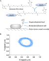
(A) Schematic diagram of bead assay. Flagellin containing a cysteine point mutation was biotinylated by using sulfhydryl-maleimide conjugation. Bacterial cells were immobilized onto a poly-l-lysine coated coverslip, and a streptavidin-coated bead was attached to the biotinylated filament stub. (B) A two-dimensional XY signal plot, showing the trajectory of the bead image.
FIG 3
FIG 3
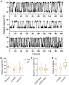
(A) Typical time traces of rotation velocities for the wild-type strain and the two stator mutants (from top to bottom are the wild-type, ΔmotAB, and ΔmotCD strains). A hyphen before “50” indicates CCW direction. (B) CW and CCW rotation speeds of the wild-type, ΔmotAB, and ΔmotCD strains. The speed of the ΔmotAB strain is nearly 40% lower than that of the wild type, and the ΔmotCD strain shows a speed comparable to that of the wild type. (C) The speed increases per stator for MotAB and MotCD are almost the same. (D) The number of stators in the ΔmotCD strain is more than in the ΔmotAB strain. In the box-whisker plot, the box represents the middle 50% of the data. The median value is shown as a line in the box, and the whiskers denote the data range of the 5th and 95th percentiles.
FIG 4
FIG 4
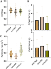
(A) CW biases of the wild-type, ΔmotAB, and ΔmotCD strains are all close to 0.5. (B) CW and CCW intervals of the wild-type, ΔmotAB, and ΔmotCD strains. Compared to the case with the wild-type strains, both the CW and CCW intervals lengthened for the ΔmotAB strain, whereas they shortened for the ΔmotCD strain. (C) Switching rates of the wild-type, ΔmotAB, and ΔmotCD strains.
FIG 5
FIG 5
Typical traces of motor speed in a steady state for motors of the ΔmotAB (upper) and ΔmotCD (lower) strains. The blue lines are the average speeds found by the step-finding algorithm.
FIG 6
FIG 6
Behavior of the flagellar motor for the wild-type, ΔmotAB, and ΔmotCD strains under different load conditions. (A) The switching rates of the three strains under different load conditions. (B and C) The CCW (B) and CW (C) rotation speeds of the three strains under different load conditions. (Inset) Viscosities of the motility buffer with different Ficoll concentrations at room temperature.
FIG 7
FIG 7
(A) The expansion of the three strains on a swimming plate. (Upper portion) Scatterplot of the expansion radius for the wild-type, ΔmotAB, and ΔmotCD strains. (Lower portion) Photo of a swimming plate with three strains. The plate was photographed at 18 h after inoculation. (B) The relationship between mean squared displacement (MSD) and the time lag of the wild-type, ΔmotAB, and ΔmotCD strains (upper portion, linear coordinates; lower portion, double logarithmic coordinates), all exhibiting superdiffusive behavior, while the wild type is most diffusive.

References

    1. Jarrell KF, McBride MJ. 2008. The surprisingly diverse ways that prokaryotes move. Nat Rev Microbiol 6:466–476. 10.1038/nrmicro1900. - DOI - PubMed
    1. Kearns DB. 2010. A field guide to bacterial swarming motility. Nat Rev Microbiol 8:634–644. 10.1038/nrmicro2405. - DOI - PMC - PubMed
    1. Macnab RM. 2003. How bacteria assemble flagella. Annu Rev Microbiol 57:77–100. 10.1146/annurev.micro.57.030502.090832. - DOI - PubMed
    1. Berg HC. 2003. The rotary motor of bacterial flagella. Annu Rev Biochem 72:19–54. 10.1146/annurev.biochem.72.121801.161737. - DOI - PubMed
    1. Minamino T, Imada K, Namba K. 2008. Molecular motors of the bacterial flagella. Curr Opin Struct Biol 18:693–701. 10.1016/j.sbi.2008.09.006. - DOI - PubMed

Publication types

MeSH terms

LinkOut - more resources