Review
. 2021 Sep 14;54(9):1933-1947.
doi: 10.1016/j.immuni.2021.08.023.

The neuroimmune response during stress: A physiological perspective

Affiliations
Review

The neuroimmune response during stress: A physiological perspective

Hedva Haykin et al. Immunity. .

Abstract

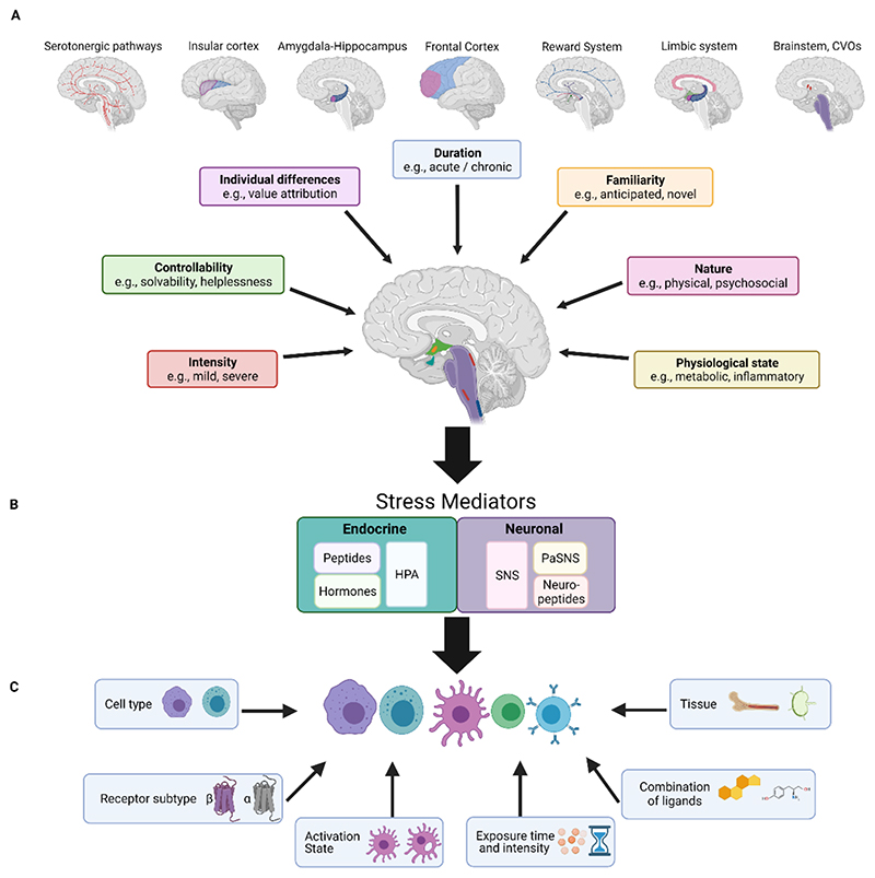
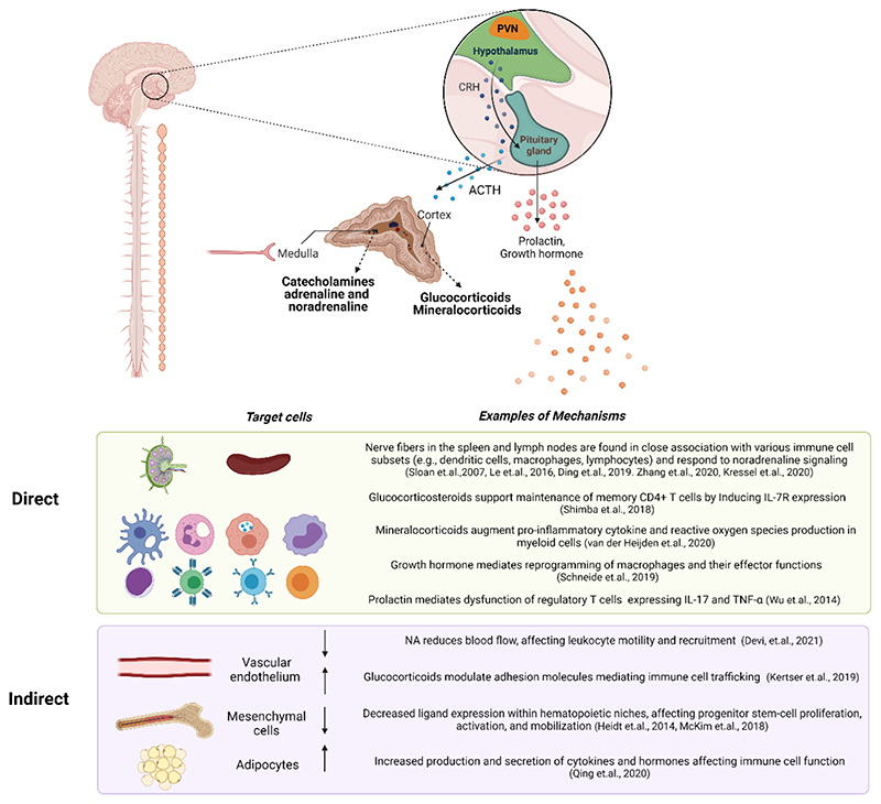
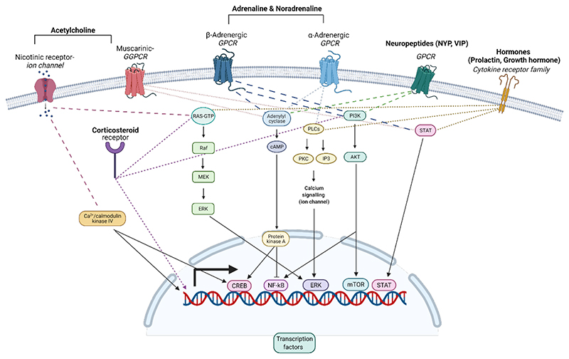
Stress is an essential adaptive response that enables the organism to cope with challenges and restore homeostasis. Different stressors require distinctive corrective responses in which immune cells play a critical role. Hence, effects of stress on immunity may vary accordingly. Indeed, epidemiologically, stress can induce either inflammation or immune suppression in an organism. However, in the absence of a conceptual framework, these effects appear chaotic, leading to confusion. Here, we examine how stressor diversity is imbedded in the neuroimmune axis. Stressors differ in the brain patterns they induce, diversifying the neuronal and endocrine mediators dispatched to the periphery and generating a wide range of potential immune effects. Uncovering this complexity and diversity of the immune response to different stressors will allow us to understand the involvement of stress in pathological conditions, identify ways to modulate it, and even harness the therapeutic potential embedded in an adaptive response to stress.


Conflict of interest statement

Declaration of interests The authors declare no competing interests.

Figures

Figure 1
Figure 1. An overview of the variables influencing the effects of stress on the immune system.
The stress response can be regulated at three levels by numerous variables, all of which ultimately alter the immune response and the immunophenotype produced. A. The characteristics of the stressful stimulus. Key stress characteristics are shown in boxes, along with their associated brain regions. Some brain regions are involved in various forms of stress, such as the reward system, which is attuned to predictability, duration, and controllability and the frontal cortex, which is receptive to predictability in addition to the controllability and duration, shown here. The brainstem, CVOs, and limbic system are sensitive to the nature of the stressful stimulus and to the physiological state of the organism. The overall activity of the brain effectively builds a specific neuromatrix for each stressor. The information reaching the brain is integrated at two main regions – the hypothalamic PVN nucleus and the brainstem nuclei (e.g., LC, NST, RVLM). These regions produce both endocrine and neuronal outputs. B. The neuronal or endocrine mediators. Two main stress-mediating systems, the endocrine (turquoise) and the neuronal (purple) responses, are represented in boxes. Listed within the boxes are the major components com rising each system, all of which participate in the mediation of stress, based on the input received from the brain. C. Characteristics of the responding immune populations. Factors that influence immune-cell responsiveness are noted in boxes. The combined effects of these factors will determine the resultant stress-induced immune response. CVOs: circumventricular organs; HPA: hypothalamus- pituitary- adrenal axis; SNS: sympathetic nervous system; PaSNS: parasympathetic nervous system; LC: Locus coeruleus; NST: nucleus of the solitary tract; RVLM: rostral ventrolateral medulla.
Figure 2
Figure 2. Examples of direct and indirect effects of stress on the immune system.
Endocrine and neuronal signals can affect immune-system function, by acting either directly on immune cells or indirectly via the surrounding tissue. The autonomic nervous system innervates the adrenal medulla, inducing it to secrete catecholamines into the bloodstream. Within the lymph nodes and spleen, various immune subsets remain in close association with sympathetic fibers. Signals received by immune cells from nerve fibers in these compartments may affect immune-cell gene expression, maturation, migration, proliferation, differentiation, activation, and other functions. The indirect effects of stress on immune cells are mediated by the cells comprising the various organs. For example, stress modulates the expression of adhesion molecules on endothelial cells, thereby affecting immune cell trafficking. Stress also reduces blood flow and interrupts the locomotion of leukocytes into tissues via calcium signaling. Within hematopoietic niches, stromal cells affect immune cell maturation and mobilization through the expression of specific ligands. Upon exposure to stress, adipocytes secrete IL-6 which affects immune cell activity and recruitment. PVN: paraventricular nucleus; CRH: corticotrophin hormone; ACTH: adrenocorticotropin hormone.
Figure 3
Figure 3. Intracellular immune cell-signaling by stress-induced mediators.
Representative examples of receptors and their corresponding pathways that can be activated by stress-induced mediators. Immune cells express nicotinic and muscarinic receptors for acetylcholine, α- and β-adrenergic receptors for both adrenaline and noradrenaline, and members of the cytokine receptor family for hormones. Receptors for neuropeptides include mainly GPCR (e.g NPY, VIP), although exceptions exist, and the receptor for substance P, for example, is a member of the cytokine receptor family. Downstream signaling of these receptors regulates gene expression via transcription factors such as NF-κB, CREB, ERK, STAT, mTOR etc. Most of the receptors are expressed on the cell membrane, while corticosteroid receptors reside mostly within the cytosol. GPCR: G-protein-coupled receptor; cAMP: cyclic adenosine monophosphate; PLC: Phospholipase C; PKC: Protein Kinase C; IP3: Inositol trisphosphate; PI3K: Phosphoinositide 3-kinases; Akt: protein kinase B; NF-κB: nuclear factor kappa-light-chain-enhancer of activated B cell; CREB: cAMP response element-binding protein; ERK: extracellular signal-regulated kinases; STAT: signal transducer and activator of transcription; mTOR: mechanistic target of rapamycin.

References

    1. Abe C, Inoue T, Inglis MA, Viar KE, Huang L, Ye H, Rosin DL, Stornetta RL, Okusa MD, Guyenet PG. C1 neurons mediate a stress-induced anti-inflammatory reflex in mice. Nat Neurosci. 2017;20:700–707. - PMC - PubMed
    1. Agac D, Estrada LD, Maples R, Hooper LV, Farrar JD. The beta2-adrenergic receptor controls inflammation by driving rapid IL-10 secretion. Brain Behav Immun. 2018;74:176–185. - PMC - PubMed
    1. Akil H, Shiomi H, Matthews J. Induction of the intermediate pituitary by stress: synthesis and release of a nonopioid form of beta-endorphin. Science. 1985;227:424–426. - PubMed
    1. Amat J, Baratta MV, Paul E, Bland ST, Watkins LR, Maier SF. Medial prefrontal cortex determines how stressor controllability affects behavior and dorsal raphe nucleus. Nat Neurosci. 2005;8:365–371. - PubMed
    1. Babb JA, Masini CV, Day HE, Campeau S. Sex differences in activated corticotropin-releasing factor neurons within stress-related neurocircuitry and hypothalamic-pituitary-adrenocortical axis hormones following restraint in rats. Neuroscience. 2013;234:40–52. - PMC - PubMed

Publication types

MeSH terms