. 2021 Sep 21;118(38):e2020385118.
doi: 10.1073/pnas.2020385118.

Annexin A1 attenuates cardiac diastolic dysfunction in mice with inflammatory arthritis

Affiliations

Annexin A1 attenuates cardiac diastolic dysfunction in mice with inflammatory arthritis

Jianmin Chen et al. Proc Natl Acad Sci U S A. .

Abstract

Rheumatoid arthritis (RA) carries a twofold increased incidence of heart failure with preserved ejection fraction, accompanied by diastolic dysfunction, which can lead to death. The causes of diastolic dysfunction are unknown, and there are currently no well-characterized animal models for studying these mechanisms. Current medications for RA do not have marked beneficial cardio-protective effects. K/BxN F1 progeny and KRN control mice were analyzed over time for arthritis development, monitoring left ventricular diastolic and systolic function using echocardiography. Excised hearts were analyzed by flow cytometry, qPCR, and histology. In pharmacological experiments, K/BxN F1 mice were treated with human recombinant AnxA1 (hrAnxA1, 1 μg/mouse) or vehicle daily. K/BxN F1 mice exhibited fully developed arthritis with normal cardiac function at 4 wk; however, by week 8, all mice displayed left ventricular diastolic dysfunction with preserved ejection fraction. This dysfunction was associated with cardiac hypertrophy, myocardial inflammation and fibrosis, and inflammatory markers. Daily treatment of K/BxN F1 mice with hrAnxA1 from weeks 4 to 8 halted progression of the diastolic dysfunction. The treatment reduced cardiac transcripts of proinflammatory cytokines and profibrotic markers. At the cellular level, hrAnxA1 decreased activated T cells and increased MHC IIlow macrophage infiltration in K/BxN F1 hearts. Similar effects were obtained when hrAnxA1 was administered from week 8 to week 15. We describe an animal model of inflammatory arthritis that recapitulates the cardiomyopathy of RA. Treatment with hrAnxA1 after disease onset corrected the diastolic dysfunction through modulation of both fibroblast and inflammatory cell phenotype within the heart.

Keywords: HFpEF; annexin A1; arthritis; cardiomyopathy; diastolic dysfunction.


Conflict of interest statement

Competing interest statement: M.P. is on the Scientific Advisory Board of ResoTher Pharma AS, which is interested in the development of AnxA1-derived peptides for cardiovascular settings. M.P. consults for Bristol Myers Squibb.

Figures
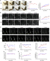
Fig. 1.
Fig. 1.
Arthritic K/BxN F1 mice display diastolic dysfunction but normal systolic function as assessed by echocardiography. (A) K/BxN F1 mice develop marked arthritis from week 4 with no significant change in body weight. Clinical severity of arthritis was assessed visually using a defined severity scoring system (max score 12). (B and C) K/BxN F1 mice fully develop diastolic dysfunction from week 8. (B) Representative B-mode four-chamber echocardiograms and increased LA area in arthritic mice. (C) Representative mitral flow patterns from pulsed-wave color Doppler echocardiography, decreased E/A ratio, decreased E wave velocity, and increased deceleration time in arthritic mice. (D) K/BxN F1 mice show no significant change in percentages of EF using M-mode echocardiograms. (E) K/BxN F1 mice develop concentric cardiac hypertrophy at week-15 indicated by an increase in interventricular septum thickness with no difference in LV internal diastolic dimensions. Data are mean ± SEM. KRN group: n = 5; K/BxN F1 group: n = 6. *P < 0.05, **P < 0.01, ***P < 0.001, ****P < 0.0001 versus nonarthritic KRN group (two-way ANOVA followed by Bonferroni’s test). K/BxN: K/BxN F1.
Fig. 2.
Fig. 2.
Arthritic K/BxN F1 mice develop cardiac hypertrophy and fibrosis. (A) Histological and structural analyses in arthritic hearts (week 15). A, i: Macroscopic view of the arthritic mouse heart stained with hematoxylin–eosin. (Scale bars, 2.5 mm.) A, ii: Representative LV sections stained with hematoxylin–eosin. A, iii: Sirius red from arthritic and nonarthritic hearts. (Scale bars, 100 μm.) KRN: n = 3; K/BxN F1: n = 6. (B and C) Quantification of galectin-3–positive cells in LV. Representative images are shown. (Scale bars, 100 μm.) n = 3 to 4 per group. (D) Quantification of fibroblast populations with representative dot plots and (E) VCAM-1 expression for KRN n = 4 and K/BxN F1 n = 5 at week 8. Data are mean ± SEM (A, D, and E) *P < 0.05, **P < 0.01, ***P < 0.001 versus nonarthritic KRN group (unpaired Student’s t test); (C) *P < 0.05, **P < 0.01 versus nonarthritic 15-wk-old KRN or 6-wk-old K/BxN F1 group (one-way ANOVA followed by Bonferroni’s test). K/BxN: K/BxN F1.
Fig. 3.
Fig. 3.
Modulated gene signature and increased infiltration of activated T cells in K/BxN F1 arthritic hearts at 8 wk. (A) Ct values of different genes analyzed by qPCR in K/BxN F1 arthritic hearts at week 8. (B) Differential gene expression in arthritic hearts at week 8. Ct values were normalized using Hprt1 as housekeeping gene, and fold change was calculated relative to age-matched KRN controls. x-axis shows fold change (in log2 scale), with continued line (X = 1) showing KRN mean and dotted lines presenting technical variance: ± average SD of replicated PCR measures. y-axis shows the P value (in log10 scale) from t test statistical analysis, with dotted line at P = 0.05; KRN group: n = 4; K/BxN group: n = 6. (C and D) CD4+ (C) or CD8+ (D) T cells were subdivided into non-proinflammatory CD44CD62L+ naïve T cells (Tn), CD44+CD62L effector memory T cells (Tem), CD44+CD62L+ central memory (Tcm), and CD44CD62L transition status (Ttrans). Data are mean values. KRN group: n = 4; K/BxN F1 group: n = 5. ***P < 0.001 versus nonarthritic KRN group (unpaired Student’s t test). K/BxN: K/BxN F1.
Fig. 4.
Fig. 4.
hrAnxA1 treatment (week 4 to week 8) halts the development of diastolic dysfunction and attenuates cardiac hypertrophy in K/BxN F1 mice. (A) Schematic indicating treatment and experimental regime. (B) Representative B-mode four-chamber echocardiograms indicating LA area and LA atrial area in mice treated with hrAnxA1 or vehicle. (C) Representative mitral flow patterns from pulsed-wave color Doppler echocardiography. (D) E/A ratio and deceleration time as assessed by echocardiography in arthritic mice treated with hrAnxA1 or vehicle. Data are mean ± SEM. K/BxN F1 + vehicle group: n = 5; K/BxN F1+ hrAnxA1 group: n = 5. *P < 0.05, **P < 0.01 versus vehicle group (two-way ANOVA followed by Bonferroni’s test). Data are mean ± SEM. K/BxN F1+ vehicle group: n = 4; K/BxN F1 + hrAnxA1 group: n = 5. *P < 0.05 versus vehicle group (unpaired Student’s t test). Veh: vehicle; hAnxA1: hrAnxA1. Echocardiography, flow cytometry, and qPCR were all analyzed blindly.
Fig. 5.
Fig. 5.
hrAnxA1 treatment (week 4 to week 8) reduces fibroblast populations and activated T cell infiltration and increases MHC IIlow macrophages in the hearts of K/BxN F1 mice. K/BxN F1 mice were treated with hrAnxA1 [1 µg/mouse daily subcutaneously (s.c.) from week 4 to week 8]. Hearts were enzymatically digested, and cardiac cell phenotype assessed and quantified by flow cytometry. (A) Representative dot plots showing cardiac monocytic fibroblasts (CD45+CD34+Thy1.2+) and cardiac structural fibroblasts (CD45CD34Thy1.2+) in mice receiving vehicle or hrAnxA1. Cumulative data indicating percentage cardiac monocytic fibroblasts and cardiac structural fibroblasts in mice treated with vehicle or hrAnxA1. (B) VCAM-1 expression on cardiac fibroblast subtypes. (C and D) Representative dot plots and cumulative data illustrating CD4 (C) and CD8 (D) noninflammatory CD44CD62L+ naïve T cells (Tn), CD44+CD62L effector memory T cells (Tem), CD44+CD62L+ central memory T cells (Tcm), and CD44CD62L transition status T cells (Ttrans). (E) Representative dot plots illustrating F/80+MHC IIlow and F/80+MHC IIhigh macrophages K/BxN F1 hearts receiving vehicle or hrAnxA1. Cumulative data indicating total numbers of F/80+MHC IIlow and F/80+MHC IIhigh macrophages. n = 4 to 5 per group. All data are mean ± SEM *P < 0.05, ***P < 0.001 versus K/BxN F1+ vehicle group (unpaired Student’s t test). Veh: vehicle; hAnxA1: hrAnxA1.
Fig. 6.
Fig. 6.
Impact of hrAnxA1 treatment (week 4 to week 8) on cardiac gene expression in K/BxN F1 mice. mRNA expression of (A) Proinflammatory cytokines: Il6 and Il1b; (B) Cardiac fibrosis markers: Tgfb, Col1a1, Acta, and Mmp1a; (C) AnxA1 receptor: Fpr2; (D) Melanocortin receptor 1: Mc1r; (E) 15-lipoxygenase: Alox15; and (F) Galectin-1: Lgals1; Galectin-3: Lgals3; TIM-3 (receptor of Galectin-9): Havcr2. Ct values were normalized using Hprt1 as housekeeping gene. Data are mean ± SEM. KRN group: n = 4; K/BxN F1 + vehicle group: n = 6; K/BxN F1 + hrAnxA1 group: n = 6. Fold change was calculated relative to age-matched nonarthritic KRN controls (mean value = 1), with dotted line showing technical variance indicated by ± average SD of replicated PCR measures for each gene. *P < 0.05, **P < 0.01, ***P < 0.001 versus vehicle group (unpaired Student’s t test).
Fig. 7.
Fig. 7.
hrAnxA1 treatment (week 8 to week 15) reverses the established diastolic dysfunction and attenuates cardiac remodeling in K/BxN F1 mice. K/BxN F1 mice were treated with either hrAnxA1 (1 μg/mouse, daily subcutaneous injection from week 8 to week 15) or vehicle (100 μL saline). Echocardiography was performed in K/BxN F1 at 8 wk (baseline) before hrAnxA1 or saline administration and at 10, 13, and 15 wk. (A) Representative B-mode four-chamber echocardiograms indicating LA area and cumulative data of LA area in mice treated with hrAnxA1 or vehicle. (B) Representative mitral flow patterns from pulsed-wave color Doppler echocardiography. (C) E/A ratio and deceleration time as assessed by echocardiography in arthritic mice treated with hrAnxA1 or vehicle. (D) hrAnxA1 reduced interventricular septum thickness in arthritic mice at 15 wk. Data are mean ± SEM. K/BxN F1 + vehicle group: n = 6; K/BxN F1+ hrAnxA1 group: n = 6. *P < 0.05, ***P < 0.001 versus vehicle group (two-way ANOVA followed by Bonferroni’s test). (E) Representative LV sections stained with hematoxylin–eosin and Sirius red from arthritic hearts received hrAnxA1 or vehicle and respective cumulative data. (Scale bars, 100 μm.) (F) Quantification of galectin-3–positive cells in LV. Representative images are also shown. (Scale bars, 100 μm.) (G) Lung wet-to-dry weight ratio. *P < 0.05, **P < 0.01, and ***P < 0.001 versus vehicle group (unpaired Student’s t test). Veh: vehicle; hAnxA1: hrAnxA1.
Fig. 8.
Fig. 8.
hrAnxA1 treatment (week 8 to week 15) reduces fibroblast populations and activated T cell infiltration in the hearts of K/BxN F1 mice. K/BxN F1 mice were treated with either hrAnxA1 (1 μg/mouse, daily subcutaneous injection from week 8 to week 15) or vehicle (100 μL saline). Hearts were enzymatically digested, and cardiac cell phenotype assessed and quantified by flow cytometry. (A) Representative dot plots showing cardiac monocytic fibroblasts (CD45+CD34+Thy1.2+) and cardiac structural fibroblasts (CD45CD34Thy1.2+) in mice receiving vehicle or hrAnxA1. Cumulative data indicating percentage cardiac monocytic fibroblasts and cardiac structural fibroblasts in mice treated with vehicle or hrAnxA1. (B) VCAM-1 expression on cardiac fibroblast subtypes. (C and D) Representative dot plots and cumulative data illustrating CD4 (C) and CD8 (D) noninflammatory CD44CD62L+ naïve T cells (Tn), CD44+CD62L effector memory T cells (Tem), CD44+CD62L+ central memory T cells (Tcm), and CD44CD62L transition status T cells (Ttrans). n = 6 per group. All data are mean ± SEM. *P < 0.05, **P < 0.01 versus K/BxN F1+ vehicle group (unpaired Student’s t test). Veh: vehicle; hAnxA1: hrAnxA1.

References

    1. Aviña-Zubieta J. A., et al. ., Risk of cardiovascular mortality in patients with rheumatoid arthritis: A meta-analysis of observational studies. Arthritis Rheum. 59, 1690–1697 (2008). - PubMed
    1. Lazúrová I., Tomáš Ľ., Cardiac impairment in rheumatoid arthritis and influence of anti-TNFα treatment. Clin. Rev. Allergy Immunol. 52, 323–332 (2017). - PubMed
    1. Nicola P. J., et al. ., Contribution of congestive heart failure and ischemic heart disease to excess mortality in rheumatoid arthritis. Arthritis Rheum. 54, 60–67 (2006). - PubMed
    1. Mantel Ä., Holmqvist M., Andersson D. C., Lund L. H., Askling J., Association between rheumatoid arthritis and risk of ischemic and nonischemic heart failure. J. Am. Coll. Cardiol. 69, 1275–1285 (2017). - PubMed
    1. Błyszczuk P., Szekanecz Z., Pathogenesis of ischaemic and non-ischaemic heart diseases in rheumatoid arthritis. RMD Open 6, e001032 (2020). - PMC - PubMed

Publication types

MeSH terms