Review
. 2021 Sep 15;41(37):7698-7711.
doi: 10.1523/JNEUROSCI.0619-21.2021.

The Glymphatic System: A Novel Component of Fundamental Neurobiology

Affiliations
Review

The Glymphatic System: A Novel Component of Fundamental Neurobiology

Lauren M Hablitz et al. J Neurosci. .

Abstract

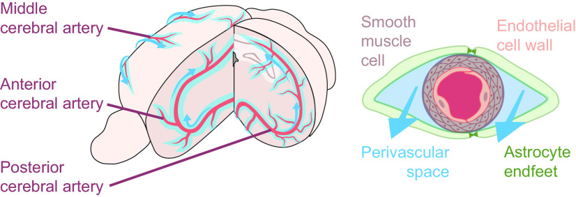
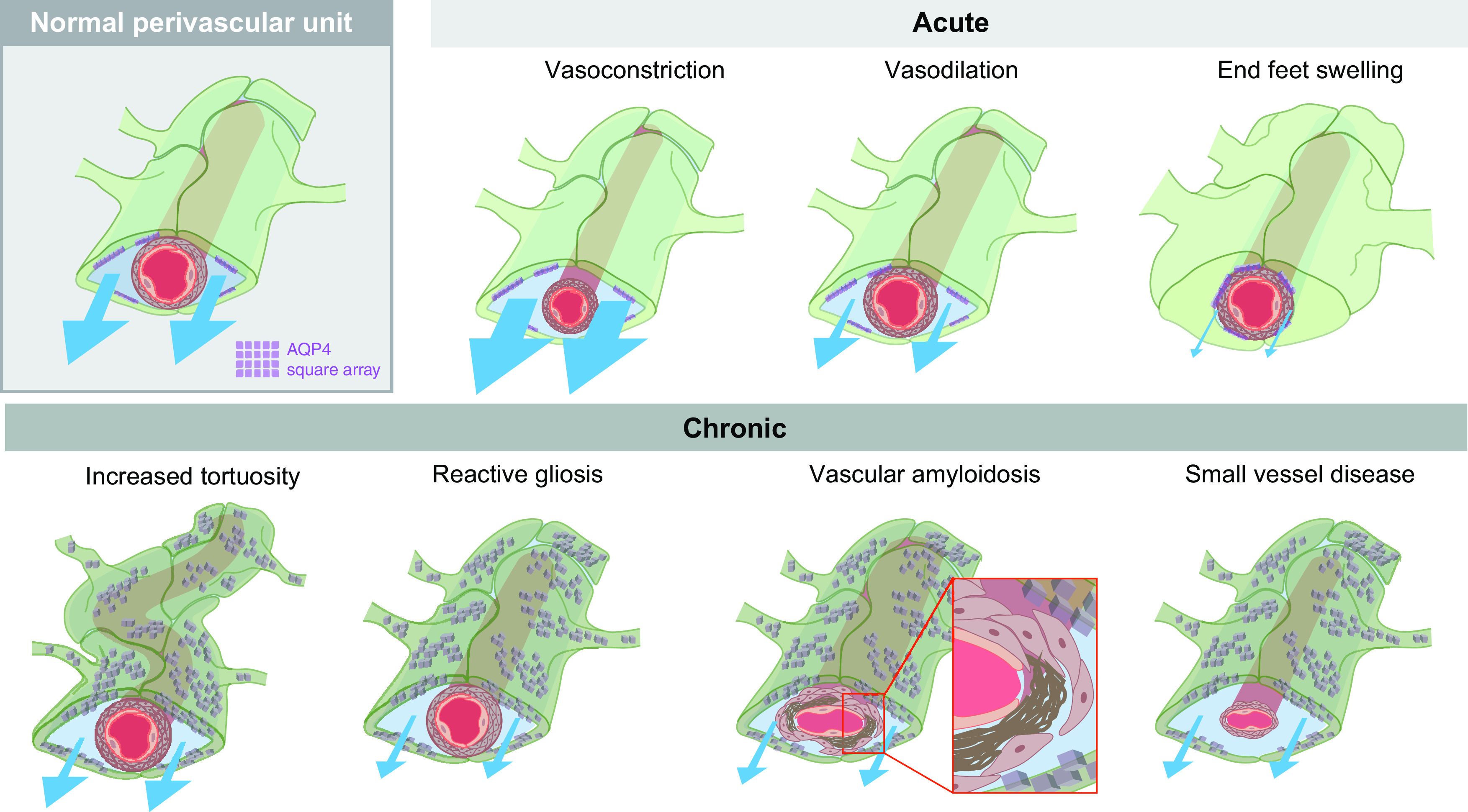
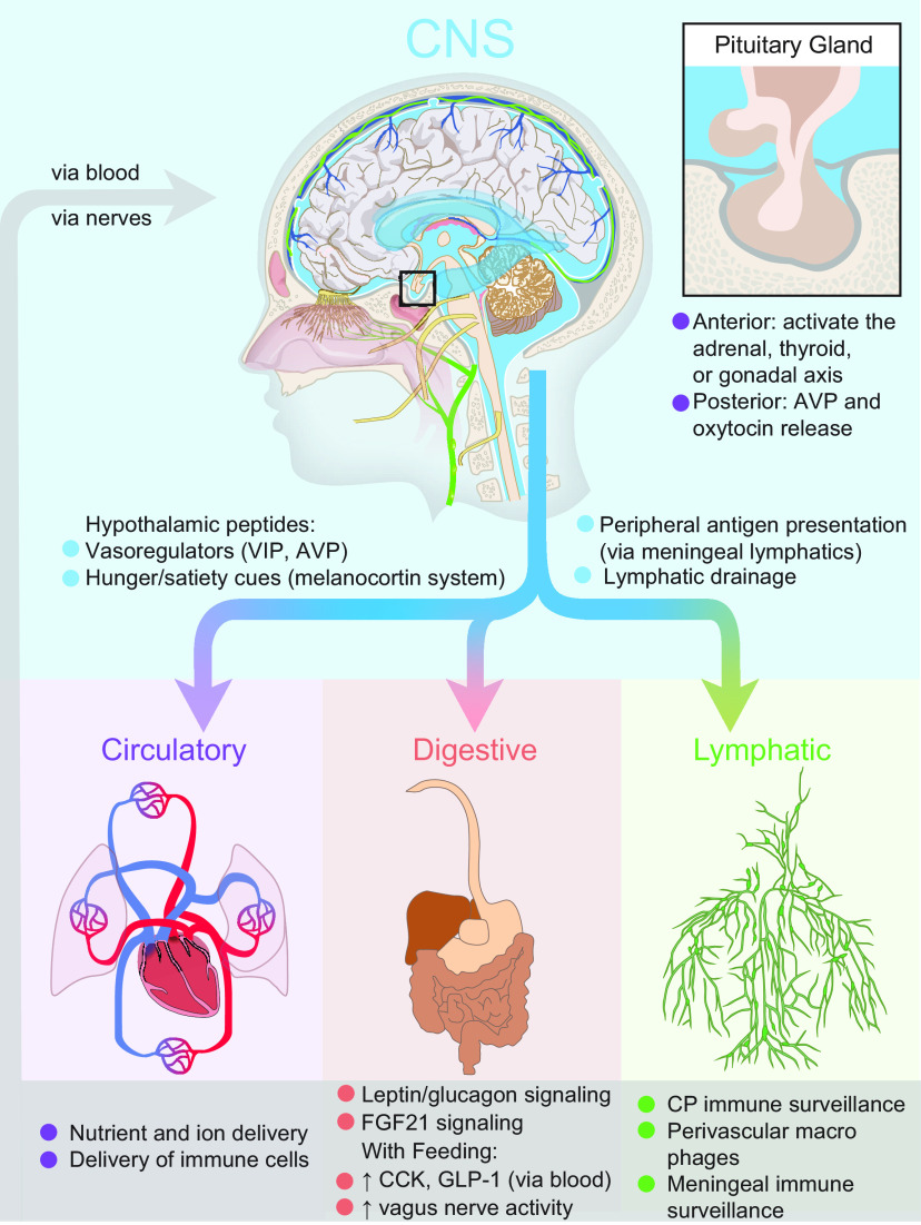
Throughout the body, lymphatic fluid movement supports critical functions including clearance of excess fluid and metabolic waste. The glymphatic system is the analog of the lymphatic system in the CNS. As such, the glymphatic system plays a key role in regulating directional interstitial fluid movement, waste clearance, and, potentially, brain immunity. The glymphatic system enables bulk movement of CSF from the subarachnoid space along periarterial spaces, where it mixes with interstitial fluid within the parenchyma before ultimately exiting from the parenchyma via perivenous spaces. This review focuses on important questions about the structure of this system, why the brain needs a fluid transport system, and unexplored aspects of brain fluid transport. We provide evidence that astrocytes and blood vessels determine the shape of the perivascular space, ultimately controlling the movement of perivascular fluid. Glymphatic fluid movement has the potential to alter local as well as global transport of signaling molecules and metabolites. We also highlight the evidence for cross talk among the glymphatic system, cardiovascular system, gastrointestinal tract, and lymphatic system. Much remains to be studied, but we propose that the glymphatic/lymphatic system acts as a cornerstone in signaling between the brain and body.

Keywords: astrocyte; cerebrospinal fluid; choroid plexus; glymphatic; peptides; perivascular space.


Figures

Figure 1.
Figure 1.
The vascular network is a scaffold for glymphatic fluid transport along the perivascular spaces. Glymphatic fluid (light blue) enters the brain via the perivascular space of the major arteries (red; left). Arteries and veins are lined by perivascular spaces, where astrocyte end feet (green) cover smooth muscle cells (gray) and the endothelial wall of the vasculature (pink; right). This perivascular unit is a critical component of the glymphatic system, and its geometry is biologically optimized to promote fluid movement (blue arrows).
Figure 2.
Figure 2.
The perivascular space (PVS) can be modulated by changes to both astrocytes and the vasculature. The normal perivascular unit is composed of astrocyte end feet (green) covering smooth muscle cells (tan) and endothelial cell walls (pink) of the vascular network, promoting CSF (blue) movement along these channels. AQP4 (purple) is located in square arrays on the vascular-adjacent end feet of astrocytes. Acute changes to either the vasculature or astrocyte end feet can alter glymphatic fluid movement. Vasoconstriction increases the PVS, increasing flow (indicated by blue arrows; Mestre et al., 2020b). This is in contrast to vasodilation that is expected to decrease flow. Swelling of astrocytic end feet can alter the size of the PVS space in the setting of pathology (e.g., spreading depression; Schain et al., 2017), but it is possible that changes in the vascular end feet of astrocytes are a physiological mechanism by which glymphatic function is controlled. Chronic pathologic changes may also impair CSF influx (bottom). We hypothesize that vasculature changes such as increased tortuosity with aging alter fluid flow. Reactive gliosis (shown as a dark green color and mislocalized AQP4), is a common hallmark of neuropathology (Ikeshima-Kataoka, 2016; Verkhratsky et al., 2016; Wang and Parpura, 2016; Kovacs et al., 2018), which will, most likely decrease flow. Vascular amyloidosis, characterized by amyloid-β plaques (brown) accumulating between the smooth muscle cells and the endothelial cell wall, and small-vessel disease, characterized by altered vascular shape and enlarged perivascular spaces, both decrease glymphatic flow.
Figure 3.
Figure 3.
Anatomical localization of key brain regions is strategically placed around CSF reservoirs. The hypothalamus is located along the third ventricle and base of the brain above the basal cisterns, a prime position for CSF signaling. It contains the suprachiasmatic nucleus (red), arcuate nucleus (orange), paraventricular nucleus (yellow), ventromedial hypothalamus (green), and the supraoptic nucleus (navy), which is a hub of peptidergic signaling that controls basic biological functions such as circadian timing, reproduction, feeding, hydration, and more. The nucleus basalis of Meynert (blue), dorsal raphe nucleus (purple), and locus coeruleus (gray) are also primed for brain-wide CSF-mediated cholinergic, serotonergic, and noradrenaline signaling.
Figure 4.
Figure 4.
The glymphatic system as an interface between the brain and body. The brain can communicate to the circulatory, digestive, and lymphatic systems by secreting signaling molecules to CSF, driving fluid to the meningeal lymphatics for antigen presentation, and ultimately draining to the lymph nodes of the lymphatic system. A unique feature of the brain is the hypothalamic signaling to the pituitary gland, where neurons can either induce neuroendocrine signaling in the anterior pituitary, or directly release peptides such as AVP and oxytocin into the blood vessels of the posterior pituitary. Feedback between these systems is potentially bidirectional when considering blood composition, feeding and fasting metabolites, and immune surveillance. The pituitary gland is uniquely shielded from direct interaction with the CSF pool and direct CNS signaling by its location in an indentation of the skull covered by the diaphragma sellae, a dural membrane.

References

    1. Abbott NJ, Pizzo ME, Preston JE, Janigro D, Thorne RG (2018) The role of brain barriers in fluid movement in the CNS: is there a “glymphatic” system? Acta Neuropathol 135:387–407. 10.1007/s00401-018-1812-4 - DOI - PubMed
    1. Abramova O, Zorkina Y, Ushakova V, Zubkov E, Morozova A, Chekhonin V (2020) The role of oxytocin and vasopressin dysfunction in cognitive impairment and mental disorders. Neuropeptides 83:102079. 10.1016/j.npep.2020.102079 - DOI - PubMed
    1. Achariyar TM, Li B, Peng W, Verghese PB, Shi Y, McConnell E, Benraiss A, Kasper T, Song W, Takano T, Holtzman DM, Nedergaard M, Deane R (2016) Glymphatic distribution of CSF-derived apoE into brain is isoform specific and suppressed during sleep deprivation. Mol Neurodegener 11:74. 10.1186/s13024-016-0138-8 - DOI - PMC - PubMed
    1. Afridi R, Lee WH, Suk K (2020) Microglia gone awry: linking immunometabolism to neurodegeneration. Front Cell Neurosci 14:246. 10.3389/fncel.2020.00246 - DOI - PMC - PubMed
    1. Agnati LF, Fuxe K, Zoli M, Ozini I, Toffano G, Ferraguti F (1986) A correlation analysis of the regional distribution of central enkephalin and beta-endorphin immunoreactive terminals and of opiate receptors in adult and old male rats. Evidence for the existence of two main types of communication in the central nervous system: the volume transmission and the wiring transmission. Acta Physiol Scand 128:201–207. 10.1111/j.1748-1716.1986.tb07967.x - DOI - PubMed

Publication types