. 2021 Aug 21:19:4854-4867.
doi: 10.1016/j.csbj.2021.08.030. eCollection 2021.

Subtypes of tail spike proteins predicts the host range of Ackermannviridae phages

Affiliations

Subtypes of tail spike proteins predicts the host range of Ackermannviridae phages

Anders Nørgaard Sørensen et al. Comput Struct Biotechnol J. .

Abstract

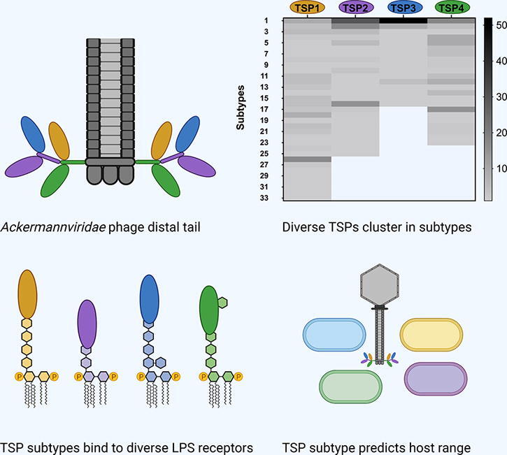
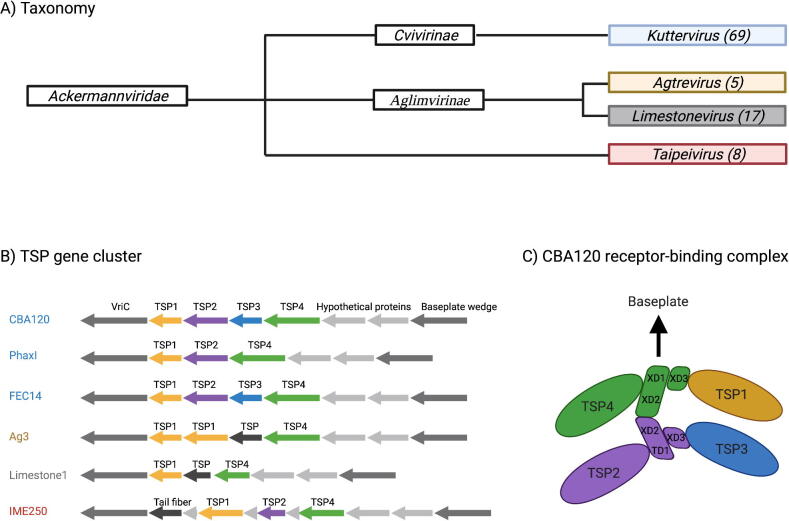
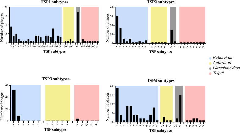
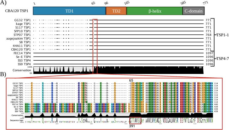
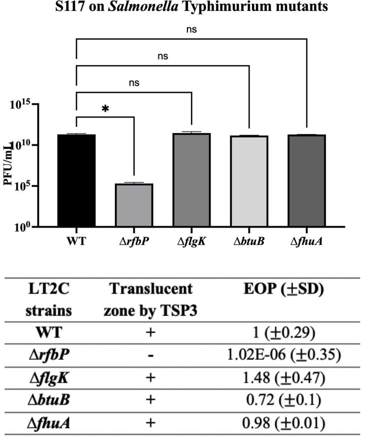
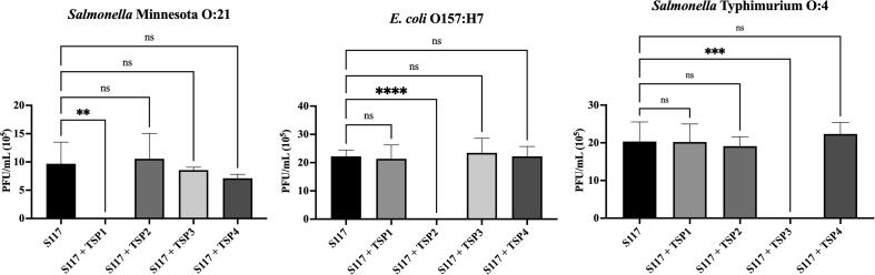
Phages belonging to the Ackermannviridae family encode up to four tail spike proteins (TSPs), each recognizing a specific receptor of their bacterial hosts. Here, we determined the TSPs diversity of 99 Ackermannviridae phages by performing a comprehensive in silico analysis. Based on sequence diversity, we assigned all TSPs into distinctive subtypes of TSP1, TSP2, TSP3 and TSP4, and found each TSP subtype to be specifically associated with the genera (Kuttervirus, Agtrevirus, Limestonevirus, Taipeivirus) of the Ackermannviridae family. Further analysis showed that the N-terminal XD1 and XD2 domains in TSP2 and TSP4, hinging the four TSPs together, are preserved. In contrast, the C-terminal receptor binding modules were only conserved within TSP subtypes, except for some Kuttervirus TSP1s and TSP3s that were similar to specific TSP4s. A conserved motif in TSP1, TSP3 and TSP4 of Kuttervirus phages may allow recombination between receptor binding modules, thus altering host recognition. The receptors for numerous uncharacterized phages expressing TSPs in the same subtypes were predicted using previous host range data. To validate our predictions, we experimentally determined the host recognition of three of the four TSPs expressed by kuttervirus S117. We confirmed that S117 TSP1 and TSP2 bind to their predicted host receptors, and identified the receptor for TSP3, which is shared by 51 other Kuttervirus phages. Kuttervirus phages were thus shown encode a vast genetic diversity of potentially exchangeable TSPs influencing host recognition. Overall, our study demonstrates that comprehensive in silico and host range analysis of TSPs can predict host recognition of Ackermannviridae phages.

Keywords: ANI, Average nucleotide identity; Ackermannviridae family; Bacteriophage; CPS, Capsular polysaccharide; EOP, Efficiency of plating; Escherichia coli O:157; Host range; LB, Luria-Bertani; LPS, Lipopolysaccharide; NCBI, National Center for Biotechnology Information; O-antigen; ORF, Open reading frame; PFU, Plaque formation unit; RBP, Receptor binding protein; Receptor-binding proteins; Salmonella; TSP, Tail spike protein; Tail spike proteins; VriC, Virulence-associated protein.


Conflict of interest statement

The authors declare that they have no known competing financial interests or personal relationships that could have appeared to influence the work reported in this paper.

Figures

None
Graphical abstract
Fig. 1
Fig. 1
Taxonomy of the Ackermannviridae family and the tail spike protein (TSP) gene clusters. A) Taxonomy of the Ackermannviridae family with the subfamilies and genera. The number of phages analyzed in this study within the respective genera are presented in the parentheses. B) The organization of the TSP gene clusters in different Ackermannviridae phages. Blue: Kuttervirus phages, olive-yellow: Agtrevirus phage, grey: Limestonevirus phage, red: Taipeivirus phages. TSP genes were colored according to annotation in GeneBank. TSP1: Yellow, TSP2: Purple, TSP3: Blue, TSP4: Green, Not specified: Black. B) Receptor-binding complex consisting of the four TSPs in kuttervirus CBA120 proposed by Plattner et al. Abbreviation: TSP, tail spike protein and VriC, Virulence-associated protein.
Fig. 2
Fig. 2
TSP subtypes in the Ackermannviridae family correlate with the phage genus. TSP genes were identified in the phage genomes and grouped into TSP1, TSP2, TSP3 and TSP4 and aligned. TSPs that were 75% or more identical were grouped into subtypes. The number of phages grouped into the individual TSP subtypes are illustrated on the y-axis. Blue: TSP subtypes expressed by Kuttervirus phages, Olive-yellow: TSP subtypes expressed by Agtrevirus phages, Grey: TSP subtypes expressed by Limestonevirus phages and Red: TSP subtypes expressed by Taipeivirus phages. Asterisk: TSPs in the TSP4-14 subtype are expressed by Agtrevirus and Limestonevirus phages. The figure was generated using Prism 9.
Fig. 3
Fig. 3
The receptor binding domains can be swapped between TSPs in the Kuttervirus phages. A) All TSP1s belonging to subtype 1–1 were aligned with all TSP4-7. B) Zoom in on the start positions of similarity of the TSPs. The alignment shows a conserved sequence motif that starts at the same amino acid position 65 and 391 for TSP1s and TSP4s, respectively. The structural domains of CBA120 TSP1 was used as a reference.
Fig. 4
Fig. 4
TSP3-1 was not able to produce a translucent zone on an LPS mutant (ΔrfbP). Phage S117 and TSP3-1 were spotted on Salmonella. Typhimurium (LT2c) mutant strains lacking known phage receptors; O-Ag (rfbP), flagella (flgK), ferrichrome transporter (fhuA) and vitamin B12 transporter (btuB). The experiment was carried out in triplicates and the error bars represent the standard deviation. The graph was generated in prism9 where the p-values were calculated using the ordinary one-way ANOVA. +: appearance of a translucent zone, -: no zone.
Fig. 5
Fig. 5
TSPs can inhibit the infection of phage S117 of their respective hosts. The infectivity of phage S117 in the presence of TSP1-1, TSP2-1, TSP3-1, TSP4-1 or nothing mixed with the bacterial hosts were measured using a double layered plaque assay. 0.5 mg/mL of the TSPs were able to block the infection of phage S117 on their respective hosts when a phage titer of 105 PFU/mL was used. The experiment was carried out in triplicates and the error bars represent the standard deviation. The graphs were generated in prism9 where the p-values were calculated using the ordinary one-way ANOVA.

References

    1. Xing S., Ma T., Zhang X., Huang Y., Mi Z., Sun Q. First complete genome sequence of a virulent bacteriophage infecting the opportunistic pathogen Serratia rubidaea. Arch Virol. 2017;162(7):2021–2028. doi: 10.1007/s00705-017-3300-x. - DOI - PubMed
    1. Barbirz S., Müller J.J., Uetrecht C., Clark A.J., Heinemann U., Seckler R. Crystal structure of Escherichia coli phage HK620 tailspike: Podoviral tailspike endoglycosidase modules are evolutionarily related. Mol Microbiol. 2008;69:303–316. doi: 10.1111/j.1365-2958.2008.06311.x. - DOI - PubMed
    1. Greenfield J., Shang X., Luo H., Zhou Y., Linden S.B., Heselpoth R.D. Structure and function of bacteriophage CBA120 ORF211 (TSP2), the determinant of phage specificity towards E. coli O157:H7. Sci Rep. 2020;10(1) doi: 10.1038/s41598-020-72373-0. - DOI - PMC - PubMed
    1. Nobrega F.L., Vlot M., de Jonge P.A., Dreesens L.L., Beaumont H.J.E., Lavigne R. Targeting mechanisms of tailed bacteriophages. Nat Rev Microbiol. 2018;16(12):760–773. doi: 10.1038/s41579-018-0070-8. - DOI - PubMed
    1. Liu B, Furevi A, Perepelov A V., Guo X, Cao H, Wang Q, et al. Structure and genetics of escherichia coli O antigens. FEMS Microbiol Rev 2020;44:655–83. https://doi.org/10.1093/femsre/fuz028. - PMC - PubMed

LinkOut - more resources