

Hepatoid adenocarcinoma of the stomach with PIK3Ca mutation during pregnancy: A case report with molecular profile
- PMID: 34527251
- PMCID: PMC8436274
- DOI: 10.1093/omcr/omab078
Hepatoid adenocarcinoma of the stomach with PIK3Ca mutation during pregnancy: A case report with molecular profile
Abstract
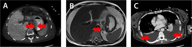
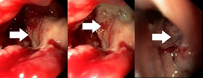
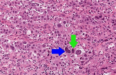
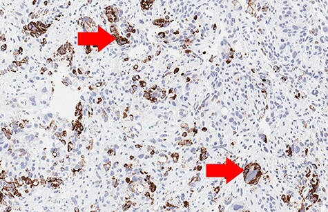
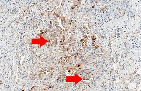
Hepatoid adenocarcinoma is an extremely aggressive special subtype of gastric tumors. It can be lethal as no standard treatment options for this type of gastric cancer exist. Here, we describe a very rare case of a young female on her 21st week of pregnancy who was diagnosed with stage IV hepatoid adenocarcinoma of the stomach with elevated α fetoprotein (AFP) level. Gene mutation analysis performed by next-generation sequencing identified somatic mutations in the PIK3CA gene. Despite the treatment, patient died 2 months after the initial disease presentation. To our best knowledge, this case represents the first report of pregnancy-associated hepatoid gastric adenocarcinoma with the PIK3CA gene mutations, which can provide further clues for the understanding of molecular features of this type of tumor that can reflect biological behavior and may lead to further effective treatment options.
Keywords: PIK3CA, NGS; gastric cancer; hepatoid adenocarcinoma; pregnancy.
© The Author(s) 2021. Published by Oxford University Press.
Figures

References
-
- Niknam R, Haghighat S, Mokhtari M. The pregnancy-associated gastric cancer: a case report and review of the literature. J Obstet Gynaecol. 2020;20:1–2. - PubMed
Publication types
LinkOut - more resources
Full Text Sources
Miscellaneous
