Review
. 2021 Aug;13(8):5159-5175.
doi: 10.21037/jtd.2020.03.94.

Foreign body aspiration

Affiliations
Review

Foreign body aspiration

Divyansh Bajaj et al. J Thorac Dis. 2021 Aug.

Abstract

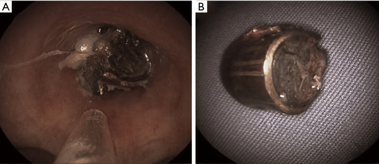
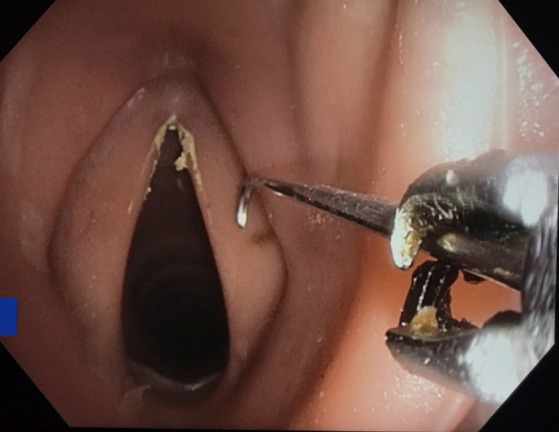
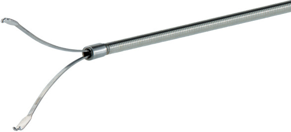
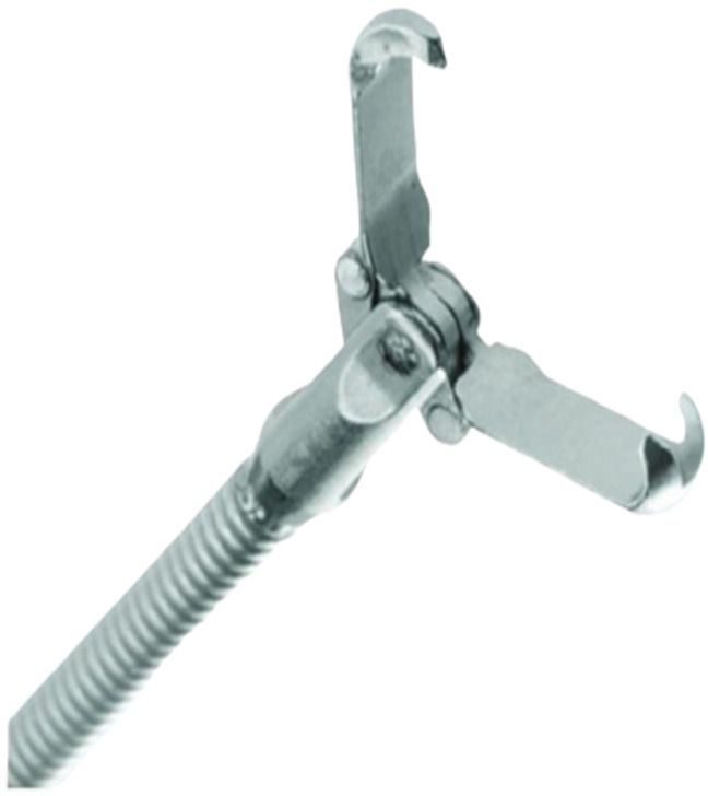
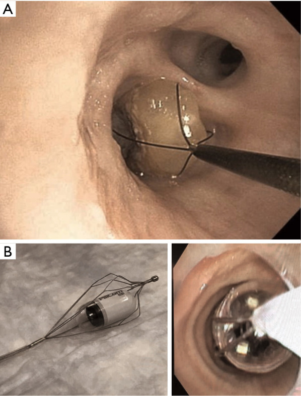
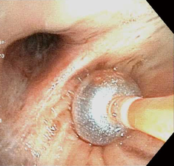
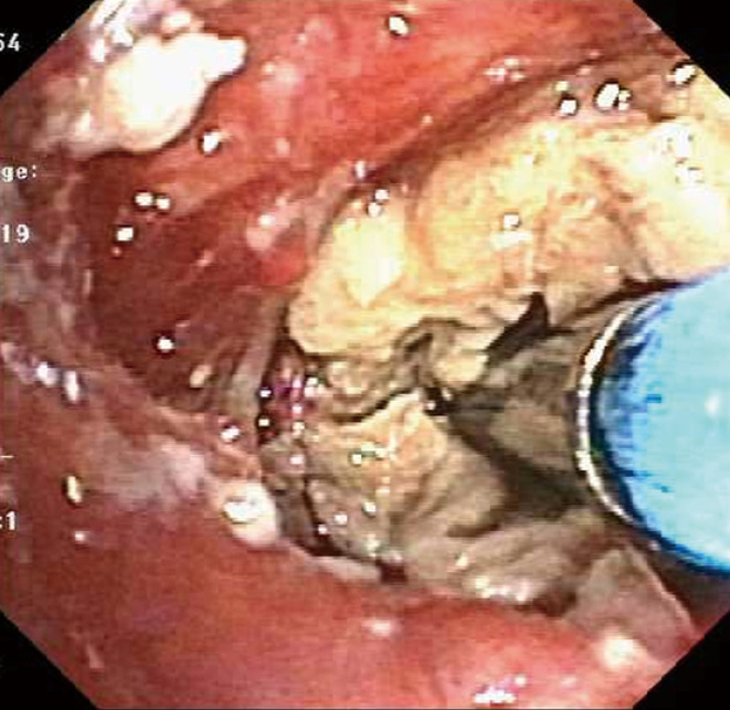
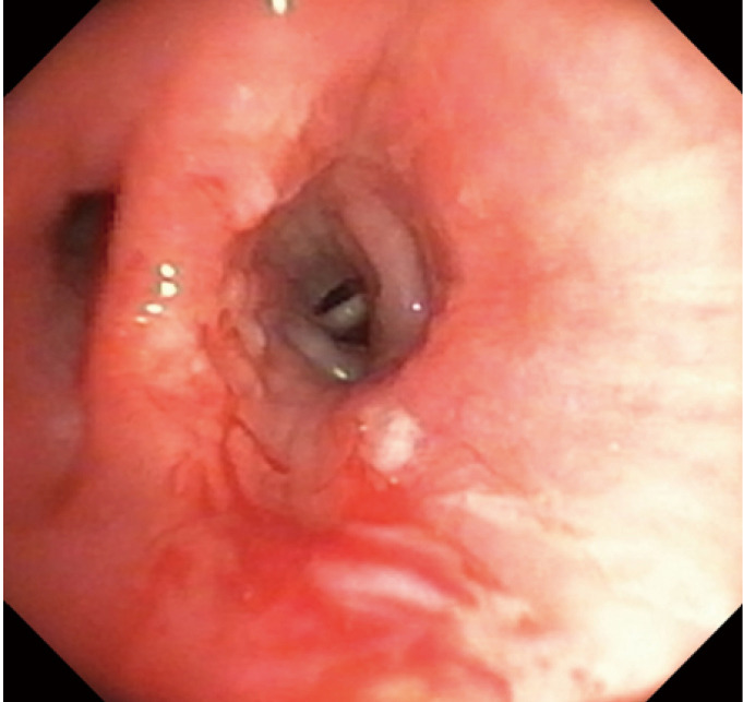
The clinical manifestations of foreign body (FB) aspiration can range from an asymptomatic presentation to a life-threatening emergency. Patients may present with acute onset cough, chest pain, breathlessness or sub-acutely with unexplained hemoptysis, non-resolving pneumonia and at times, as an incidental finding on imaging. Patients with iatrogenic FB such as an aspirated broken tooth during difficult intubation or a broken instrument are more common scenarios in the intensive care unit (ICU). Patients with post-obstructive pneumonia with or without sepsis, or variable degree of hemoptysis often require ICU level of care and bronchoscopic interventions. Rigid bronchoscopy has traditionally been the modality of choice; however, with the innovation in instrumentation and wider availability of flexible bronchoscopes, most of the FB removal is now successfully performed using flexible bronchoscopy. Proceduralists choose instruments in accordance with their training and expertise. We describe the use of most common instruments including forceps, balloon catheters, and baskets. Role of cryoprobe and LASER in FB removal is reviewed as well. In general, larger working channel bronchoscopes are preferred; however, smaller working channel bronchoscopes may be used in situations when the patients are intubated with a smaller diameter endotracheal or tracheostomy tubes. Large size FB are removed en bloc with the grasping tool, bronchoscope, and endotracheal or tracheostomy tube, requiring preparation to safely re-establish the airway. After FB removal, bronchoscopy is re-performed to identify any residual FB, assess any injury to the airway, suction post-obstructive secretions or pus, control any active bleeding and remove granulation tissue that may be obstructing the airway. Additional interventions like balloon dilatation may be required to dislodge an impacted FB or to maintain patency of bronchial lumen. If bronchoscopic methods fail, surgery may be required for retrieval of FB in symptomatic patients or to resect suppurative or necrotizing lung process. Multidisciplinary approach involving intensivists, surgeons, and anesthesiologists is the key to optimal patient outcomes.

Keywords: Foreign body (FB) aspiration; bronchoscopy; intensive care.


Conflict of interest statement

Conflicts of Interest: All authors have completed the ICMJE uniform disclosure form (available at: http://dx.doi.org/10.21037/jtd.2020.03.94). The series “Interventional Pulmonology in the Intensive Care Unit Environment” was commissioned by the editorial office without any funding or sponsorship. Ashutosh Sachdeva served as the unpaid Guest Editor of the series. The authors have no other conflicts of interest to declare.

Figures

Figure 1
Figure 1
A 46-year old man, never smoker, with history of Asthma presented with non-resolving cough and hemoptysis. He received two prior antibiotic treatments for presumed pneumonia a month apart with mild improvement. Chest imaging revealed findings concerning for necrotizing pneumonia as result of lingular obstruction (A-C). Computed tomography scans revealed high density foreign body (B) and post obstructive bronchiectasis (C). 3-dimensional reconstruction of the tracheobronchial tree was performed to localize the obstruction (D). After receiving intravenous antibiotics for 72 hours, patient was intubated electively given concern for “spill-over” of pus from relief of obstruction. A significant amount of pus was suctioned post-extraction of the foreign body (E-G). Foreign body was determined to be a corn kernel (H). Patient completed a course of antibiotics and had no symptoms on a follow up visit.
Figure 2
Figure 2
An 80-year-old gentleman presented with acute respiratory failure requiring intubation and mechanical ventilation, a few hours after a witnessed choking episode with the first bite of a “hot-dog”. Cryotherapy extraction of the foreign body was performed and the patient was extubated the following morning. He received a short course of antibiotics to manage post-obstructive pneumonia.
Figure 3
Figure 3
Examples of iatrogenic foreign body aspiration. (A) A 78-year-old gentleman was emergently intubated for acute respiratory failure and a tooth was dislodged into the bronchus intermedius. Bronchoscopy was performed for hemoptysis a few days after the intubation and the diagnosis of iatrogenic foreign body impaction was made. (B) Image of a broken disposable bronchoscope (AMBU®) that was inadvertently introduced via “Murphy eye” of a size 8.0 endotracheal tube. This resulted in iatrogenic foreign body that required removal using a forceps. One jaw of the forceps was engaged in the lumen of the broken scope and grasped to extract in its entirety.
Figure 4
Figure 4
A 13-year-old boy presented with history of whistling sound while breathing. A history of aspirating components of a plastic whistle 6 weeks back was elicited. (A) Chest X-ray demonstrates hyper-lucency of left lung with opacity (red arrow) in left hilar region. (B) Computed tomography showed a reverse C shaped artifact (yellow arrow) in left main bronchus with hyperinflation of left lung fields. A plastic whistle component was removed using sharktooth forceps that revealed significant granulation of the surrounding mucosa that was managed conservatively. Repeat bronchoscopy after 2 months showed normal lumen with mild inflammatory changes.
Figure 5
Figure 5
Impacted foreign body in the right lower lobe in a patient presenting with mandibular fractures as a result of firearm injury. (A) Foreign body accessed via a 12-mm Bryan-Dumon rigid bronchoscope; (B) A 9-mm bullet shell was extracted from bronchus intermedius using rigid forceps. Interestingly, there were no other associated injuries in the bullet path likely due to loss of velocity at the site of its impact in the mandibular bone.
Figure 6
Figure 6
Rat tooth forceps being used for the retrieval of airway foreign body in a simulation session.
Figure 7
Figure 7
V-shape grasping forceps.
Figure 8
Figure 8
Rat tooth grasping forceps.
Figure 9
Figure 9
Shark tooth grasping forceps.
Figure 10
Figure 10
Rubber tip grasping forceps.
Figure 11
Figure 11
Mini three-prong grasping forceps.
Figure 12
Figure 12
Mini grasping basket.
Figure 13
Figure 13
Grasping basket.
Figure 14
Figure 14
Zero tip airway retrieval basket
Figure 15
Figure 15
Fishnet basket. Courtesy of Steris Endoscopy. Unauthorized use not permitted.
Figure 16
Figure 16
Foreign body extraction with two different type of baskets. (A) Zero tip (Boston Scientific™) basket encasing a corn kernel from right lower lobe; (B) Olympus™ “flower basket” used to extract a “Pillcam” from right lower lobe.
Figure 17
Figure 17
Fogarty balloon is used to tamponade bleeding airway mucosa after retrieval of foreign body.
Figure 18
Figure 18
Fogarty balloon use to dislodge an impacted foreign body and bring it to the central airway is demonstrated. (A) Impacted foreign body; (B) Fogarty balloon passed beyond the foreign body; (C) Inflated Fogarty balloon being pulled back to dislodge the foreign body; (D) Granulation tissue at the site of foreign body impaction post-removal.
Figure 19
Figure 19
The cryoprobe is impaled into the foreign body (mucus cast) and freeze cycle is initiated. After, adequate cryoadhesion is visibly noticed, the foreign body is extracted by co-axial movement of the scope, the cryoprobe, and the adhered FB making sure that the cold probe doesn’t catch the airway mucosa.
Figure 20
Figure 20
Airway mucosal injury resulting in bleeding during retrieval of an impacted foreign body. This was managed by using argon plasma coagulation.
Video 1
Video 1
Simulation bronchoscopy demonstrating the retrieval of an airway foreign body (peanut) using Zero Tip basket.

References

    1. Salih AM, Alfaki M, Alam-Elhuda DM. Airway foreign bodies: A critical review for a common pediatric emergency. World J Emerg Med 2016;7:5-12. 10.5847/wjem.j.1920-8642.2016.01.001 - DOI - PMC - PubMed
    1. Zöllner F. Gustav Killian, father of bronchoscopy. Arch Otolaryngol 1965;82:656-9. 10.1001/archotol.1965.00760010658020 - DOI - PubMed
    1. Rafanan AL, Mehta AC. Adult airway foreign body removal. What's new? Clin Chest Med 2001;22:319-30. 10.1016/S0272-5231(05)70046-0 - DOI - PubMed
    1. Baharloo F, Veyckemans F, Francis C, et al. Tracheobronchial foreign bodies: presentation and management in children and adults. Chest 1999;115:1357-62. 10.1378/chest.115.5.1357 - DOI - PubMed
    1. Blanco Ramos M, Botana-Rial M, Garcia-Fontan E, et al. Update in the extraction of airway foreign bodies in adults. J Thorac Dis 2016;8:3452-6. 10.21037/jtd.2016.11.32 - DOI - PMC - PubMed