Review
. 2021 Aug;13(8):5277-5296.
doi: 10.21037/jtd-19-3728.

An overview of percutaneous endoscopic gastrostomy tube placement in the intensive care unit

Affiliations
Review

An overview of percutaneous endoscopic gastrostomy tube placement in the intensive care unit

Margaret Wei et al. J Thorac Dis. 2021 Aug.

Abstract

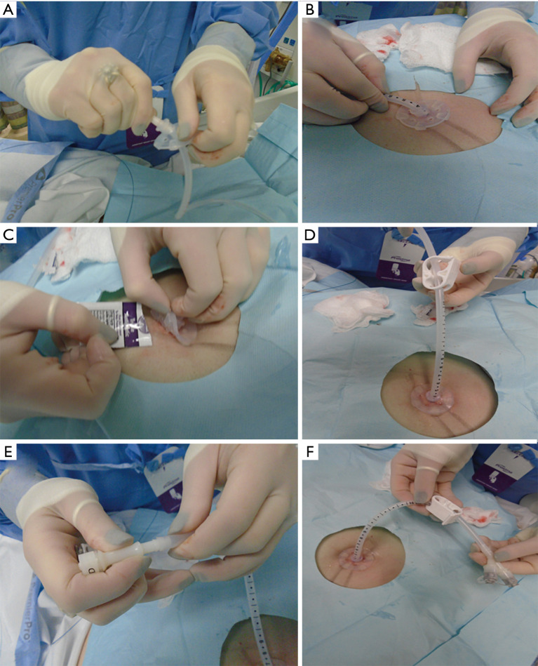
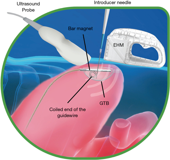
Critically ill patients are at increased risk for malnutrition as they often have underlying acute and chronic illness, stress related catabolism, decreased appetite, trauma and ongoing inflammation. Malnutrition is recognized as a leading cause of adverse outcomes, higher mortality, and increased hospital costs. Percutaneous endoscopic gastrostomy (PEG) tubes provide a safe and effective route to provide supplemental enteral nutrition to these patients. PEG placement has essentially replaced surgical gastrostomy as the modality of choice for longer term feeding in patients. This is a highly prevalent procedure with 160,000 to 200,000 PEG procedures performed each year in the United States. The purpose of this review is to provide an overview of current knowledge and practice standards with regards to placement of PEG tube in the Intensive Care Unit (ICU). When a patient is considered for a PEG tube, it is important to evaluate the treatment alternatives and identify the best option for each patient. In this review, we provide the advantages and disadvantages of various feeding modalities and devices. We review the indications and contraindications for PEG tube placement as well as the risks of this procedure. We then describe in detail the per-oral pull, per-oral push, and direct percutaneous techniques for PEG tube placement. Additionally, we review the feasibility of having interventional pulmonologists place PEG tubes in the ICU.

Keywords: Intensive Care Unit (ICU); Percutaneous endoscopic gastrostomy (PEG) placement; interventional pulmonology.


Conflict of interest statement

Conflicts of Interest: All authors have completed the ICMJE uniform disclosure form, available at: http://dx.doi.org/10.21037/jtd-19-3728. The series “Interventional Pulmonology in the Intensive Care Unit Environment” was commissioned by the editorial office without any funding sponsorship. The authors have no other conflicts of interest to declare.

Figures

Figure 1
Figure 1
Boston Scientific EndoVive™ 20 French Safety PEG kit, Pull technique.
Figure 2
Figure 2
Pre-procedural evaluation for PEG tube placement. (A) Patient’s abdomen marked with a surgical pen prior to the procedure; (B) endoscope retroflexed, evaluating the gastric cardia for lesions; (C) evaluating the duodenum for evidence of post-pyloric obstruction; (D) evaluating the esophagus for lesions.
Figure 3
Figure 3
Insertion of the guidewire into the stomach. (A) Finger indentation at the site of trans-illumination; (B) indentation visualized from inside the stomach; (C) lidocaine with epinephrine administered at 90-degree angle to the surgical site; (D) small skin incision made with a scalpel at 90-degree angle; (E) insertion of the needle-catheter combination (trocar) through the incision at 90-degree angle; (F) visualization of the trocar from inside the stomach; (G) catheter positioned inside the stomach with the needle withdrawn; (H) snare system is passed through the endoscope and catches the wire loop.
Figure 4
Figure 4
Placement of the PEG tube via the pull method. (A) Guidewire with wire loop pulled out of the mouth; (B) lubricating gel applied to the PEG tube prior to insertion; (C) PEG tube looped through the guidewire; (D) PEG tube attached to the guidewire via a knot; (E) firm pressure applied while pulling the PEG tube through the abdominal wall; (F) direct visualization of the PEG tube bumper from inside the stomach. PEG, percutaneous endoscopic gastrostomy.
Figure 5
Figure 5
Completion of the PEG tube procedure. (A) External bolster placed onto the PEG tube; (B) skin marking on the external bolster kept at 2–5 cm depending on the patient’s body habitus, making sure that the bolster is neither too tight nor too loose; (C) application of antibiotic ointment at the surgical site; (D) clamp positioned onto the PEG tube; (E) external port attached to the PEG tube; (F) successful placement of the PEG tube. PEG, percutaneous endoscopic gastrostomy.
Figure 6
Figure 6
Illustration of the percutaneous ultrasound gastrostomy technique.

References

    1. Sharma K, Mogensen KM, Robinson MK. Pathophysiology of Critical Illness and Role of Nutrition. Nutr Clin Pract 2019;34:12-22. 10.1002/ncp.10232 - DOI - PubMed
    1. Mogensen KM, Robinson MK, Casey JD, et al. Nutritional Status and Mortality in the Critically Ill. Crit Care Med 2015;43:2605-15. 10.1097/CCM.0000000000001306 - DOI - PubMed
    1. Gauderer MW, Ponsky JL, Izant RJ, Jr. Gastrostomy without laparotomy: a percutaneous endoscopic technique 1980. Nutrition 1998;14:736-8. - PubMed
    1. Kalfarentzos F, Kehagias J, Mead N, et al. Enteral nutrition is superior to parenteral nutrition in severe acute pancreatitis: results of a randomized prospective trial. Br J Surg 1997;84:1665-9. 10.1046/j.1365-2168.1997.02851.x - DOI - PubMed
    1. Braunschweig CL, Levy P, Sheean PM, et al. Enteral compared with parenteral nutrition: a meta-analysis. Am J Clin Nutr 2001;74:534-42. 10.1093/ajcn/74.4.534 - DOI - PubMed