. 2021 Nov;48(11):7127-7140.
doi: 10.1002/mp.15231. Epub 2021 Sep 25.

CARes-UNet: Content-aware residual UNet for lesion segmentation of COVID-19 from chest CT images

Affiliations

CARes-UNet: Content-aware residual UNet for lesion segmentation of COVID-19 from chest CT images

Xinhua Xu et al. Med Phys. 2021 Nov.

Abstract

Purpose: Coronavirus disease 2019 (COVID-19) has caused a serious global health crisis. It has been proven that the deep learning method has great potential to assist doctors in diagnosing COVID-19 by automatically segmenting the lesions in computed tomography (CT) slices. However, there are still several challenges restricting the application of these methods, including high variation in lesion characteristics and low contrast between lesion areas and healthy tissues. Moreover, the lack of high-quality labeled samples and large number of patients lead to the urgency to develop a high accuracy model, which performs well not only under supervision but also with semi-supervised methods.

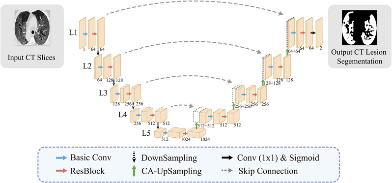
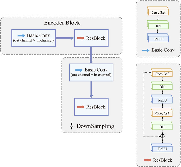
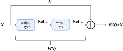
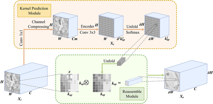
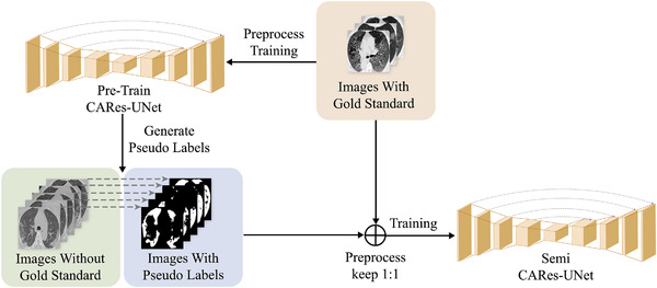
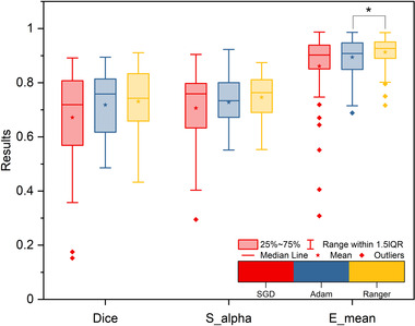
Methods: We propose a content-aware lung infection segmentation deep residual network (content-aware residual UNet (CARes-UNet)) to segment the lesion areas of COVID-19 from the chest CT slices. In our CARes-UNet, the residual connection was used in the convolutional block, which alleviated the degradation problem during the training. Then, the content-aware upsampling modules were introduced to improve the performance of the model while reducing the computation cost. Moreover, to achieve faster convergence, an advanced optimizer named Ranger was utilized to update the model's parameters during training. Finally, we employed a semi-supervised segmentation framework to deal with the problem of lacking pixel-level labeled data.

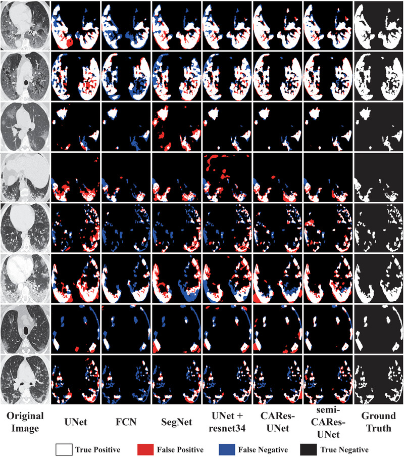
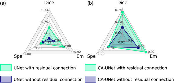
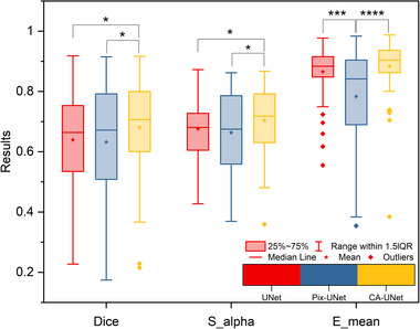
Results: We evaluated our approach using three public datasets with multiple metrics and compared its performance to several models. Our method outperforms other models in multiple indicators, for instance in terms of Dice coefficient on COVID-SemiSeg Dataset, CARes-UNet got the score 0.731, and semi-CARes-UNet further boosted it to 0.776. More ablation studies were done and validated the effectiveness of each key component of our proposed model.

Conclusions: Compared with the existing neural network methods applied to the COVID-19 lesion segmentation tasks, our CARes-UNet can gain more accurate segmentation results, and semi-CARes-UNet can further improve it using semi-supervised learning methods while presenting a possible way to solve the problem of lack of high-quality annotated samples. Our CARes-UNet and semi-CARes-UNet can be used in artificial intelligence-empowered computer-aided diagnosis system to improve diagnostic accuracy in this ongoing COVID-19 pandemic.

Keywords: computed tomography (CT) image; content-aware residual UNet; coronavirus disease 2019 (COVID-19); deep learning; segmentation.


Conflict of interest statement

The authors have no conflicts to disclose.

Figures

FIGURE 1
FIGURE 1
Network architecture of content‐aware residual UNet (CARes‐UNet)
FIGURE 2
FIGURE 2
The structure of encoder block and downsampling block. Both of them consist of one basic convolution operation and one resblock. The meanings of colored arrows are the same as those in Figure 1
FIGURE 3
FIGURE 3
Residual connection
FIGURE 4
FIGURE 4
Content‐aware upsampling. Kernel prediction module generates reassembly kernels. Reassemble module upsamples the feature map using predicted kernels. Unfold returns a view of the original tensor which contains all slices of size from tensor in the dimension and the cross‐product sign represents the matrix product of two tensors
FIGURE 5
FIGURE 5
Overview of our semi‐supervised framework
FIGURE 6
FIGURE 6
Qualitative analysis of different models. The first column on the left is the original computed tomography (CT) slices, and the first column on the right is their corresponding ground truth masks. Columns between them are images of lesion areas predicted by different models. White, red, blue, and black regions identify true positive, false positive, false negative, and true negative regions, respectively
FIGURE 7
FIGURE 7
Comparison of models with and without residual connection. (a) UNet. (b) CA‐UNet. All metrics are the higher the better. Models with residual connection are better than ones without residual connection
FIGURE 8
FIGURE 8
Comparison of UNet (without residual connection) with different upsampling blocks. Different colors represent different upsampling blocks and all metrics are the higher the better. The number of asterisks indicates the statistical significance calculated without outliers
FIGURE 9
FIGURE 9
Comparison of CARes‐UNet with different optimizers (learning rate is set to 5 × 10⁻⁴). Different colors represent different optimizers, and all metrics are the higher the better. The number of asterisks indicates the statistical significance calculated without outliers. The test results were recorded after the models converged given enough training period
FIGURE 10
FIGURE 10
Comparison of convergence using different optimization algorithms (a) training curve of three optimizers. (b) Testing curve of three optimizers. Different colors represent different optimizers. Ranger has a faster convergence and less variance than SGD and Adam optimizer

Similar articles

Cited by

References

    1. Wang C, Horby PW, Hayden FG, Gao GF. A novel coronavirus outbreak of global health concern. Lancet. 2020;395(10223):470‐473. - PMC - PubMed
    1. Huang C, Wang Y, Li X, et al. Clinical features of patients infected with 2019 novel coronavirus in Wuhan, China. Lancet. 2020;395(10223):497‐506. - PMC - PubMed
    1. Fang Y, Zhang H, Xie J, et al. Sensitivity of chest CT for COVID‐19: comparison to RT‐PCR. Radiology. 2020;296(2):200432. - PMC - PubMed
    1. Ai T, Yang Z, Hou H, et al. Correlation of chest CT and RT‐PCR testing in coronavirus disease 2019 (COVID‐19) in China: a report of 1014 cases. Radiology. 2020;296(2):E32‐E40. - PMC - PubMed
    1. Lessmann N, Sánchez CI, Beenen L, et al. Automated assessment of COVID‐19 reporting and data system and chest CT severity scores in patients suspected of having COVID‐19 using artificial intelligence. Radiology. 2021;298(1):E18‐E28. - PMC - PubMed