. 2021 Oct;20(10):e13434.
doi: 10.1111/acel.13434. Epub 2021 Sep 16.

p38 MAPK-mediated loss of nuclear RNase III enzyme Drosha underlies amyloid beta-induced neuronal stress in Alzheimer's disease

Affiliations

p38 MAPK-mediated loss of nuclear RNase III enzyme Drosha underlies amyloid beta-induced neuronal stress in Alzheimer's disease

Haidong Xu et al. Aging Cell. 2021 Oct.

Abstract

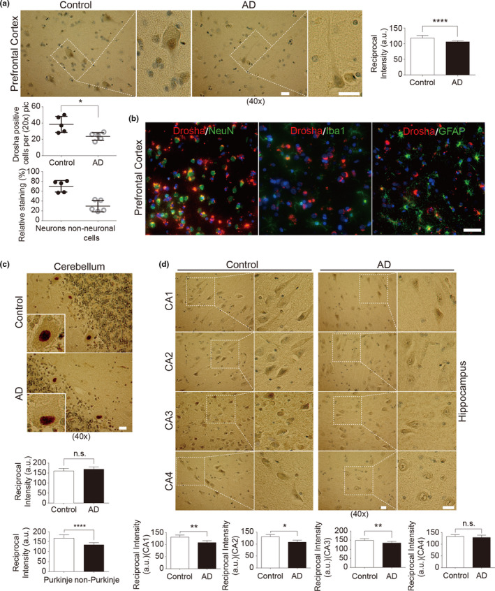
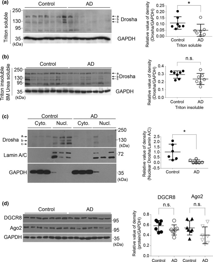
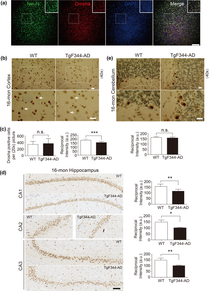
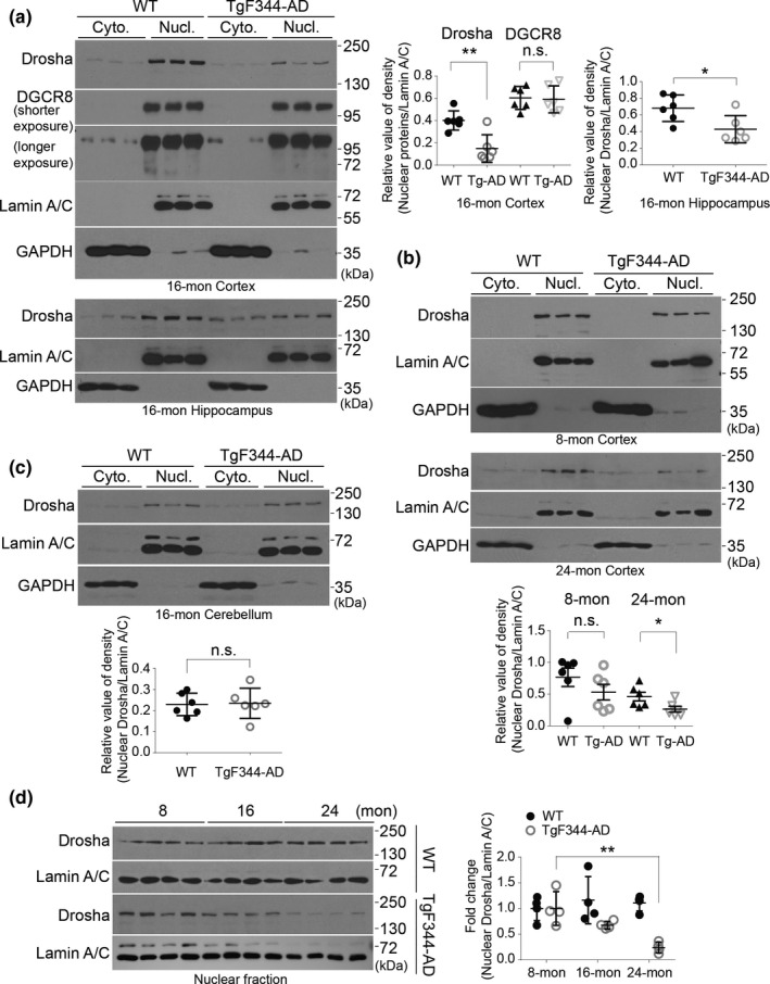
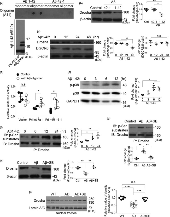
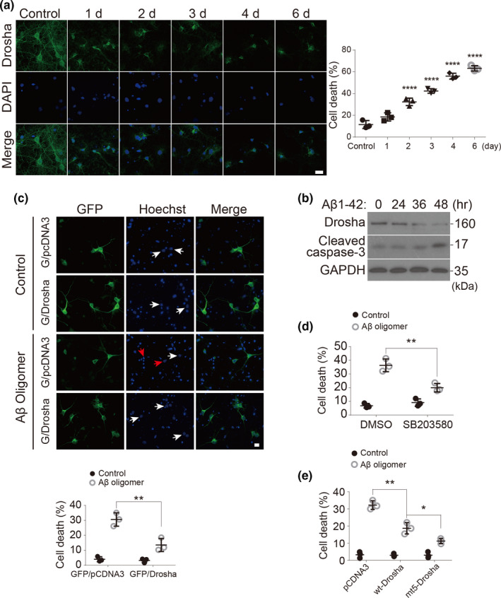
MicroRNAs (miRNAs) are small noncoding RNAs ubiquitously expressed in the brain and regulate gene expression at the post-transcriptional level. The nuclear RNase III enzyme Drosha initiates the maturation process of miRNAs in the nucleus. Strong evidence suggests that dysregulation of miRNAs is involved in many neurological disorders including Alzheimer's disease (AD). Dysfunction of miRNA biogenesis components may be involved in the processes of those diseases. However, the role of Drosha in AD remains unknown. By using immunohistochemistry, biochemistry, and subcellular fractionation methods, we show here that the level of Drosha protein was significantly lower in the postmortem brain of human AD patients as well as in the transgenic rat model of AD. Interestingly, Drosha level was specifically reduced in neurons of the cortex and hippocampus but not in the cerebellum in the AD brain samples. In primary cortical neurons, amyloid-beta (Aβ) oligomers caused a p38 MAPK-dependent phosphorylation of Drosha, leading to its redistribution from the nucleus to the cytoplasm and a decrease in its level. This loss of Drosha function preceded Aβ-induced neuronal death. Importantly, inhibition of p38 MAPK activity or overexpression of Drosha protected neurons from Aβ oligomers-induced apoptosis. Taken together, these results establish a role for p38 MAPK-Drosha pathway in modulating neuronal viability under Aβ oligomers stress condition and implicate loss of Drosha as a key molecular change in the pathogenesis of AD.

Keywords: Alzheimer's disease; Drosha; amyloid beta (Aβ); neuronal death; p38 MAPK.


Conflict of interest statement

The authors declare that they have no conflicts of interest with the contents of this article.

Figures

FIGURE 1
FIGURE 1
Drosha is decreased in the prefrontal cortex and hippocampus of AD patients. (a) Drosha immunohistochemistry in the prefrontal cortex. Areas in white boxes are shown at higher magnification on the right. The slices were counterstained with hematoxylin, and the nuclei number and Drosha‐positive cells (Figure S2a) were counted. The relative percentage staining in identified neurons and non‐neuronal cells in the prefrontal cortex of control was also quantified (at least 300 positive staining cells were counted). (b) Representative immunofluorescent images of prefrontal cortex slices of human control stained with Drosha and NeuN, or Iba1, or GFAP, respectively. Scale bar, 100 μm. (c–d) Drosha immunohistochemistry in the cerebellum (c) and hippocampus (d). Insets are enlarged images of a single cell (c) and areas in white boxes are shown at higher magnification on the right (d). The reciprocal intensities of DAB in each brain region as well as Drosha signals in Purkinje cells and other morphological non‐Purkinje cells (c) were analyzed by ImageJ (n = 5 control or AD cases for each area); Scale bar, 20 μm (a, c, and d). *p < 0.05, **p < 0.01, and ****p < 0.0001 versus control group. Error bars show mean ± SD
FIGURE 2
FIGURE 2
Soluble Drosha is decreased in the nucleus of human AD brain. (a–b) Triton X‐100 soluble and 8 M urea soluble (Triton insoluble) lysates were immunoblotted with Drosha antibody. GAPDH was used as a loading control. Right panels show quantitative analysis of the protein levels of Drosha (n = 7). (c) Cytosol and nuclear fractions were prepared from human prefrontal cortex and analyzed as shown (n = 6). (d) Triton soluble lysates prepared from human prefrontal cortex were blotted (n = 7). Data showed here are the representative blots from three independent experiments. *p < 0.05 and n.s. (not significant) versus the control groups. Error bars show mean ± SD
FIGURE 3
FIGURE 3
Drosha is decreased in the brains of transgenic rat model of AD, TgF344‐AD. (a) Representative immunofluorescent images of rat cortical brain slices stained with NeuN (green), Drosha (red), and DAPI (blue). (b–e) Drosha immunohistochemistry in the cortex (b), hippocampus (d), and cerebellum (e). Areas in white boxes are shown at higher magnification. Arrows (b) indicated Drosha staining in the nucleus. The number of Drosha‐positive cells and signal intensity in (b) were quantified (c). Scale bar, 200 μm (a), 20 μm (b and e), and 100 μm (d). *p < 0.05, **p < 0.01, ***p < 0.005, and n.s. (not significant) versus WT groups. Data were acquired from four animals (n = 4). Error bars show mean ± SD
FIGURE 4
FIGURE 4
Nuclear Drosha is decreased in the brain of TgF344‐AD rat. (a–b) The protein levels of Drosha and DGCR8 in the cortex and hippocampus (a), as well as Drosha level in the cerebellum (b) of 16‐month WT and TgF344‐AD rats were examined by Western blot. Graphs are the quantitative analysis of the indicated proteins (n = 6). (c) The protein levels of Drosha in the cortex of 8‐ and 24‐month WT and TgF344‐AD rats were examined by Western blot. Quantitative analysis is shown below (n = 6). (d) Nuclear Drosha changes with aging. Levels of nuclear Drosha in the cortical tissue of animals at different ages were analyzed and quantified (n = 4). Data showed here are the representative blots from three independent experiments. *p < 0.05, **p < 0.01, and n.s. (not significant) versus WT groups. Error bars show mean ± SD
FIGURE 5
FIGURE 5
Aβ oligomers reduce Drosha level in a p38 MAPK‐dependent manner in primary cortical neurons. (a) Aβ oligomers were confirmed by dot blot with A11 antibody and by Western blot with 6E10 antibody. (b) Rat primary cortical neurons at 14 DIV were treated with 1 μM of Aβ 1‐42 or 42‐1 oligomers for 36 h, and the protein levels of Drosha and β‐actin were examined by Western blot and quantified. (c) Rat primary cortical neurons at 14 DIV were treated with 1 μM of Aβ 1‐42 oligomers for the indicated time, and the protein levels of Drosha, DGCR8, and β‐actin were examined by Western blot and quantified on the right. (d) Rat primary cortical neurons at 14 DIV were transfected with empty vector control or Pri‐let‐7a‐1 or Pri‐miR‐16‐1 reporters for 24 h and treated with 1 μM of Aβ 1‐42 oligomers for another 24 h. The dual‐luciferase assays were performed. RL‐luciferase activities were normalized with FF‐luciferase, and the percentage of relative enzyme activity compared with the control (vector reporter) was plotted. Error bars represent mean ± SD from four replicates. (e) Rat primary cortical neurons were treated with Aβ 1‐42 oligomers (1 μM) for the indicated time, and the phosphorylated p38, p38, and GAPDH were blotted and quantified. (f) Primary cortical neurons were treated with Aβ oligomers for the indicated time and Drosha was immunoprecipitated with anti‐Drosha antibody and the phosphorylated Drosha was examined with phospho‐Ser substrate antibody and quantified. (g) Primary cortical neurons were treated with Aβ 1‐42 oligomers (1 μM), or Aβ 1‐42 oligomers with SB203580 (10 μM) for 12 h. Drosha immunoprecipitated from lysates was blotted with the anti‐phospho‐Ser antibody and quantified below. (h) Drosha level from the primary cortical neurons treated with Aβ 1‐42 oligomers (1 μM) for 36 h in the presence of SB203580 or not was blotted and quantified. (n = 3 for b–h). (i) Twelve‐month‐old WT or TgF344‐AD rats were injected intraperitoneally (i. p.) with either DMSO or SB203580 (2 μg/g body weight). After three days, the nuclear fraction prepared from the cortex was blotted and quantified on the right (n = 6). Data showed here are the representative blots from at least three independent experiments. *p < 0.05, **p < 0.01, ***p < 0.005, ****p < 0.0001, and n.s. (not significant) versus the indicated groups. Error bars show mean ± SD
FIGURE 6
FIGURE 6
Aβ oligomers decrease Drosha levels and induce apoptosis in neurons and Drosha overexpression prevents Aβ oligomers‐induced neuronal death. (a) Primary cortical neurons at 14 DIV were treated with Aβ 1‐42 oligomers (1 μM) or its control (Aβ 42‐1) for 1–6 days. The neurons were stained with Drosha and DAPI for fluorescence microscopy analysis. Scale bar, 20 μm. Quantitative analysis of the neurons with condensed or fragmented nuclei (over 200 neurons were counted, n = 3). (b) Rat primary cortical neurons at 14 DIV were treated with Aβ 1‐42 oligomers (1 μM) for the indicated time and the levels of Drosha, cleaved caspase‐3 and GAPDH were blotted. Data showed here are the representative one from three independent experiments. (c) Primary cortical neurons at 14 DIV were co‐transfected with GFP/pcDNA3 or GFP/Drosha for 12 h and treated with Aβ 1‐42 oligomers (1 μM) or its control (Aβ 42‐1) for 48 h. The neurons were stained with Hoechst and fixed with paraformaldehyde for fluorescence microscopy analysis. The white arrows indicate the nucleus of neurons transfected with plasmids, and the red arrows indicate the abnormal nuclear morphology of transfected neurons. Quantitative analysis of the transfected neurons with condensed nuclei was shown below (over 300 neurons were counted). Scale bar, 5 μm. (d) Primary cortical neurons were treated with Aβ 1‐42 oligomers (1 μM) for 48 h in the presence of SB203580 (10 μM) or not, and the neuronal death based on nuclear morphology were performed and quantified. (e) Primary cortical neurons at 14 DIV were transfected with pcDNA3, wt‐Drosha, or mt‐Drosha (five putative p38 phosphorylation sites mutated to alanine) for 12 h and treated with Aβ 1‐42 oligomers or its control (Aβ 42‐1) for another 48 h. Neuronal death based on nuclear morphology were performed and quantified. (n = 3 for d, e). Data showed here are the representative one from three independent experiments. Quantitative analysis of the neuronal death based on nuclear morphology were performed as in (c) *p < 0.05, **p < 0.01, and ****p < 0.0001 versus the indicated groups

References

    1. Babiarz, J. E. , Hsu, R. , Melton, C. , Thomas, M. , Ullian, E. M. , & Blelloch, R. (2011). A role for noncanonical microRNAs in the mammalian brain revealed by phenotypic differences in Dgcr8 versus Dicer1 knockouts and small RNA sequencing. RNA, 17(8), 1489–1501. 10.1261/rna.2442211 - DOI - PMC - PubMed
    1. Bernstein, E. , Kim, S. Y. , Carmell, M. A. , Murchison, E. P. , Alcorn, H. , Li, M. Z. , Mills, A. A. , Elledge, S. J. , Anderson, K. V. , & Hannon, G. J. (2003). Dicer is essential for mouse development. Nature Genetics, 35(3), 215–217. 10.1038/ng1253 - DOI - PubMed
    1. Boissonneault, V. , Plante, I. , Rivest, S. , & Provost, P. (2009). MicroRNA‐298 and microRNA‐328 regulate expression of mouse beta‐amyloid precursor protein‐converting enzyme 1. Journal of Biological Chemistry, 284(4), 1971–1981. 10.1074/jbc.M807530200 - DOI - PMC - PubMed
    1. Burger, K. , & Gullerova, M. (2015). Swiss army knives: Non‐canonical functions of nuclear Drosha and Dicer. Nature Reviews Molecular Cell Biology, 16(7), 417–430. 10.1038/nrm3994 - DOI - PubMed
    1. Cherubini, E. , & Miles, R. (2015). The CA3 region of the hippocampus: how is it? What is it for? How does it do it? Front Cell Neurosci, 9, 19. 10.3389/fncel.2015.00019 - DOI - PMC - PubMed

Publication types

Substances