Review
. 2022 Jan 20;34(1):72-102.
doi: 10.1093/plcell/koab225.

Fifteen compelling open questions in plant cell biology

Affiliations
Review

Fifteen compelling open questions in plant cell biology

Adrienne H K Roeder et al. Plant Cell. .

Abstract

As scientists, we are at least as excited about the open questions-the things we do not know-as the discoveries. Here, we asked 15 experts to describe the most compelling open questions in plant cell biology. These are their questions: How are organelle identity, domains, and boundaries maintained under the continuous flux of vesicle trafficking and membrane remodeling? Is the plant cortical microtubule cytoskeleton a mechanosensory apparatus? How are the cellular pathways of cell wall synthesis, assembly, modification, and integrity sensing linked in plants? Why do plasmodesmata open and close? Is there retrograde signaling from vacuoles to the nucleus? How do root cells accommodate fungal endosymbionts? What is the role of cell edges in plant morphogenesis? How is the cell division site determined? What are the emergent effects of polyploidy on the biology of the cell, and how are any such "rules" conditioned by cell type? Can mechanical forces trigger new cell fates in plants? How does a single differentiated somatic cell reprogram and gain pluripotency? How does polarity develop de-novo in isolated plant cells? What is the spectrum of cellular functions for membraneless organelles and intrinsically disordered proteins? How do plants deal with internal noise? How does order emerge in cells and propagate to organs and organisms from complex dynamical processes? We hope you find the discussions of these questions thought provoking and inspiring.


Figures

Figure 1
Figure 1
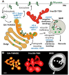
Vesicular trafficking and plant endosomes. A, Diagram showing the main vesicular trafficking pathways involving the TGN and MVEs. The main coats components and factors involved in vesicle formation are in blue. The two “zones” of the Golgi-associated TGN/early endosome (GA-TGN/EE) are characterized by either AP-1, Epsin, VAMP727, and clathrin (exocytosis/secretion trafficking zone) or APP-4, VAMP727, MTV1 (vacuolar trafficking zone). Small green hexagons depict soluble vacuolar cargo transported through the vacuolar trafficking zone of the TGN to MVEs and to the vacuole. At MVEs, intralumenal vesicles containing endocytosed PM proteins targeted for vacuolar degradation are formed by the action of ESCRT proteins and recycling vesicles bud into the cytoplasm, likely coated with the retromer complex. B, Electron tomographic reconstructions of a Golgi-associated TGN/early endosome (GA-TGN/EE), a free/Golgi-independent TGN (free/GI-TGN), and an MVE from a root plant cell. Scale bar = 50 nm.
Figure 2
Figure 2
Cortical microtubules as mechanosensors. A, Regulation of the alignment and density of cortical microtubules can impact their ability to sense mechanical signals. Modification of cortical microtubules (blue) by microtubule-associated proteins and tubulin posttranslational modifications provides a potential mechanism to locally control force-sensitivity of cortical microtubules. B, Disruption of the microtubule lattice by mechanical stress (red arrow) might affect protein interactions to directly transduce force into a biochemical signal. In this example, a hypothetical protein MAP-x preferentially binds to unstretched microtubules, whereas a hypothetical protein MAP-y preferentially binds to stretched microtubules. For simplicity, microtubules are shown as a single protofilament of tubulin dimers. C, Sensors are proposed to perceive severe cortical microtubule perturbations such as detachment from the PM and extensive depolymerization or fragmentation to initiate cortical microtubule integrity signaling.
Figure 3
Figure 3
Potential links between intracellular and extracellular cell wall regulation in growing plant cells. Transcription of cell wall-related genes in the nucleus, followed by mRNA export and translation on the rough ER surface, leads to the production of soluble and membrane proteins that enter the anterograde membrane trafficking system. In the Golgi, matrix polysaccharides including pectins and hemicelluloses are synthesized by large suites of glycosyltransferases (GTs) and other enzymes. Matrix polysaccharides and cellulose synthesis complexes (CSCs) are trafficked from the Golgi to the cell surface, where matrix polysaccharides (purple) are exocytosed into the apoplast and CSCs extrude cellulose (green) into the wall. These polymers and glycoproteins assemble into a strong, flexible wall that can expand anisotropically (more in one direction than another, see red dashed arrows), and matrix polysaccharides are modified in the wall and can be degraded by glycosyl hydrolases/lyases (GH). Wall polymer fragments can bind to wall receptors, which initiate intracellular signaling cascades that can change gene expression or modulate wall synthesis and/or assembly through trafficking and posttranslational modification of relevant proteins. Compartments/proteins are labeled with normal text and processes are highlighted in white ovals; arrows can entail multiple events/processes. MAPK, mitogen-activated protein kinase.
Figure 4
Figure 4
Plasmodesmata are dynamic intercellular connections. A, Plasmodesmata connect neighboring cells and close and open via regulation of callose synthesis and degradation. Adapted from (Maule et al., 2012) under CC BY-NC 3.0 https://creativecommons.org/licenses/by-nc/3.0/#. B, When plasmodesmata close, it limits the intercellular movement of molecules, but we do not understand how this contributes to the success of responses. In these images, GFP (green) is being synthesized in the cells marked with an asterisk. When the plasmodesmata are closed, the GFP is restricted to the site of synthesis (left image) whereas when the plasmodesmata are open, the GFP moves through plasmodesmata into the surrounding cells (right image). Cell outlines are in magenta and the scale bar is 20 μm. Images are maximum projections of confocal z-stacks of Arabidopsis leaf epidermal cells.
Figure 5
Figure 5
Mutations of a vacuolar regulator cause the arrest of mitotic division during female gametogenesis and the disruption of the stem cell niche (SCN) in the RAM. A–D, Confocal laser scanning microscopy (CLSM) of a wild-type (A and B) or mutant ovule (C and D) at Stage 3-I (A and C) or Stage 3-III (B and D). V, vacuole; CN, chalazal nucleus; FM, functional megaspore; MN, micropylar nucleus. The arrowhead points at the nucleus, which failed to undergo mitosis. E and F, CLSM of wild-type (E) or mutant (F) SCN. Mutations of a vacuolar regulator cause abnormal divisions of the quiescent center. Roots were stained with PI. Bars = 5 μm.
Figure 6
Figure 6
The PAM in a Medicago truncatula root cortical cell. Visualized as a consequence of a phosphate transporter-GFP fusion protein located in the membrane. A, GFP image. B, GFP and bright-field images merged. The PAM closely outlines the fungal arbuscule, which occupies a substantial proportion of the cortical cell. The name “arbuscule” is derived from the Latin for small tree.
Figure 7
Figure 7
Cell edges in morphogenesis. A, At the subcellular scale, edges can provide directional information to establish anisotropy at cell faces, for example, through nucleation or local stabilization of microtubules (red arrows). B, Cell edges can accumulate stresses (red arrows) arising at the cellular and supracellular scale through differential growth in adjacent cells. C, Cell edges can act as persistent polarity landmarks during organogenesis: For example, periclinal cell edges established in early lateral root development (red line, left) retain their relative position in the tissue during subsequent organ development (red line, right).
Figure 8
Figure 8
A cell decides where it divides. A, Cell division could occur symmetrically (1, 3) or asymmetrically (2, 4, 5). The orientation of the division plane could also vary. B, PPB marks the future division site prior to spindle assembly (arrowheads). Phragmoplasts and the associated cell plate expand toward the marked site. However, the division site can be defined without PPB at least in some cell types (bottom). How this is achieved is largely unknown. One possibility is that the division site is marked without the aid of microtubular PPB (dotted arrowheads). The formation of MTOC in the absence of PPB has been observed in moss gametophores. Microtubules are colored green; chromosomes are in gray.
Figure 9
Figure 9
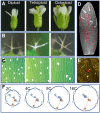
Ploidy induces nucleotypic changes in size. A, Arabidopsis flowers increase in size with polyploidy. B, Arabidopsis sepal trichomes increase in size and degree of branching with polyploidy. C, Sepal abaxial epidermal cell area increases, and density of guard cells (white) decreases, with ploidy. D, Within a single organ (sepal epidermis), cell size varies dramatically due to differences in ploidy resulting from programmed endoreduplication (16C sepal giant cells shown in red, diploid guard cells appear as white ovals). E, Nuclear volume increases with increasing cell size induced by endoreduplication. Yellow and red arrows indicate the nuclei of a diploid and an endopolyploid epidermal cell, respectively, in the sepal epidermis (cell walls stained with propidium iodide shown in yellow, nuclei expressing ML1:H2B-GFP, an epidermis-specific nuclear marker, shown in green). F, Simplified diagrams of nuclei from diploid, tetraploid, octoploid, and endopolyploid 16C cells (left to right), illustrating nucleotypic changes in nuclear size, density of nuclear pores (blue cylinders on nuclear surface), chromatin compaction (a strand of DNA in the nuclear interior shown as a line with nucleosomes shown as blue circles), and distances from sites of transcription to nuclear pores (orange arrows). The effects of polyploidy on chromatin accessibility can vary from locus to locus. A–C, Images within panels are shown at the same scale. A–E, Adapted from Robinson et al. (2018), Figure 3. Copyright American Society of Plant Biologists.
Figure 10
Figure 10
Altered patterning and fates upon compression in sunflower capitulum. Left: A young capitulum is constrained for several days with a vice. Scale bar: 5 mm. Right: Observation of floret with the symmetry of a central floret, but with the dimension of a ray floret, from a constrained capitulum. Scale bar: 3 mm. Adapted from Figure 7, A and D in Hernandez and Green (1993). Copyright American Society of Plant Biologists.
Figure 11
Figure 11
Regeneration of a whole plant from a single leaf mesophyll protoplast. A, A protoplast isolated from a leaf mesophyll cell. B, Protoplast cells that have undergone several rounds of cell division. C, Shoot meristem formation from protoplast-derived callus. D, A regenerated plant. Arrowheads in (A) mark a chloroplast. Scale bar = 10 mm (A), 50 mm (B), 1 mm (C), 2 mm (D) (Photos courtesy of Yuki Sakamoto).
Figure 12
Figure 12
De novo polarization of Equisetum spores. A, Unpolarized germinating spore with no obvious polar axis. The nucleus is the white disc at the cell center with two dark nucleoli. The light source was at the top of the figure. B, Organelles (mainly chloroplasts) are cleared from the shaded (lower) side of the cell. C, The nucleus is located in the shaded (lower side) of the cell from which other organelles are largely excluded. D, Mitotic spindle located in the shaded (lower) side of the cell. E, After cytokinesis a larger cell is located on the illuminated (upper) side and a smaller cell located on the shaded (lower) side of the two-celled sporeling. The light source was located at the top of the figure. Modified from Nienburg (1924) Die Wirkung des Lichtes auf die Keimung der Equisetum spore. Ber Deutsch Bot Ges 42, 95–99 with permission. © 1924 Deutsche Botanische Gesellschaft/German Botanical Society.
Figure 13
Figure 13
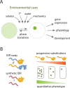
Examining the drivers and functions of IDP phase separation. A, IDPs undergo reversible liquid–liquid phase separation to form MLOs under different environmental cues, which likely function to facilitate a range of cellular, developmental, and physiological processes under changing environmental conditions. B, Two methods that can be used to assess the specific function of IDP phase separation: (left) swapping the IDR from a native IDP with either a heterologous or synthetic IDR that is known to phase separate and (right) quantitatively tuning IDP phase separation behavior and testing if it quantitatively tunes the respective phenotypic output.
Figure 14
Figure 14
How do plants deal with internal noise? A, Tissue with cells that stochastically express a gene of interest; transcription is on in green-filled nuclei and off in empty blue nuclei. B and C, Histograms of two traits with low (B) or high (C) standard deviation (hypothetical histograms for traits quantified over 1,000 individual plants). Does cell-to-cell variability (A) lead to variable phenotypes (B) or to precise phenotypes (C)? Examples of variable traits include germination time, number of petals in Cardamine, and number of secondary stems in Arabidopsis. Examples of robust traits include sepal size and number of petals in Arabidopsis. Traits such as plant height or rosette diameter in Arabidopsis may be variable or robust, depending on growth conditions.
Figure 15
Figure 15
Barrenness is an emergent phenotype resulting from downward causality and competition. Maize phenotypes observed in agriculture production fields managed to attain record yields in Nebraska. White arrow indicates dominated and barren plant.

Comment in

  • Back to the roots: A focus on plant cell biology.
    Weijers D, Bezanilla M, Jiang L, Roeder AHK, Williams M. Weijers D, et al. Plant Cell. 2022 Jan 20;34(1):1-3. doi: 10.1093/plcell/koab278. Plant Cell. 2022. PMID: 34755878 Free PMC article. No abstract available.

References

    1. Abu-Abied M, Rogovoy Stelmakh O, Mordehaev I, Grumberg M, Elbaum R, Wasteneys GO, Sadot E (2015) Dissecting the contribution of microtubule behaviour in adventitious root induction. J Exp Bot 66: 2813–2824 - PMC - PubMed
    1. Alamos S, Reime, r A, N, iyogi KK, Garcia HG (2021) Quantitative imaging of RNA polymerase II activity in plants reveals the single-cell basis of tissue-wide transcriptional dynamics. Nat Plants 7: 1037–1049 - PMC - PubMed
    1. Albert R (2007) Network inference, analysis, and modeling in systems biology. Plant Cell 19: 3327–3338 - PMC - PubMed
    1. Almonacid M, Terret MÉ, Verlhac MH (2014) Actin-based spindle positioning: new insights from female gametes. J Cell Sci 127: 477–483 - PubMed
    1. Ambrose C, Wasteneys GO (2011) Cell edges accumulate gamma tubulin complex components and nucleate microtubules following cytokinesis in Arabidopsis thaliana. PLoS One 6: e27423. - PMC - PubMed

Publication types