

CERMEP-IDB-MRXFDG: a database of 37 normal adult human brain [18F]FDG PET, T1 and FLAIR MRI, and CT images available for research
- PMID: 34529159
- PMCID: PMC8446124
- DOI: 10.1186/s13550-021-00830-6
CERMEP-IDB-MRXFDG: a database of 37 normal adult human brain [18F]FDG PET, T1 and FLAIR MRI, and CT images available for research
Abstract
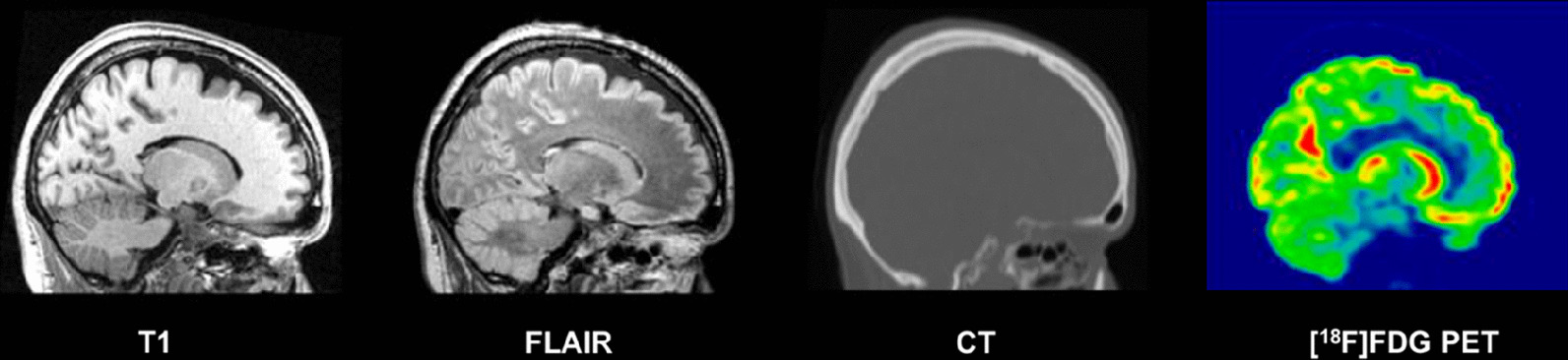
We present a database of cerebral PET FDG and anatomical MRI for 37 normal adult human subjects (CERMEP-IDB-MRXFDG). Thirty-nine participants underwent static [18F]FDG PET/CT and MRI, resulting in [18F]FDG PET, T1 MPRAGE MRI, FLAIR MRI, and CT images. Two participants were excluded after visual quality control. We describe the acquisition parameters, the image processing pipeline and provide participants' individual demographics (mean age 38 ± 11.5 years, range 23-65, 20 women). Volumetric analysis of the 37 T1 MRIs showed results in line with the literature. A leave-one-out assessment of the 37 FDG images using Statistical Parametric Mapping (SPM) yielded a low number of false positives after exclusion of artefacts. The database is stored in three different formats, following the BIDS common specification: (1) DICOM (data not processed), (2) NIFTI (multimodal images coregistered to PET subject space), (3) NIFTI normalized (images normalized to MNI space). Bona fide researchers can request access to the database via a short form.
Keywords: CT; Database sharing; Healthy subjects; MRI; Neuroimaging; PET-FDG.
© 2021. The Author(s).
Conflict of interest statement
The authors have no relevant financial or non-financial interests to disclose.
Figures


References
-
- Yaakub S, McGinnity C, Beck K, Merida I, Dunston E, Muffoletto M, et al. Brain PET-MR attenuation correction with deep learning. J Cereb Blood Flow Metab. 2019;39:600–601.
Grants and funding
LinkOut - more resources
Full Text Sources
Medical
