. 2021 Nov;190(2):255-264.
doi: 10.1007/s10549-021-06388-0. Epub 2021 Sep 16.

Hyperglycemic conditions proliferate triple negative breast cancer cells: role of ornithine decarboxylase

Affiliations

Hyperglycemic conditions proliferate triple negative breast cancer cells: role of ornithine decarboxylase

Caleb C Capellen et al. Breast Cancer Res Treat. 2021 Nov.

Abstract

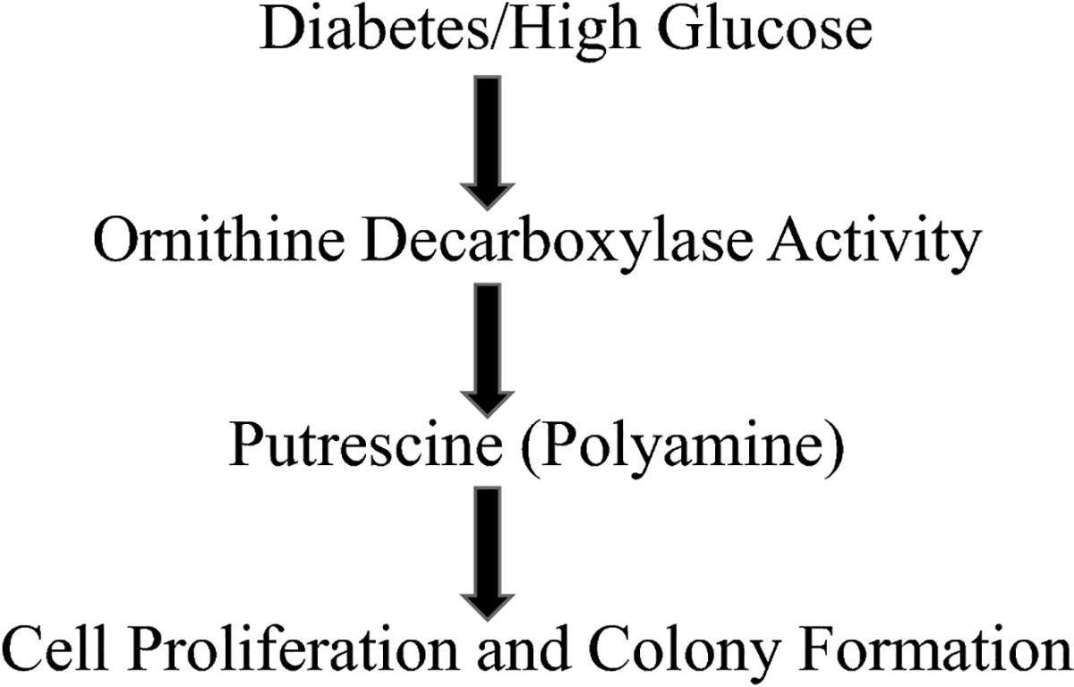
Purpose: Several cancer subtypes (pancreatic, breast, liver, and colorectal) rapidly advance to higher aggressive stages in diabetes. Though hyperglycemia has been considered as a fuel for growth of cancer cells, pathways leading to this condition are still under investigation. Cellular polyamines can modulate normal and cancer cell growth, and inhibitors of polyamine synthesis have been approved for treating colon cancer, however the role of polyamines in diabetes-mediated cancer advancement is unclear as yet. We hypothesized that polyamine metabolic pathway is involved with increased proliferation of breast cancer cells under high glucose (HG) conditions.

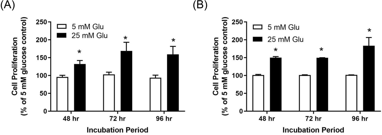
Methods: Studies were performed with varying concentrations of glucose (5-25 mM) exposure in invasive, triple negative breast cancer cells, MDA-MB-231; non-invasive, estrogen/progesterone receptor positive breast cancer cells, MCF-7; and non-tumorigenic mammary epithelial cells, MCF-10A.

Results: There was a significant increase in proliferation with HG (25 mM) at 48-72 h in both MDA-MB-231 and MCF-10A cells but no such effect was observed in MCF-7 cells. This was correlated to higher activity of ornithine decarboxylase (ODC), a rate-limiting enzyme in polyamine synthesis pathway. Inhibitor of polyamine synthesis (difluoromethylornithine, DFMO, 5 mM) was quite effective in suppressing HG-mediated cell proliferation and ODC activity in MDA-MB-231 and MCF-10A cells. Polyamine (putrescine) levels were significantly elevated with HG treatment in MDA-MB-231 cells. HG exposure also increased the metastasis of MDA-MB-231 cells.

Conclusions: Our cellular findings indicate that polyamine inhibition should be explored in patient population as a target for future chemotherapeutics in diabetic breast cancer.

Keywords: Breast cancer; Diabetes; High glucose; Ornithine decarboxylase; Polyamine; Putrescine.


Conflict of interest statement

Declarations of interest: none

Conflicts of interest/Competing interests: None

Figures

Figure 1.
Figure 1.
Schematic of polyamine pathway in animal cells. Primary pathway for polyamine synthesis is presented in the middle which involves the action of ornithine decarboxylase to produce the polyamines (putrescine, PUT, spermidine, SPD, and spermine, SPM) from L-ornithine. In addition, low levels of SPD or SPM also trigger input of these metabolites from a parallel alternate pathway through the decarboxylation of S-adenosylmethionine (S-AdM). Catabolism of SPD and SPM is mediated through the enzymes spermine/spermidine acetyltransferase (SSAT), and polyamine oxidase (PAOX). ODC, the first enzyme in the pathway, can be endogenously inhibited by ODC antizyme or exogenously using DFMO.
Figure 2.
Figure 2.
Dose response of proliferation with glucose treatment in MCF-7, MDA-MB-231, and MCF-10A cells treated with 5mM, 10mM, 25mM glucose, or mannitol (20mM with 5mM glucose) for 72h; *p<0.05 vs. respective 5mM glucose). Values are presented as %mean ± SEM compared to respective 5mM Glu treatment at that time point, n=3, each treatment with replicates.
Figure 3.
Figure 3.
Time response of proliferation with glucose treatment in (A) MDA-MB-231, and (B) MCF-10A cells. LG (5mM Glu) and HG (25mM Glu); *p<0.05 vs. respective 5mM glucose. Values are presented as %mean ± SEM compared to respective 5mMGlu treatment at that time point, n=3, each treatment with replicates.
Figure 4.
Figure 4.
(A) Cell proliferation of MDA-MB-231 and MCF-10A cells in LG (5mM Glu) and HG (25mM Glu), in presence or absence of polyamine inhibitor, DFMO (5mM) for 72h (*p<0.05 vs. 5mM glucose, #p<0.05 vs. 25mM glucose). Values are mean ± SEM, n=3, each treatment with replicates. (B) Colony forming assay (clonogenic) for MCF-7, MDA-MB-231, and MCF-10A cells in LG (5mM Glu) and HG (25mM Glu), with and without polyamine inhibitor, DFMO (2mM) for 72h (*p<0.05 vs. 5mM glucose, #p<0.05 vs. 25mM glucose). Survival fraction (SF) represents the number of colonies formed based on number of cells plated and cells survived after treatments. Colonies with ≥ 50 cells were counted for calculations. Values are mean ± SEM, n=3, each treatment with replicates.
Figure 5.
Figure 5.
ODC activity assay in cells treated with varying concentrations of glucose (5mM, 25mM) and/or DFMO for 72h. Values are mean±SEM, *p<0.05 vs 5mM glucose, #p<0.05 vs. 25mM glucose.
Figure 6.
Figure 6.
Scratch wound healing assay in MDA-MB-231 cells treated with glucose and/or DFMO for 0, 24, 48h. Images were taken using phase contrast microscopy, and area of wound (% of total area) was calculated using ImageJ (NIH) software. Multiple images were taken for each treatment, and averaged. All treatments were performed in replicates. Values are mean±SEM, *p<0.05 vs 5mM glucose.
Figure 7:
Figure 7:
Link between glucose and polyamine through the aspartate-argininosuccinate shunt. Glucose is metabolized through glycolysis (cytosol) and citric acid cycle (mitochondria). Oxaloacetate, a product of citric acid cycle can be shunted to form aspartate through transaminase which is further metabolized to argininosuccinate, a part of the urea cycle. Argininosuccinate is converted to arginine and then ornithine in the urea cycle, which are upstream substrates for conversion to polyamines.
Figure 8:
Figure 8:
Schematic of proposed model for diabetes-mediated aggression of breast cancer states

References

    1. Onitilo AA, Engel JM, Glurich I, Stankowski RV, Williams GM, and Doi SA, Diabetes and cancer I: risk, survival, and implications for screening. Cancer Causes Control, 2012. 23(6): p. 967–81. - PMC - PubMed
    1. Onitilo AA, Stankowski RV, Berg RL, Engel JM, Glurich I, Williams GM, and Doi SA, Breast cancer incidence before and after diagnosis of type 2 diabetes mellitus in women: increased risk in the prediabetes phase. Eur J Cancer Prev, 2014. 23(2): p. 76–83. - PubMed
    1. Giovannucci E, Harlan DM, Archer MC, Bergenstal RM, Gapstur SM, Habel LA, Pollak M, Regensteiner JG, and Yee D, Diabetes and cancer: a consensus report. CA Cancer J Clin, 2010. 60(4): p. 207–21. - PubMed
    1. Chowdhury TA, Diabetes and cancer. QJM, 2010. 103(12): p. 905–15. - PubMed
    1. Xu CX, Zhu HH, and Zhu YM, Diabetes and cancer: Associations, mechanisms, and implications for medical practice. World J Diabetes, 2014. 5(3): p. 372–80. - PMC - PubMed