. 2021 Sep 16;17(9):e1009804.
doi: 10.1371/journal.ppat.1009804. eCollection 2021 Sep.

Identification of immune correlates of fatal outcomes in critically ill COVID-19 patients

Affiliations

Identification of immune correlates of fatal outcomes in critically ill COVID-19 patients

Jonathan Youngs et al. PLoS Pathog. .

Abstract

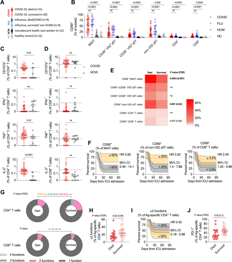
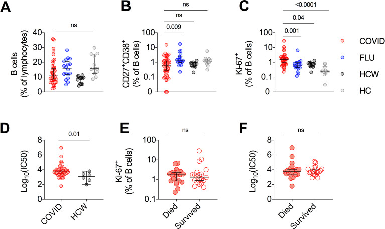
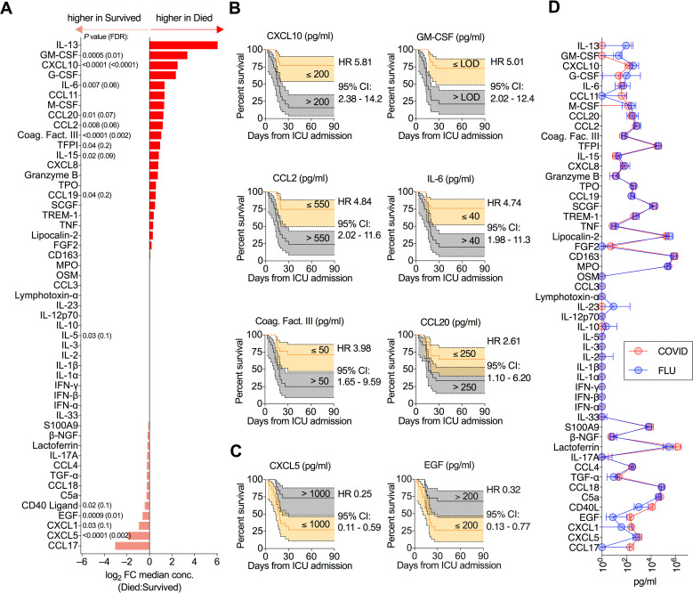
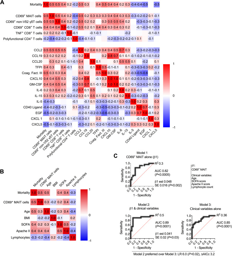
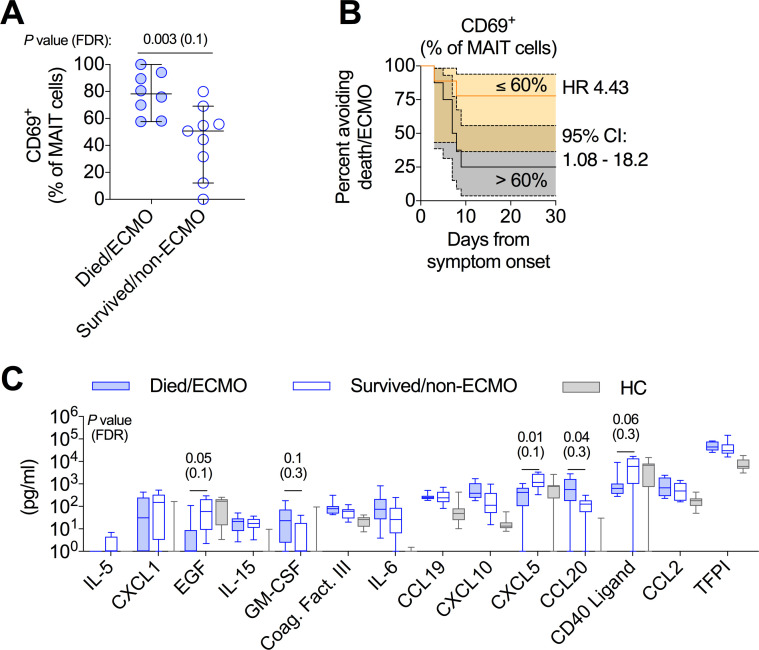
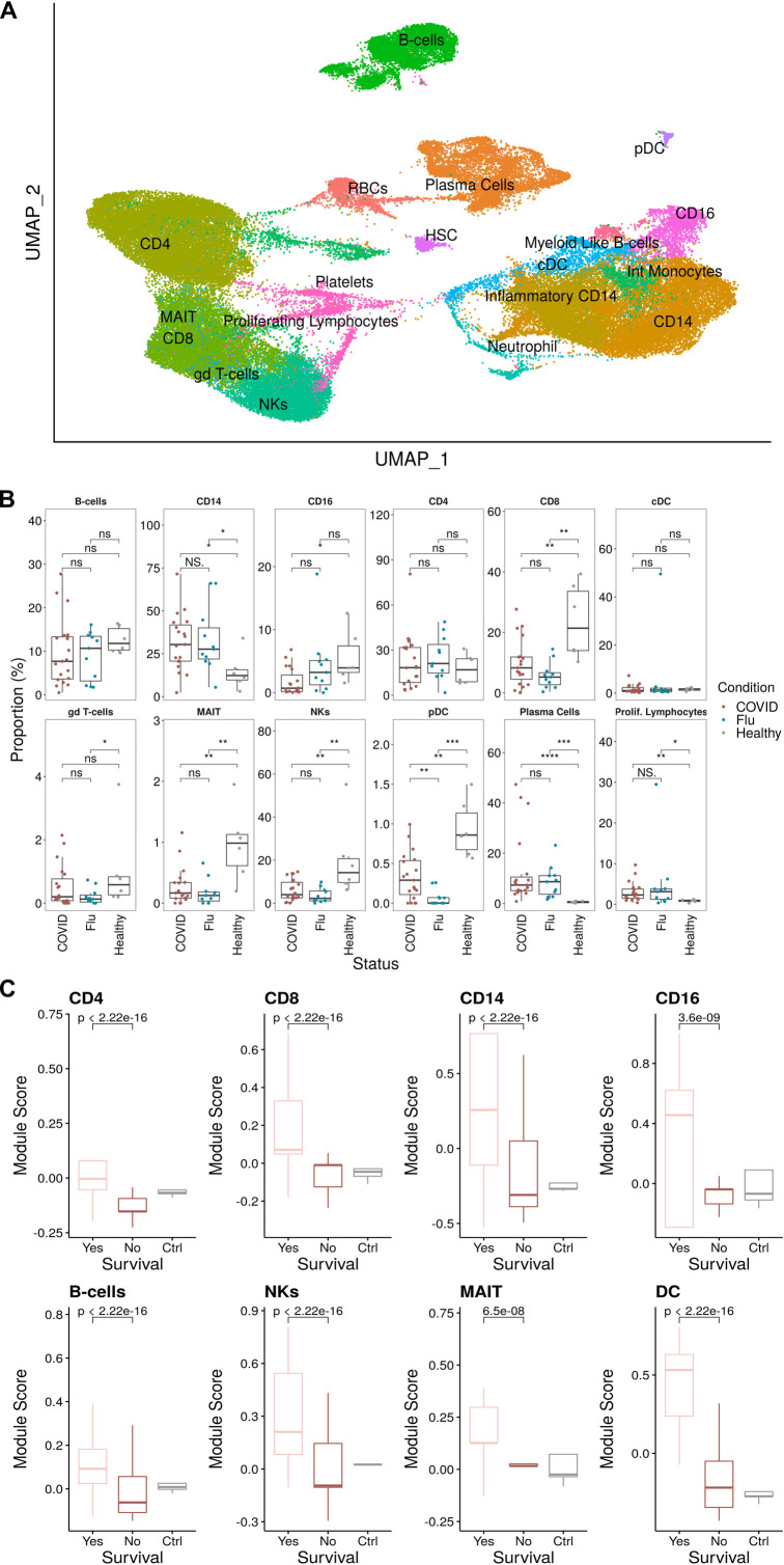
Prior studies have demonstrated that immunologic dysfunction underpins severe illness in COVID-19 patients, but have lacked an in-depth analysis of the immunologic drivers of death in the most critically ill patients. We performed immunophenotyping of viral antigen-specific and unconventional T cell responses, neutralizing antibodies, and serum proteins in critically ill patients with SARS-CoV-2 infection, using influenza infection, SARS-CoV-2-convalescent health care workers, and healthy adults as controls. We identify mucosal-associated invariant T (MAIT) cell activation as an independent and significant predictor of death in COVID-19 (HR = 5.92, 95% CI = 2.49-14.1). MAIT cell activation correlates with several other mortality-associated immunologic measures including broad activation of CD8+ T cells and non-Vδ2 γδT cells, and elevated levels of cytokines and chemokines, including GM-CSF, CXCL10, CCL2, and IL-6. MAIT cell activation is also a predictor of disease severity in influenza (ECMO/death HR = 4.43, 95% CI = 1.08-18.2). Single-cell RNA-sequencing reveals a shift from focused IFNα-driven signals in COVID-19 ICU patients who survive to broad pro-inflammatory responses in fatal COVID-19 -a feature not observed in severe influenza. We conclude that fatal COVID-19 infection is driven by uncoordinated inflammatory responses that drive a hierarchy of T cell activation, elements of which can serve as prognostic indicators and potential targets for immune intervention.


Conflict of interest statement

I have read the journal’s policy and the authors of this manuscript have the following competing interests:WH reports lecture fees from Gilead.

Figures

Fig 1
Fig 1. Relationship between T cell activation and mortality in critically ill COVID-19 patients.
(A) Schematic of the study cohort. All COVID-19 and influenza patients were critically ill. Convalescent health care workers had mild disease. Samples from healthy controls were collected pre-pandemic. (B) Direct ex vivo measurement of general activation of each T cell subset. (C-D) Quantification of spike-specific CD4+ T cells (C) and CD8+ T cells (D) in acute critically ill COVID-19 patients and health care workers. Dashed line indicates the upper 95% confidence interval for responses detected in pre-pandemic healthy controls. (E) Median ex vivo activation level in each T cell subset of critically ill COVID-19 patients who died or survived. (F) Kaplan-Meier survival curves of critically ill COVID-19 patients based on relative CD69 expression on each T cell subset. (G) Polyfunctionality (CD107a, IFNγ, TNF, and/or IL-2) of spike-specific CD4+ T cells (top) and CD8+ T cells (bottom) between critically ill COVID-19 patients who died or survived. (H) Fraction of spike-specific CD4+ T cells that are polyfunctional (≥2 cytokines produced) in critically ill COVID-19 patients that died versus survived. (I) Kaplan-Meier survival curve of critically ill COVID-19 patients based on fraction of polyfunctional spike-specific CD4+ T cells. (J) PD-1 expression on spike-specific CD4+ T cells from critically ill COVID-19 patients who died or survived. Dots represent individual patients. Median ± 95% CI are shown. (B) Kruskal-Wallis tests with Dunn’s multiple comparison test. (A to E, G, H, and J) Mann-Whitney U-test. Benjamini-Hochberg FDR calculation was used for all statistical analyses involving associations with mortality.
Fig 2
Fig 2. No association between SARS-CoV-2 neutralizing antibodies and fatal outcome in critically ill COVID-19 patients.
(A-C) Frequency of B cells (A) CD27+CD38+ plasmablasts (B) and Ki-67+ proliferating B cells (C). (D) Log10 of the reciprocal plasma dilution that neutralized 50% of SARS-CoV-2 spike protein-expressing pseudovirus. 50% Inhibitory concentration (IC50). (E-F) Frequency of Ki-67+ B cells (E) and Log10 of the reciprocal plasma dilution that neutralized 50% of SARS-CoV-2 spike protein-expressing pseudovirus (F) with critically ill COVID-19 patients stratified by survival. Median ± 95% CI are shown. (A to C) Kruskal-Wallis tests with Dunn’s multiple comparison test. (D to F) Mann-Whitney U-test.
Fig 3
Fig 3. Mortality in critically ill COVID-19 patients is associated with perturbations in serum protein levels.
(A) Fold-change in median serum protein concentration between critically ill COVID-19 patients who died versus survived. (B-C) Kaplan-Meier survival curves of critically ill COVID-19 patients based on concentration of the indicated analyte. Only proteins where FDR<0.1 (from (A)) are plotted. (B) Kaplan-Meier survival curves for serum proteins where above median expression is associated with increased mortality. (C) Kaplan-Meier survival curves for serum proteins where above median expression is associated with decreased mortality. (D) Median serum cytokine concentration in critically ill COVID-19 patients and critically ill influenza patients. (A) Mann-Whitney U-test with Benjamini-Hochberg FDR calculation on data presented in S4A Fig.
Fig 4
Fig 4. Activation of MAIT cells is associated with worse disease outcomes in critically ill COVID-19.
(A) Pearson correlation of mortality with all statistically significant immunologic parameters. (B) Pearson correlation of mortality, MAIT cell CD69 expression, clinical measures, and timing of sampling. (C) ROC curve of MAIT cell CD69 expression (Model 1), ROC curve of MAIT cell CD69 expression combined with clinical variables (Model 2), and ROC curve of clinical variables alone (model 3). LR: likelihood ratio, ΔAICc: Difference in Akaike’s Information Criterion corrected.
Fig 5
Fig 5. Activation of MAIT cells is associated with worse disease outcomes in critically ill influenza patients.
Investigation of immunologic measures that were associated with mortality in the critically ill COVID-19 cohort, in a cohort of critically ill influenza patients. (A) MAIT cell CD69 expression on critically ill influenza patients who died or required ECMO versus those who did not. (B) Kaplan-Meier survival curve of disease outcome of critically ill influenza patients based on above or below median MAIT cell CD69 expression. (C) Examination of the concentration of 14 serum proteins in the critically ill influenza cohort and healthy controls, which were associated with mortality in the critically ill COVID-19 cohort (Fig 3). Dots represent individual patients (A), and median ± 95% CI are shown. (C) Median, IQR, and min to max are shown. (A and C) Mann-Whitney U-test with Benjamini-Hochberg FDR calculation.
Fig 6
Fig 6. Elevated type I interferon signaling across multiple cell populations in critically ill COVID-19 patients who survive.
(A) UMAP projection of the scRNA-seq cohort consisting of 75,601 PBMCs (43,687 cells from COVID, 16,616 Healthy, and 15,298 Flu) colored by manually annotated cell types. (B) Relative cell proportions within each individual separated by disease condition. Statistical tests were conducted using the Wilcoxon rank-sum test between each condition. Summary values were subsequently displayed using the boxplot. The box is equivalent to the interquartile range (IQR) with the median as the center, and whiskers correspond to the 25th percentile—1.5x IQR or the lowest value, and 75th percentile +1.5x IQR or the highest value. (C) Type I IFN module scores for each major cell type compared between COVID survival and death with healthy controls as a reference. Significance was determined using the Wilcoxon test.
Fig 7
Fig 7. Divergent cytokine signaling pathways associated with clinical outcome in critically ill COVID-19 and influenza patients.
(A,B) Curated dotplot of enriched pathways between both survival and death in critically ill COVID patients (A) and critically ill influenza patients (B). Enriched pathways were obtained via reactome pathways identified via GSEA between survival and death conditions within the COVID dataset, or ECMO non-ECMO in the influenza cohort. FDRs were calculated based on q-values obtained from the hypergeometric test applied to the geneset followed by multiple hypothesis correction using the Benjamini-Hochberg method. NES: normalized enrichment score, FDR: false discovery rate.

References

    1. Docherty AB, Harrison EM, Green CA, Hardwick HE, Pius R, Norman L, et al.. Features of 20 133 UK patients in hospital with covid-19 using the ISARIC WHO Clinical Characterisation Protocol: prospective observational cohort study. BMJ. 2020;: m1985–12. doi: 10.1136/bmj.m1985 - DOI - PMC - PubMed
    1. Yang Y, Shen C, Li J, Yuan J, Wei J, Huang F, et al.. Plasma IP-10 and MCP-3 levels are highly associated with disease severity and predict the progression of COVID-19. Journal of Allergy and Clinical Immunology. 2020;146: 119–127.e4. doi: 10.1016/j.jaci.2020.04.027 - DOI - PMC - PubMed
    1. Lucas C, Wong P, Klein J, Castro TBR, Silva J, Sundaram M, et al.. Longitudinal analyses reveal immunological misfiring in severe COVID-19. Nature. Springer US; 2020;: 1–27. doi: 10.1038/s41586-020-2588-y - DOI - PMC - PubMed
    1. Laing AG, Lorenc A, del Molino del Barrio I, Das A, Fish M, Monin L, et al.. A dynamic COVID-19 immune signature includes associations with poor prognosis. Nat Med. Nature Publishing Group; 2020;26: 1623–1635. doi: 10.1038/s41591-020-1038-6 - DOI - PubMed
    1. Hadjadj J, Yatim N, Barnabei L, Corneau A, Boussier J, Smith N, et al.. Impaired type I interferon activity and inflammatory responses in severe COVID-19 patients. Science. 2020;31: eabc6027–15. doi: 10.1126/science.abc6027 - DOI - PMC - PubMed

Publication types

MeSH terms