. 2021 Nov 14;42(43):4431-4441.
doi: 10.1093/eurheartj/ehab653.

Untangling the pathophysiologic link between coronary microvascular dysfunction and heart failure with preserved ejection fraction

Affiliations

Untangling the pathophysiologic link between coronary microvascular dysfunction and heart failure with preserved ejection fraction

Aish Sinha et al. Eur Heart J. .

Abstract

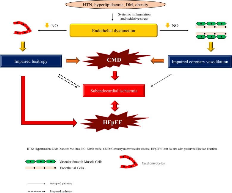
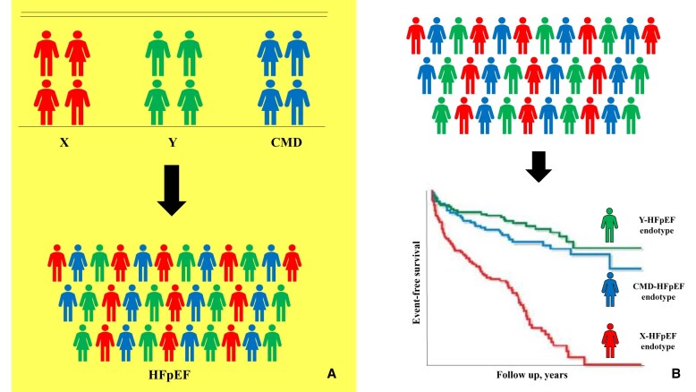
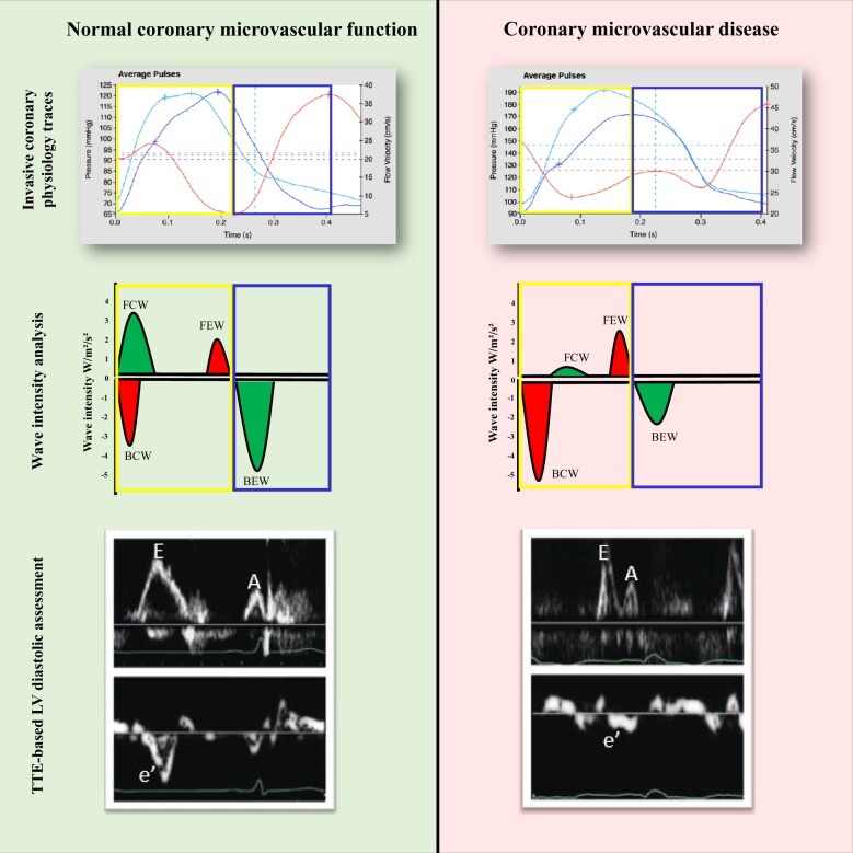
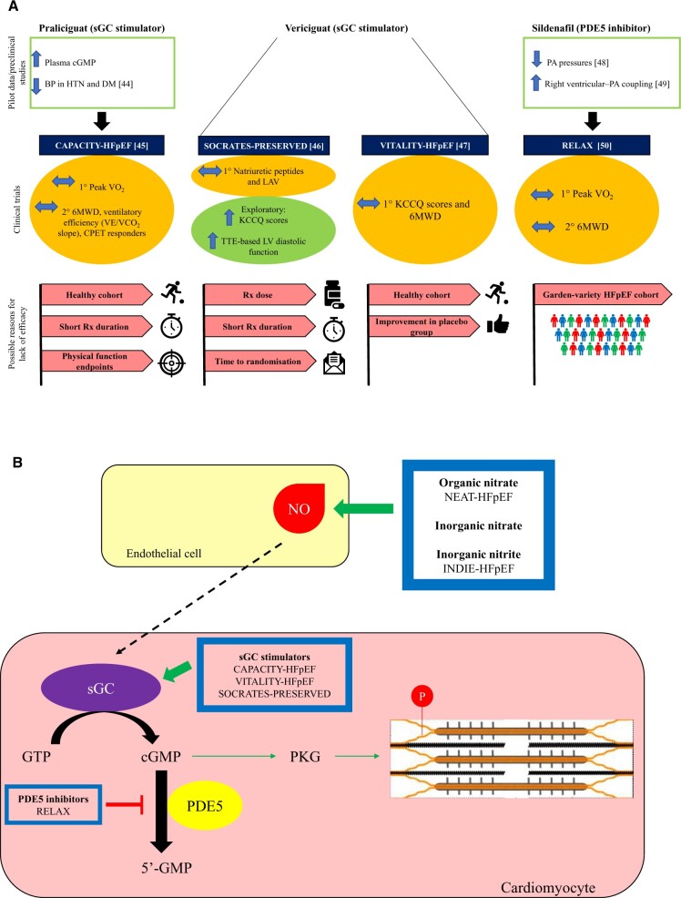
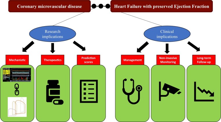
Coronary microvascular disease (CMD), characterized by impaired coronary flow reserve (CFR), is a common finding in patients with stable angina. Impaired CFR, in the absence of obstructive coronary artery disease, is also present in up to 75% of patients with heart failure with preserved ejection fraction (HFpEF). Heart failure with preserved ejection fraction is a heterogeneous syndrome comprising distinct endotypes and it has been hypothesized that CMD lies at the centre of the pathogenesis of one such entity: the CMD-HFpEF endotype. This article provides a contemporary review of the pathophysiology underlying CMD, with a focus on the mechanistic link between CMD and HFpEF. We discuss the central role played by subendocardial ischaemia and impaired lusitropy in the development of CMD-HFpEF, as well as the clinical and research implications of the CMD-HFpEF mechanistic link. Future prospective follow-up studies detailing outcomes in patients with CMD and HFpEF are much needed to enhance our understanding of the pathological processes driving these conditions, which may lead to the development of physiology-stratified therapy to improve the quality of life and prognosis in these patients.

Keywords: Coronary flow reserve; Coronary microvascular disease; Heart failure with preserved ejection fraction; Lusitropy; Subendocardial ischaemia.


Figures

None
The mechanistic link between coronary microvascular disease and heart failure with preserved ejection fraction.
Figure 1
Figure 1
This figure illustrates that heart failure with preserved ejection fraction is a heterogeneous syndrome comprising distinct endotypes, each with disparate underlying pathophysiology, therapeutic options, and outcomes. Patients can be characterized clinically according to factors such as pathophysiology (A), or they can be characterized using artificial intelligence-derived clusters that groups patients according to their clinical characteristics and clinical outcomes (B). Note: (B) is illustrative and not based on real data. CMD, coronary microvascular disease; HFpEF, heart failure with preserved ejection fraction.
Figure 2
Figure 2
The left panel represents a normal control: the backward expansion wave becomes augmented on exertion, indicating enhanced lusitropy and myocardial perfusion. The right panel represents a patient with coronary microvascular disease: The backward compression wave indicates deceleration of flow and is augmented during exertion in these patients, whereas the backward expansion wave is attenuated. Note that diastole is defined electrically and all haemodynamic traces are gated to the R wave. The traces of aortic pressure, coronary pressure, and flow velocity are ensemble-averaged waveforms in a single calibrated wave. The wave intensity values (W/m2/s2) are for illustration purposes only and do not represent real data. The transthoracic echocardiogram-derived Doppler traces demonstrate normal left ventricular diastolic function in a control patient and impaired left ventricular diastolic function in a patient with coronary microvascular disease. BCW, backward compression wave; BEW, backward expansion wave; FCW, forward compression wave; FEW, forward expansion wave; LV, left ventricular; TTE, transthoracic echocardiogram.
Figure 3
Figure 3
Acetylcholine has dual effects on coronary microvasculature. It binds to the muscarinic 3 receptor on endothelial cells and leads to an influx of intracellular calcium via the L-type calcium channels. Intracellular calcium binds to the protein calmodulin, and the calcium–calmodulin complex activates the endothelial nitric oxide synthase enzyme, which catalyzes the conversion of L-Arginine into nitric oxide. Nitric oxide then diffuses into the neighbouring vascular smooth muscle cell and activates soluble Guanylate Cyclase enzyme to catalyze the conversion of Guanosine Triphosphate into cyclic Guanosine Monophosphate. Cyclic Guanosine Monophosphate activates the protein kinase G, which, via a series of intracellular events, inactivates the calcium channels on the vascular smooth muscle cell. This reduces the intracellular influx of calcium into the vascular smooth muscle cell, therefore leading to vasodilation. Acetylcholine also binds to the muscarinic 3 receptor on the surface of vascular smooth muscle cells and, in the presence of endothelial dysfunction, leads to unopposed vasoconstriction. Calcium enters vascular smooth muscle cells via the L-type calcium channels and binds to the protein calmodulin. The calcium–calmodulin complex activates myosin light chain kinase, which phosphorylates myosin light chains. Myosin light chains are found on the myosin heads and myosin light chain phosphorylation leads to cross-bridge formation between the myosin heads and the actin filaments, leading to vascular smooth muscle contraction. Myosin light chain phosphatase dephosphorylates myosin light chain and promotes unbinding of the myosin-actin filaments, therefore leading to vasodilation. Cyclic Guanosine Monophosphate promotes myosin light chain phosphatase activity. The myosin head detaches from the actin binding site after adenosine triphosphate attaches to the myosin head. This adenosine triphosphate is then hydrolyzed to adenosine diphosphate and inorganic phosphate by the myosin head; this adenosine diphosphate and inorganic phosphate is then released by the myosin head after the power stroke. At this point, the myosin head is ready for the next adenosine triphosphate to allow detachment from the myosin head. ACh, acetylcholine; ADP, adenosine diphosphate; ATP, adenosine triphosphate; Ca2+, calcium; cGMP, cyclic Guanosine Monophosphate; eNOS, endothelial nitric oxide synthase; GTP, Guanosine Triphosphate; M3, muscarinic 3; MLCs, myosin light chains; MLCK, myosin light chain kinase; MLCP, Myosin light chain phosphatase; NO, nitric oxide; Pi, inorganic phosphate; PKG, protein kinase G; sGC, soluble Guanylate Cyclase; VSMC, vascular smooth muscle cell.
Figure 4
Figure 4
Results of therapeutic trials targeting the nitric oxide–cyclic Guanosine Monophosphate–protein kinase G pathway. (A) Summarizes the pilot data (or pre-clinical data), clinical trial data and the potential reasons for neutral outcomes, whilst (B) highlights the specific intracellular pathways that each of the novel agents act on. None of these trials met their primary endpoints. However, all of these trials are plagued by specific and generic trial design limitations. The treatment arm of CAPACITY-HFpEF and SOCRATES-PRESERVED trials lasted for only 12 weeks, which may not have been long enough to lead to sustained improvements in the study endpoints. The patient cohort recruited in the CAPACITY-HFpEF and VITALITY-HFpEF trials may represent a ‘healthier’ cohort that is not representative of the ‘real-world’ patient cohort. In the CAPACITY-HFpEF study, only 20% of patients had elevated filling pressures and a majority of patients had New York Heart Association II symptoms. Attenuation of cyclic Guanosine Monophosphate levels in patients with HFpEF is due to the loss of upstream nitric oxide rather than an excessive breakdown of cyclic Guanosine Monophosphate. It is, therefore, not unexpected that attempting to augment cyclic Guanosine Monophosphate by inhibiting its breakdown did not lead to clinically meaningful improvements in patients’ haemodynamics or exercise capacity in the RELAX trial. Furthermore, whilst the agents used in these trials target the nitric oxide–cyclic Guanosine Monophosphate–protein kinase G pathway, none of the trials directly studied physiological endpoints of impaired vascular function, such as peripheral or coronary endothelial function. Additionally, the physical functioning endpoints (6-min walk distance and change in peak VO2) may not be able to discriminate among effective therapies because patients with HFpEF usually suffer from multiple comorbidities and their impaired physical functioning may be multifactorial in nature. BP, blood pressure; cGMP, cyclic Guanosine Monophosphate; DM, diabetes mellitus; GTP, Guanosine Triphosphate; HFpEF, heart failure with preserved ejection fraction; HTN, hypertension; KCCQ: Kansas City Cardiomyopathy Questionnaire; LAV, left atrial volume; NO, nitric oxide; PA, pulmonary artery; PKG, protein kinase G; sGC, soluble Guanylate Cyclase; 6MWD, 6-min walk distance.
Figure 5
Figure 5
Clinical and research implications of the coronary microvascular disease–heart failure with preserved ejection fraction mechanistic link.

References

    1. Sara J, Widmer R, Matsuzawa Y, Lennon R, Lerman L, Lerman A. Prevalence of coronary microvascular dysfunction among patients with chest pain and nonobstructive coronary artery disease. JACC Cardiovasc Interv 2015;8:1445–1453. - PubMed
    1. Cannon R, Epstein S. “Microvascular angina” as a cause of chest pain with angiographically normal coronary arteries. Am J Cardiol 1988;61:1338–1343. - PubMed
    1. Rahman H, Ryan M, Lumley M, Modi B, McConkey H, Ellis H, Scannell C, Clapp B, Marber M, Webb A, Chiribiri A, Perera D. Coronary microvascular dysfunction is associated with myocardial ischemia and abnormal coronary perfusion during exercise. Circulation 2019;140:1805–1816. - PMC - PubMed
    1. Rahman H, Demir OM, Khan F, Ryan M, Ellis H, Mills MT, Chiribiri A, Webb A, Perera D. Physiological stratification of patients with angina due to coronary microvascular dysfunction. J Am Coll Cardiol 2020;75:2538–2549. - PMC - PubMed
    1. Bairey Merz CN, Pepine CJ, Walsh MN, Fleg JL. Ischemia and no obstructive coronary artery disease (INOCA): developing evidence-based therapies and research agenda for the next decade. Circulation 2017;135:1075–1092. - PMC - PubMed

Publication types