. 2021 Oct;20(10):e13480.
doi: 10.1111/acel.13480. Epub 2021 Sep 16.

Activation of angiotensin-converting enzyme 2/angiotensin (1-7)/mas receptor axis triggers autophagy and suppresses microglia proinflammatory polarization via forkhead box class O1 signaling

Affiliations

Activation of angiotensin-converting enzyme 2/angiotensin (1-7)/mas receptor axis triggers autophagy and suppresses microglia proinflammatory polarization via forkhead box class O1 signaling

Ruili Dang et al. Aging Cell. 2021 Oct.

Abstract

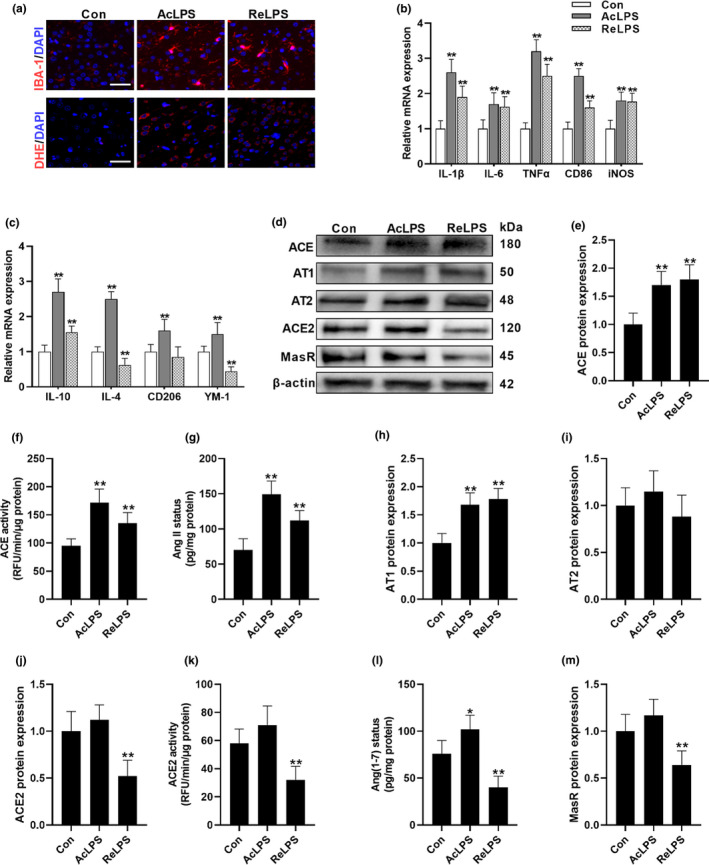
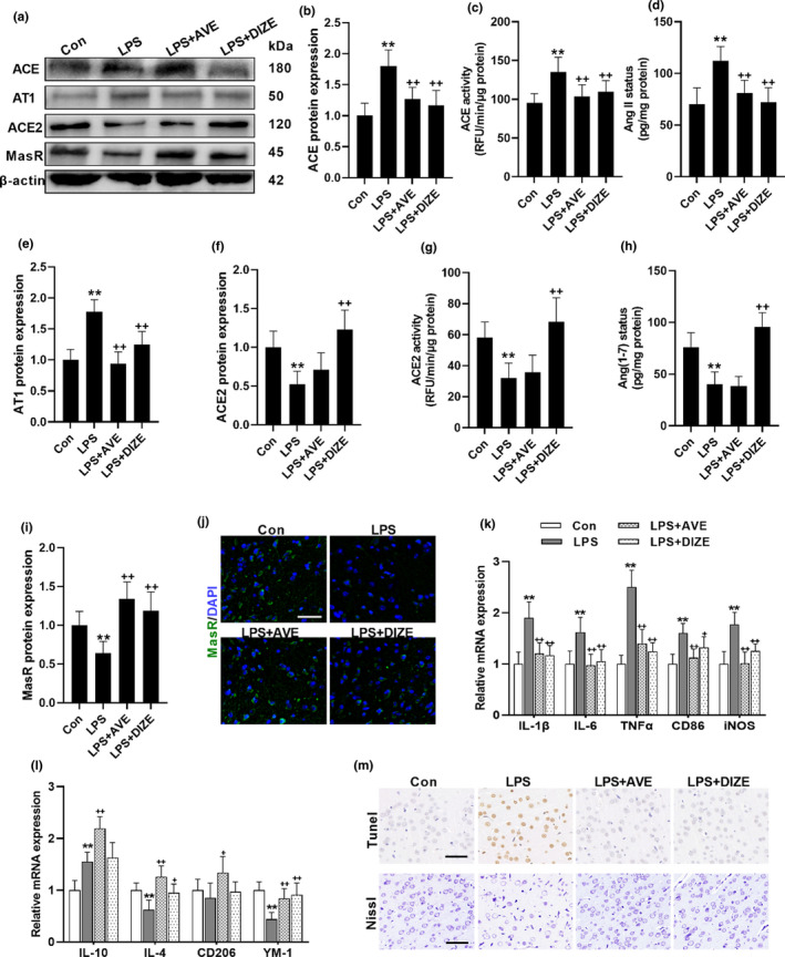
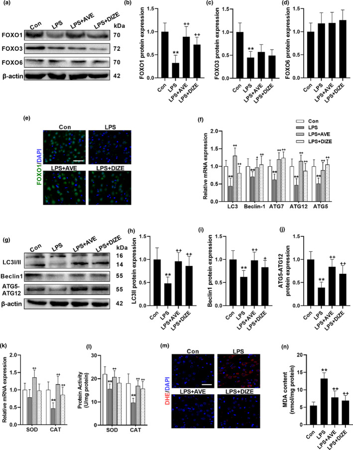
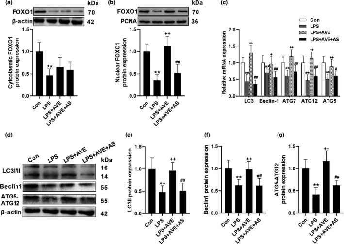
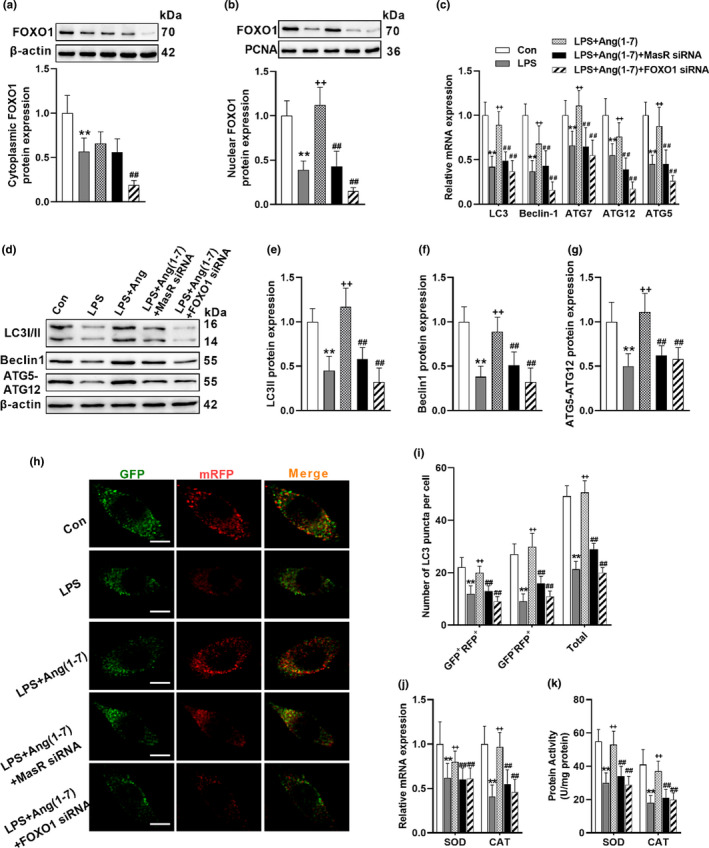
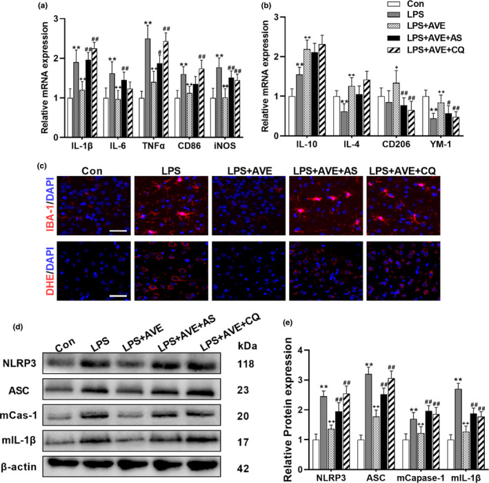
Brain renin-angiotensin (Ang) system (RAS) is implicated in neuroinflammation, a major characteristic of aging process. Angiotensin (Ang) II, produced by angiotensin-converting enzyme (ACE), activates immune system via angiotensin type 1 receptor (AT1), whereas Ang(1-7), generated by ACE2, binds with Mas receptor (MasR) to restrain excessive inflammatory response. Therefore, the present study aims to explore the relationship between RAS and neuroinflammation. We found that repeated lipopolysaccharide (LPS) treatment shifted the balance between ACE/Ang II/AT1 and ACE2/Ang(1-7)/MasR axis to the deleterious side and treatment with either MasR agonist, AVE0991 (AVE) or ACE2 activator, diminazene aceturate, exhibited strong neuroprotective actions. Mechanically, activation of ACE2/Ang(1-7)/MasR axis triggered the Forkhead box class O1 (FOXO1)-autophagy pathway and induced superoxide dismutase (SOD) and catalase (CAT), the FOXO1-targeted antioxidant enzymes. Meanwhile, knockdown of MasR or FOXO1 in BV2 cells, or using the selective FOXO1 inhibitor, AS1842856, in animals, suppressed FOXO1 translocation and compromised the autophagic process induced by MasR activation. We further used chloroquine (CQ) to block autophagy and showed that suppressing either FOXO1 or autophagy abrogated the anti-inflammatory action of AVE. Likewise, Ang(1-7) also induced FOXO1 signaling and autophagic flux following LPS treatment in BV2 cells. Cotreatment with AS1842856 or CQ all led to autophagic inhibition and thereby abolished Ang(1-7)-induced remission on NLRP3 inflammasome activation caused by LPS exposure, shifting the microglial polarization from M1 to M2 phenotype. Collectively, these results firstly illustrated the mechanism of ACE2/Ang(1-7)/MasR axis in neuroinflammation, strongly indicating the involvement of FOXO1-mediated autophagy in the neuroimmune-modulating effects triggered by MasR activation.

Keywords: FOXO1; autophagy; neuroinflammation; renin-angiotensin system.


Conflict of interest statement

No potential conflicts of interest needed to be disclosed.

Figures

FIGURE 1
FIGURE 1
LPS exposure shifts the balance of brain RAS. (a) The impacts of acute LPS (AcLPS) or repeated LPS (ReLPS) exposure on microglial activation (IBA‐1 staining) and ROS generation (DHE staining) in the brain cortex of C57BL/6 mice. (b) Biomarkers of microglial M1 phenotype mRNA expression. (c) Biomarkers of microglial M2 phenotype mRNA expression. (d–m) Alterations of ACE/AngII/AT1 and ACE2/Ang(1–7)/MasR pathways following LPS treatment. (d) Representative western blots. ACE protein expression (e) and activity (f). (g) AngII concentration. (h) AT1 protein expression. (i) AT2 protein expression. ACE2 protein expression (j) and activity (k). (L) Ang(1–7) concentration. (m) MasR protein expression. Scale bar = 50 μm. Data are means ± SD (n = 7–9). *p < 0.05, **p < 0.01 compared to control group
FIGURE 2
FIGURE 2
MasR activation restores LPS‐induced overactivation of ACE/AngII/AT1 axis and contains neuroprotective properties. (a–j) The impacts of MasR agonist, AVE, and the ACE2 activator, DIZE, on brain RAS following repeated LPS treatment. (a) Representative western blots. ACE protein expression (b) and activity (c). (d) AngII concentration. (e) AT1 protein expression. ACE2 protein expression (f) and activity (g). (h) Ang(1–7) concentration. MasR protein expression (i) and immunofluorescence staining (j). (m) Representative images of TUNEL and Nissl staining. (k, l) mRNA expression of biomarkers of microglial M1 phenotype (k) and M2 phenotype (l). Scale bar = 50 μm. Data are means ± SD (n = 7). **p < 0.01 compared to control group. + p < 0.05, ++ p < 0.01 compared to LPS group
FIGURE 3
FIGURE 3
MasR activation triggers FOXO1 signaling and promotes autophagy. (a–e) The impacts of MasR agonist, AVE, and the ACE2 activator, DIZE, on the expression of FOXOs following repeated LPS treatment. (a) Representative western blots of FOXOs. Statistical graphs of protein expression of FOXO1 (b), FOXO3 (c), and FOXO6 (d). (e) Representative images of immunofluorescence assays of FOXO1. (f–j) The impacts of MasR agonist, AVE, and the ACE2 activator, DIZE, on autophagic genes. (f) mRNA expression of autophagic genes. (g) Representative western blots of autophagic genes. Statistical graphs of protein expression of LC3II (h), Beclin1 (i), and ATG5‐ATG12 (j). mRNA expression (k) and protein activity (l) of FOXO downstream antioxidant enzymes. (m) Representative images of DHE immunofluorescence assays. (n) MDA concentration. Scale bar = 50 μm. Data are means ± SD (n = 7). **p < 0.01 compared to control group. ++ p < 0.01 compared to LPS group
FIGURE 4
FIGURE 4
MasR activation promotes autophagy via FOXO1 signaling in mice brain cortex. (a, b) The selective FOXO1 inhibitor, AS, suppressed AVE‐induced FOXO1 translocation. (c) AS abrogated AVE‐induced transcriptional status of autophagic genes. (d–g) Representative western blots (d) and statistical graphs of protein expression of LC3II (e), Beclin (f), and ATG5‐ATG12 (g). Data are means ± SD (n = 7). **p < 0.01 compared to control group. ++ p < 0.01 compared to LPS group. # p < 0.05, ## p < 0.01 compared to LPS + AVE group
FIGURE 5
FIGURE 5
Ang(1–7) promotes autophagy via FOXO1 signaling in BV2 cells. (a, b) Knockdown either MasR or FOXO1 by using siRNAs suppressed Ang(1–7)‐induced FOXO1 translocation. (c) MasR siRNA and FOXO1 siRNA both abrogated Ang(1–7)‐induced transcriptional status of autophagic genes. (d–g) Representative western blots (d) and statistical graphs of protein expression of LC3II (e), Beclin (f), and ATG5‐ATG12 (g). (h) Immunofluorescence staining of microglial cells transfected with mRFP‐GFP‐LC3 adenovirus. (i) Calculated numbers of autophagosome (GFP+RFP+ yellow puncta) and autolysosome (GFPRFP+ red puncta) numbers. (j, k) mRNA expression (j) and protein activity (k) of FOXO downstream antioxidant enzymes. Scale bar = 5 μm. Data are means ± SD (= 6). *p < 0.05, **p < 0.01 compared to control group. + p < 0.05, ++ p < 0.01 compared to LPS group. # p < 0.05, ## p < 0.01 compared to LPS+Ang(1–7) group
FIGURE 6
FIGURE 6
Inhibition of FOXO1 or autophagy compromised the antioxidative and anti‐inflammatory actions of AVE in mice. (a, b) The selective FOXO1 inhibitor, AS, and the autophagy inhibitor, CQ, abrogated the immune‐regulatory effect of AVE on microglial polarization. (c) AS and CQ both inhibited AVE‐induced alleviation of microglial activation (IBA‐1 staining) and ROS generation (DHE staining) following LPS exposure. (d, e) Representative western blots (d) and statistical graphs (e) of the major components of NLRP3 inflammasomes. Scale bar = 50 μm. Data are means ± SD (= 6–7). *p < 0.05, **p < 0.01 compared to control group. + p < 0.05, ++ p < 0.01 compared to LPS group. # p < 0.05, ## p < 0.01 compared to LPS + AVE group

References

    1. Abdul‐Muneer, P. M. , Bhowmick, S. , & Briski, N. (2018). Angiotensin II causes neuronal damage in stretch‐injured neurons: Protective effects of losartan, an angiotensin t1 receptor blocker. Molecular Neurobiology, 55(7), 5901–5912. 10.1007/s12035-017-0812-z - DOI - PubMed
    1. Alenina, N. , & Bader, M. (2019). ACE2 in brain physiology and pathophysiology: Evidence from transgenic animal models. Neurochemical Research, 44(6), 1323–1329. 10.1007/s11064-018-2679-4 - DOI - PMC - PubMed
    1. Chen, J.‐L. , Zhang, D.‐L. , Sun, Y. , Zhao, Y.‐X. , Zhao, K.‐X. , Pu, D. , & Xiao, Q. (2017). Angiotensin‐(1–7) administration attenuates Alzheimer’s disease‐like neuropathology in rats with streptozotocin‐induced diabetes via Mas receptor activation. Neuroscience, 346, 267–277. 10.1016/j.neuroscience.2017.01.027 - DOI - PubMed
    1. Cui, C. , Xu, P. , Li, G. , Qiao, Y. , Han, W. , Geng, C. , Liao, D. , Yang, M. , Chen, D. , & Jiang, P. (2019). Vitamin D receptor activation regulates microglia polarization and oxidative stress in spontaneously hypertensive rats and angiotensin II‐exposed microglial cells: Role of renin‐angiotensin system. Redox Biology, 26, 101295. 10.1016/j.redox.2019.101295 - DOI - PMC - PubMed
    1. Dang, R. , Zhou, X. , Tang, M. , Xu, P. , Gong, X. , Liu, Y. , Jiao, H. , & Jiang, P. (2018). Fish oil supplementation attenuates neuroinflammation and alleviates depressive‐like behavior in rats submitted to repeated lipopolysaccharide. European Journal of Nutrition, 57(3), 893–906. 10.1007/s00394-016-1373-z - DOI - PubMed

Publication types

MeSH terms