. 2021 Oct 21;81(20):4176-4190.e6.
doi: 10.1016/j.molcel.2021.08.024. Epub 2021 Sep 15.

Mechanism of activation and regulation of deubiquitinase activity in MINDY1 and MINDY2

Affiliations

Mechanism of activation and regulation of deubiquitinase activity in MINDY1 and MINDY2

Syed Arif Abdul Rehman et al. Mol Cell. .

Abstract

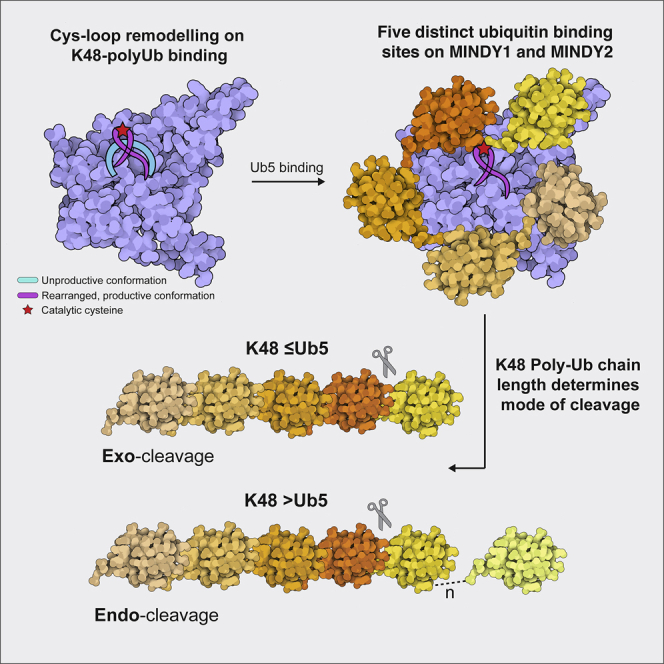
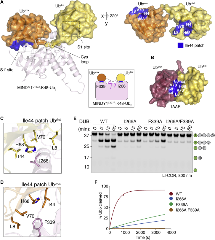
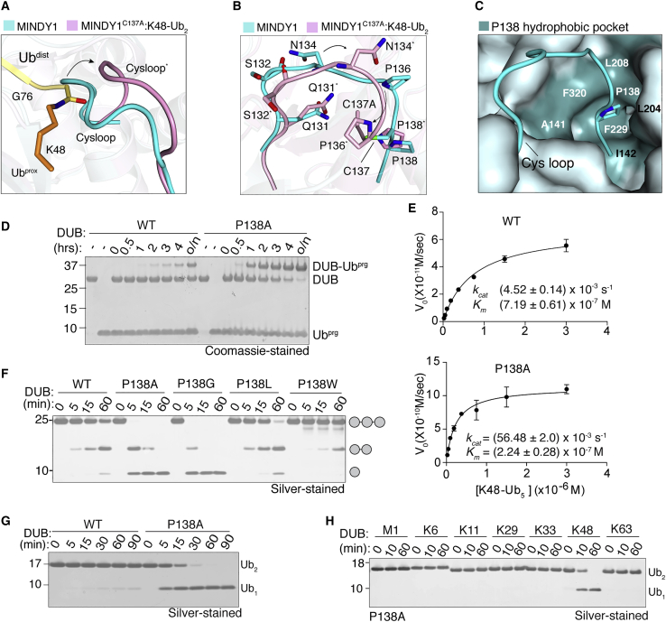
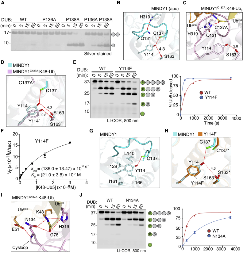
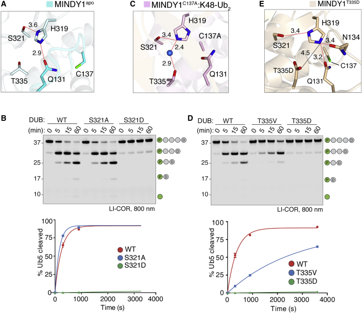
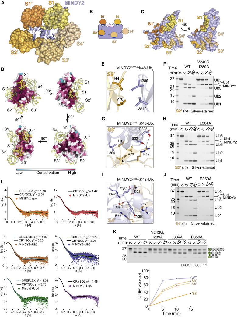
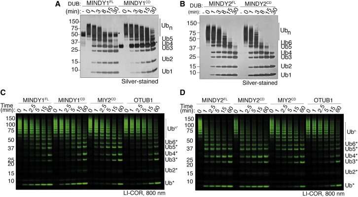
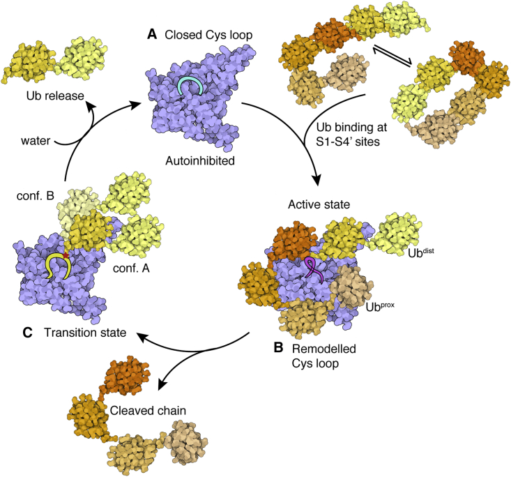
Of the eight distinct polyubiquitin (polyUb) linkages that can be assembled, the roles of K48-linked polyUb (K48-polyUb) are the most established, with K48-polyUb modified proteins being targeted for degradation. MINDY1 and MINDY2 are members of the MINDY family of deubiquitinases (DUBs) that have exquisite specificity for cleaving K48-polyUb, yet we have a poor understanding of their catalytic mechanism. Here, we analyze the crystal structures of MINDY1 and MINDY2 alone and in complex with monoUb, di-, and penta-K48-polyUb, identifying 5 distinct Ub binding sites in the catalytic domain that explain how these DUBs sense both Ub chain length and linkage type to cleave K48-polyUb chains. The activity of MINDY1/2 is inhibited by the Cys-loop, and we find that substrate interaction relieves autoinhibition to activate these DUBs. We also find that MINDY1/2 use a non-canonical catalytic triad composed of Cys-His-Thr. Our findings highlight multiple layers of regulation modulating DUB activity in MINDY1 and MINDY2.

Keywords: autoinhibition; conformational change; crystal structure; deubiquitinase; enzyme mechanism; polyubiquitin; protease; proteasome; protein degradation; ubiquitylation.


Conflict of interest statement

Declaration of interests The authors declare no competing interests.

Figures

None
Graphical abstract
Figure 1
Figure 1
Crystal structures of MINDY1 in complex with K48-linked di-ubiquitin (A) The MINDY1C137A:K48-Ub2 complex crystal structure is shown with MINDY1 in illustration (light pink). Ub molecules are depicted with transparent surfaces (tv-orange:Ubprox and yelloworange:Ubdist). I44 patches on Ub are colored blue, and an alternate view of the bound diUb rotated by 220° along the x axis is shown on the right side. Schematic representation of MINDY1C137A:K48-Ub2 complex (inset). (B) Surface representation of the closed conformation of K48-Ub2 (PDB: 1AAR) with I44 patches highlighted in blue. (C and D) Close-up views of the key residues on the MINDY1 S1 and S1′ sites and their interactions with the I44 patches on Ubdist and Ubprox. (E) DUB assay monitoring cleavage of K48-linked pentaUb, in which Ubprox is fluorescently labeled by MINDY1 and indicated mutants. (F) Quantification of pentaUb hydrolysis shown in (D). The percentage of the total intensities of Ub4, Ub3, Ub2, and Ub1 formed is shown on the y axis. n = 2; mean ± SD. See also Figures S1 and S2.
Figure 2
Figure 2
Cys loop mobility regulates DUB activity (A) Representation of the Cys loop in a superposition of MINDY1apo (cyan) and MINDY1C137A:K48-Ub2 complex (pink). The isopeptide bond between K48 of Ubprox (orange) and G76 of Ubdist (yellow) is shown in sticks. (B) Close-up view of (A) showing amino acid side chain rearrangements (side view). (C) Surface representation of the hydrophobic pocket in MINDY1apo that accommodates the Cys loop residue P138. (D) Coomassie-stained gel comparing activity of MINDY1 WT and P138A to UbPrg in a time course. (E) Steady-state kinetics of K48-linked pentaUb cleavage by MINDY1 WT and P138A mutant derived from reactions with varying concentrations of fluorescently labeled Ub5 (n = 2; means ± SDs). (F) Silver-stained gel comparing cleavage of K48-Ub3 by MINDY1 WT and indicated mutants. (G) DUB assay comparing cleavage of K48-Ub2 by MINDY1 WT and P138A mutants. (H) DUB assay monitoring cleavage of diUb of 7 different linkage types by MINDY1 P138A. See also Figure S3.
Figure 3
Figure 3
Autoinhibition and activation of MINDY1 (A) DUB assay monitoring the cleavage of K48-Ub2 by MINDY1 and indicated mutants. (B) Close-up view of catalytic residues and their interactions in MINDY1 (apo). C137 is out of plane with H139 and is hydrogen bonded with Y114 in MINDY1 (apo). Red dotted lines indicate hydrogen bonds. (C) Close-up view as in (B) for the MINDY1C137A:K48-Ub2 complex. The catalytically productive state conformation leads to the formation of new sets of bonds as shown. The oxyanion hole residue Q131, which was in contact with catalytic H319 in (B), now forms interactions with the carbonyl of the incoming scissile bond. (D) Lateral movement of Y114 and its interactions in MINDY1 (apo) and MINDY1C137A:K48-Ub2 complex. (E) DUB assays comparing cleavage of fluorescently labeled pentaUb by MINDY1 and Y114F mutant. The percentage hydrolysis of pentaUb is plotted against time (right). (F) Steady-state kinetics of K48-linked pentaUb cleavage by MINDY1 Y114F (n = 2; means ± SDs). (G) Close-up view of Y114 (phenyl ring) interactions with hydrophobic residues on adjoining secondary structure elements in MINDY1 (apo). (H) A close-up image of active site of apo MINDY1 Y114F mutant compared to WT. Hydrogen bonding of C137 to Y114 is broken in the mutant. (I) Interactions of Cys loop residue N134 in stabilizing the isopeptide bond for catalysis. (J) Cleavage of pentaUb chains fluorescently labeled on Ubprox by MINDY1 WT and N134A mutant. The panel on the right shows the quantification of the DUB assay. n = 2; mean ± SD. See also Figure S4.
Figure 4
Figure 4
MINDY1 uses a non-canonical catalytic mechanism (A) Close-up view of the catalytic site in MINDY1 apo. The dotted red lines indicate hydrogen bonds, the dotted black line the ionic bond, and the blue sphere indicates water molecule. (B) DUB assay comparing the cleavage of fluorescently labeled K48-Ub5 by MINDY1 WT, S321A, and S321D mutants. The percentage of pentaUb hydrolyzed is plotted against time (right). n = 2; mean ± SD. (C) Close-up view of the catalytic site in MINDY1C137A:K48-Ub2 complex. (D) DUB assay as in (B) comparing the chain cleavage by MINDY1 WT, T335V, and T335D mutants. The percentage of pentaUb hydrolyzed is plotted against time (right). n = 2; mean ± SD. (E) Close-up view of the catalytic site in MINDY1T335D. See also Figure S5.
Figure 5
Figure 5
Crystal structure of MINDY2 in complex with K48-linked pentaUb (A) The MINDY2C266A:K48-Ub5 complex crystal structure with MINDY2 (light blue). Ub molecules are depicted with transparent surfaces (S1: yellow orange, S1′: tv-orange, S2′: bright orange, S3′ light orange, S4′: wheat). (B) Schematic representation of MINDY2C266A:K48-Ub5 complex with Ile44 patches on Ub involved in binding shown in blue. (C) Surface representation of MINDY2, with the footprint of each Ub highlighted at each Ub-binding site. (D) Surface conservation analysis of MINDY2 from metazoan ortholog sequences. MINDY2 is shown as a surface model, rotated by 90° in each view, and the surface residues are colored by conservation score. The pentaUb chain is shown as a yellow ribbon model, and the 5 Ub binding sites on MINDY2 are annotated. (E) Close-up of key residues at the S2′ site and their interactions with Ub. (F) DUB assay comparing cleavage of K48-Ub5 by MINDY2 WT and the indicated S2′ site mutants. (G) Close-up of key residues at the S3′ site and their interactions with Ub. (H) DUB assay comparing cleavage of K48-Ub5 by MINDY2 WT and indicated S3′ site mutants. (I) Close-up of key residues at the S4′ site and their interactions with Ub. (J) DUB assay comparing cleavage of K48-Ub5 by MINDY2 WT and indicated S4′ site mutants. Dashed line: gel truncation to exclude mutants irrelevant to this study. (K) DUB assay comparing cleavage of fluorescently labeled K48-Ub5 by MINDY2 WT and S2′, S3′, and S4′ mutants. n = 3; mean ± SEM. (L) SAXS curves of MINDY2, apo molecule, and in complex with monoUb, K48-linked Ub2, Ub3, Ub4, and Ub5, respectively, and their fits computed from atomic models by CRYSOL. For Mindy2-Ub3, normal mode analysis (NMA) with SREFLEX was used for the refinement of the expected atomic models. For Mindy2-Ub2, OLIGOMER was applied on atomic models in which Ub2 occupying positions S1 and S1′ or S1 and S3′ were used to quantify their mixture. See also Figures S6 and S7.
Figure 6
Figure 6
Ubiquitin chain length determines exo- and endo-cleavage activities (A) Silver-stained gels of DUB assays monitoring cleavage of long K48-linked polyUb chains containing >6 Ub moieties by MINDY1FL and MINDY1cat. (B) As in (A), but comparing MINDY2FL and MINDY2cat. (C) DUB assay monitoring cleavage of distally labeled longer polyUb chains by MINDY1FL and MINDY1cat with 2 known endo-DUB controls: MIY2 and OTUB1. (D) As in (D), but comparing MINDY2FL and MINDY2cat.
Figure 7
Figure 7
Model summarizing the catalytic mechanism of K48-linked polyUb (A) MINDY1 and MINDY2 exist in an autoinhibited conformation in which the Cys loop is in a closed conformation that sterically interferes with Ub binding and also contributes to keeping the catalytic site inhibited. (B) In a substrate-driven mechanism, Ub interactions release inhibition and activate the DUB, resulting in chain cleavage and release of the Ub chain. (C) In the product intermediate transitional tetrahedral state, the Ub occupies the S1 site. As this is not a strong binding interface, this Ub exists in 2 different conformers. Attack by a water molecule releases the Ub and returns the DUB to an inhibited conformation. Created using Illustrate (Goodsell et al., 2019). See also Figure S8.

References

    1. Abdul Rehman S.A., Kristariyanto Y.A., Choi S.Y., Nkosi P.J., Weidlich S., Labib K., Hofmann K., Kulathu Y. MINDY-1 Is a Member of an Evolutionarily Conserved and Structurally Distinct New Family of Deubiquitinating Enzymes. Mol. Cell. 2016;63:146–155. - PMC - PubMed
    1. Altenhoff A.M., Train C.-M., Gilbert K.J., Mediratta I., Mendes de Farias T., Moi D., Nevers Y., Radoykova H.-S., Rossier V., Warwick Vesztrocy A. OMA orthology in 2021: website overhaul, conserved isoforms, ancestral gene order and more. Nucleic Acids Res. 2021;49(D1):D373–D379. - PMC - PubMed
    1. Ashkenazy H., Abadi S., Martz E., Chay O., Mayrose I., Pupko T., Ben-Tal N. ConSurf 2016: an improved methodology to estimate and visualize evolutionary conservation in macromolecules. Nucleic Acids Res. 2016;44(W1):W344–W350. - PMC - PubMed
    1. Békés M., Rut W., Kasperkiewicz P., Mulder M.P.C., Ovaa H., Drag M., Lima C.D., Huang T.T. SARS hCoV papain-like protease is a unique Lys48 linkage-specific di-distributive deubiquitinating enzyme. Biochem. J. 2015;468:215–226. - PMC - PubMed
    1. Békés M., van der Heden van Noort G.J., Ekkebus R., Ovaa H., Huang T.T., Lima C.D. Recognition of Lys48-Linked Di-ubiquitin and Deubiquitinating Activities of the SARS Coronavirus Papain-like Protease. Mol. Cell. 2016;62:572–585. - PMC - PubMed

Publication types