. 2022 Jan:39:102459.
doi: 10.1016/j.nano.2021.102459. Epub 2021 Sep 13.

Poly(2-oxazoline)-magnetite NanoFerrogels: Magnetic field responsive theranostic platform for cancer drug delivery and imaging

Affiliations

Poly(2-oxazoline)-magnetite NanoFerrogels: Magnetic field responsive theranostic platform for cancer drug delivery and imaging

Youngee Seo et al. Nanomedicine. 2022 Jan.

Abstract

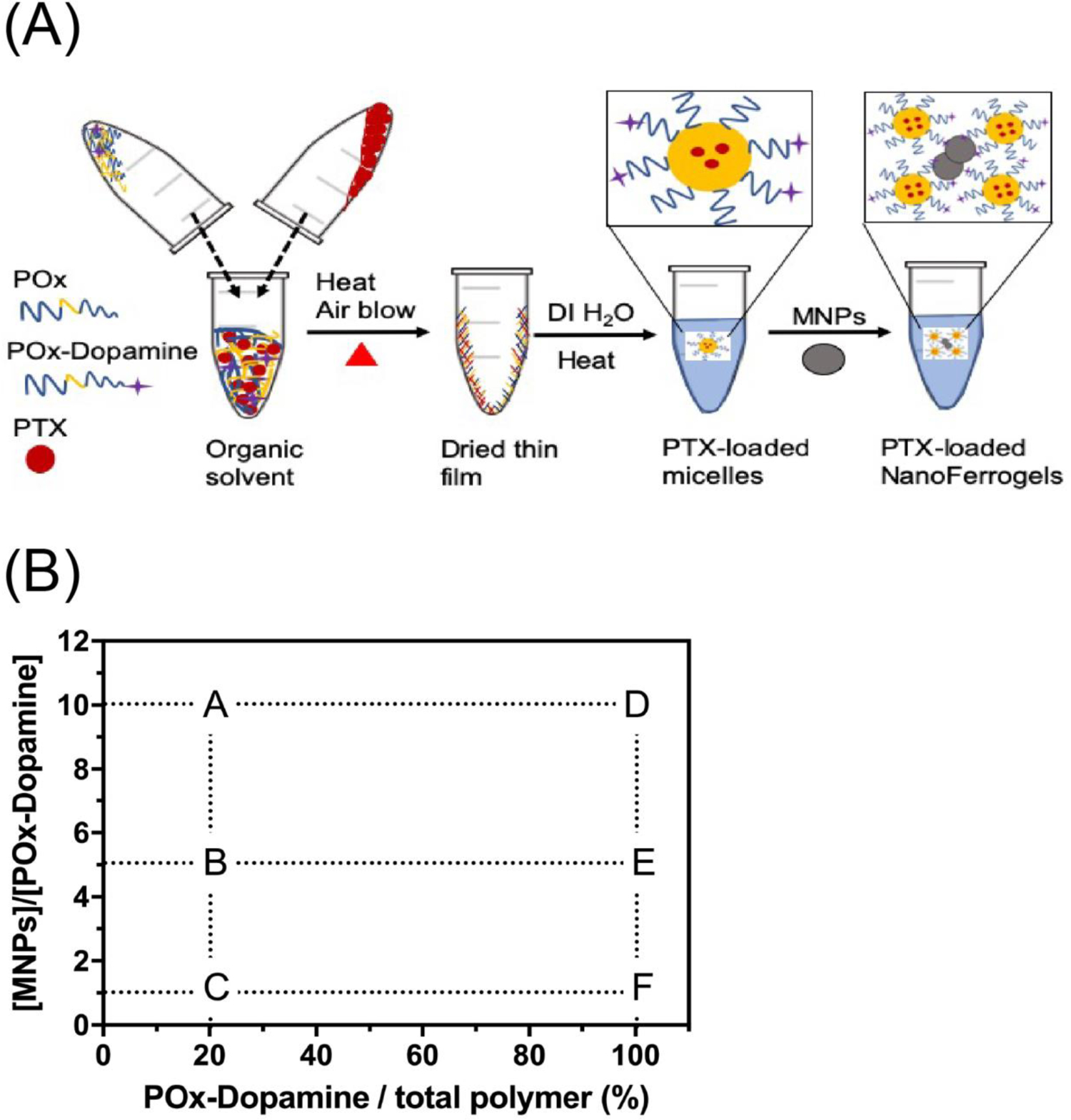
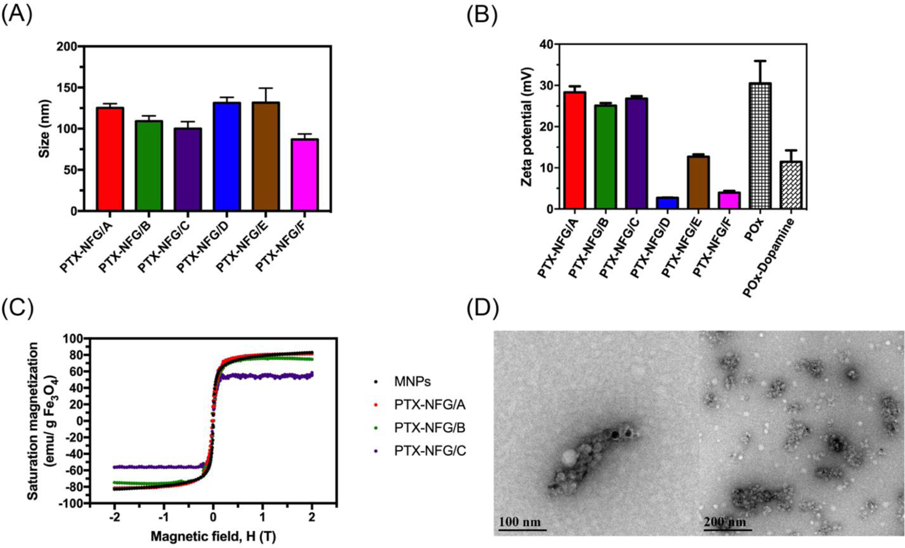
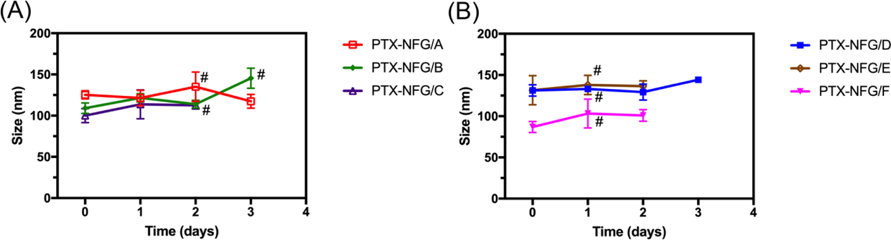
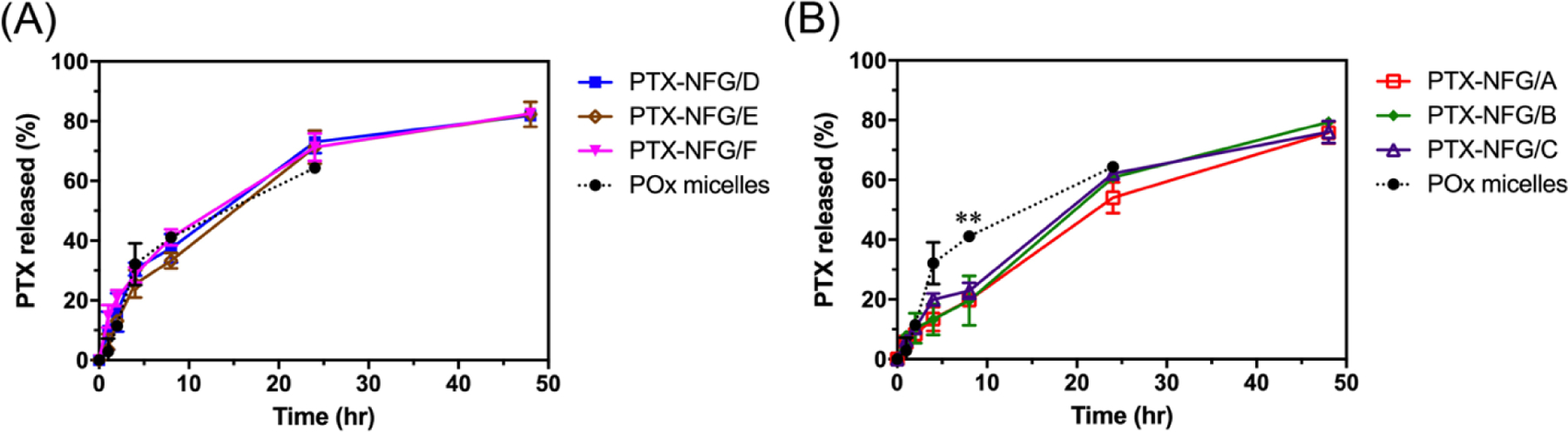
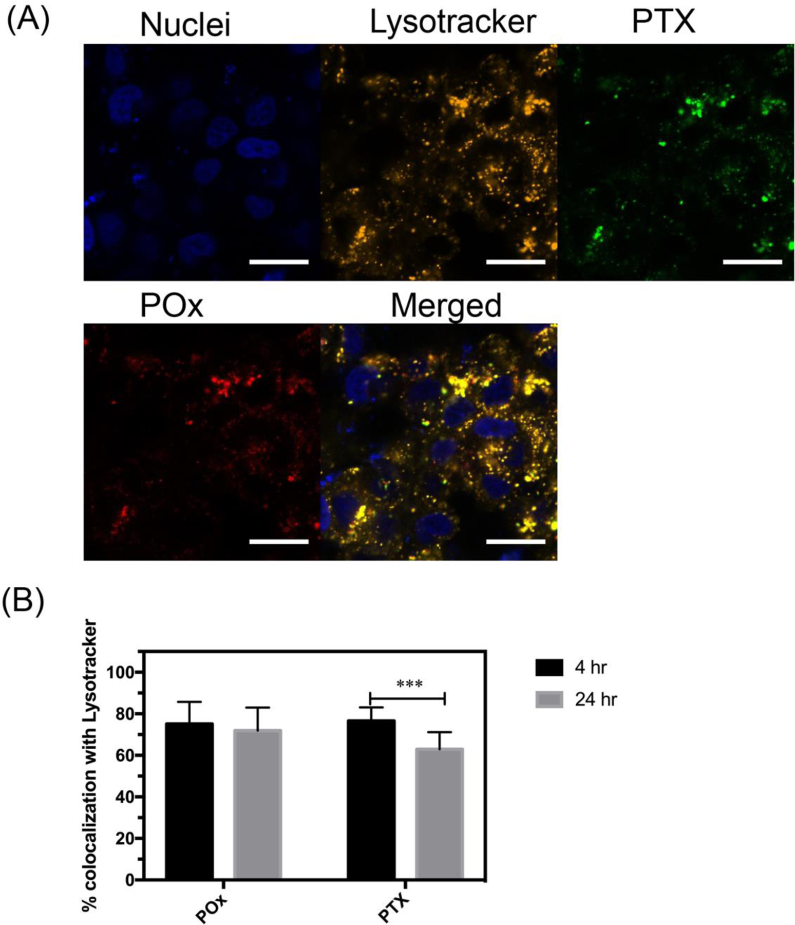
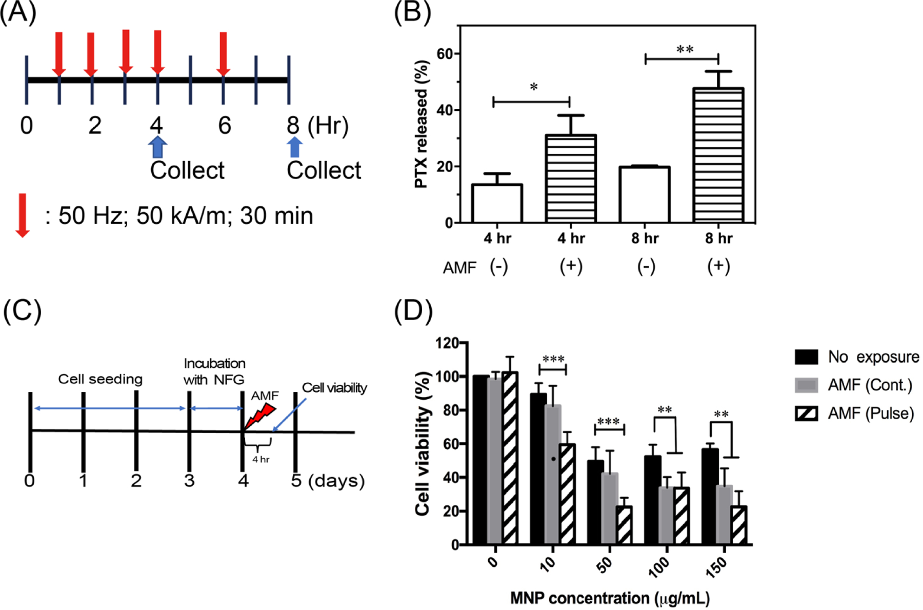
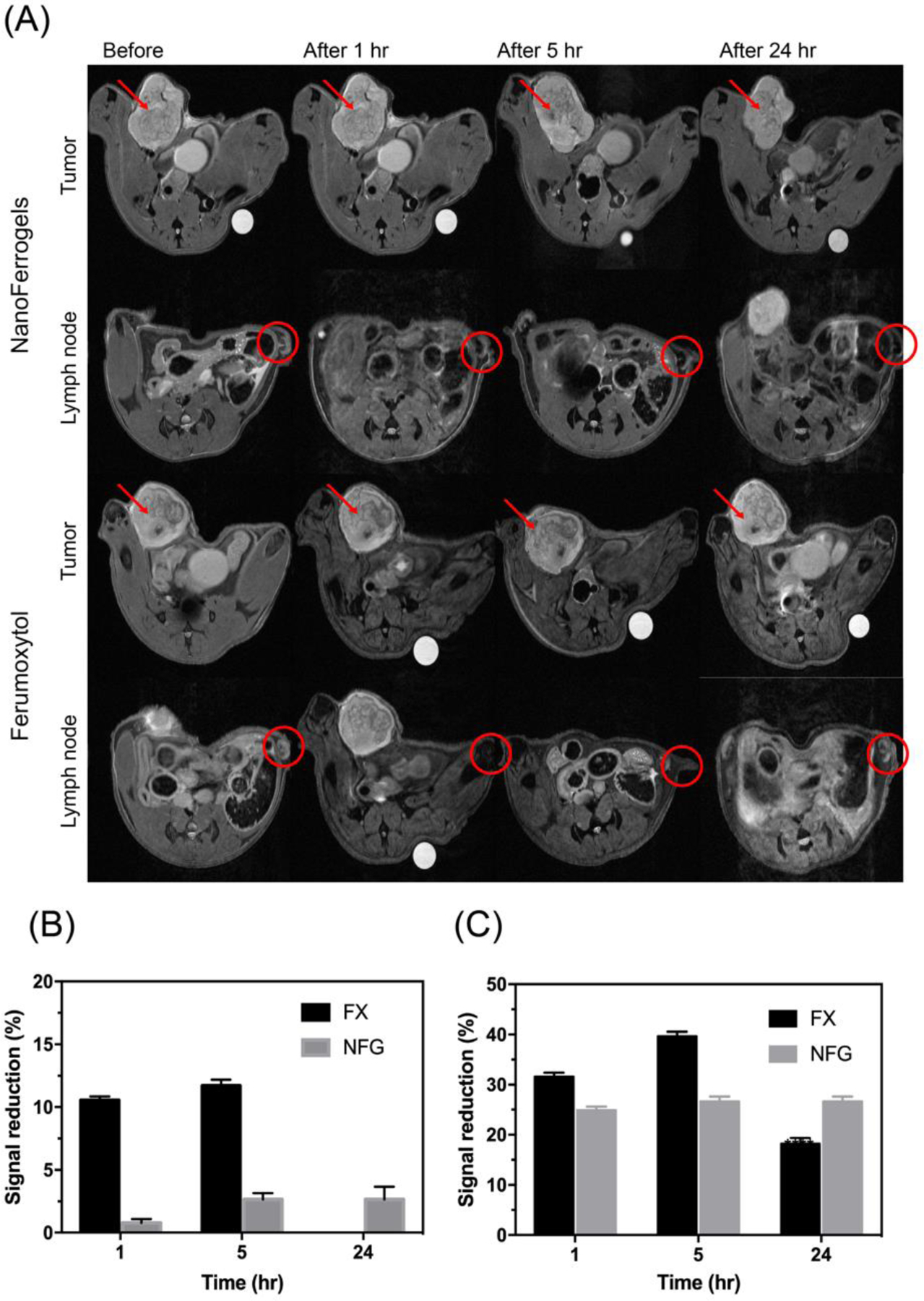
Combining diagnosis and treatment approaches in one entity is the goal of theranostics for cancer therapy. Magnetic nanoparticles have been extensively used as contrast agents for nuclear magnetic resonance imaging as well as drug carriers and remote actuation agents. Poly(2-oxazoline)-based polymeric micelles, which have been shown to efficiently solubilize hydrophobic drugs and drug combinations, have high loading capacity (above 40% w/w) for paclitaxel. In this study, we report the development of novel theranostic system, NanoFerrogels, which is designed to capitalize on the magnetic nanoparticle properties as imaging agents and the poly(2-oxazoline)-based micelles as drug loading compartment. We developed six formulations with magnetic nanoparticle content of 0.3%-12% (w/w), with the z-average sizes of 85-130 nm and ξ-potential of 2.7-28.3 mV. The release profiles of paclitaxel from NanoFerrogels were notably dependent on the degree of dopamine grafting on poly(2-oxazoline)-based micelles. Paclitaxel loaded NanoFerrogels showed efficacy against three breast cancer lines which was comparable to free paclitaxel. They also showed improved tumor and lymph node accumulation and signal reduction in vivo (2.7% in tumor; 8.5% in lymph node) compared to clinically approved imaging agent ferumoxytol (FERAHEME®) 24 h after administration. NanoFerrogels responded to super-low frequency alternating current magnetic field (50 kA m-1, 50 Hz) which accelerated drug release from paclitaxel-loaded NanoFerrogels or caused death of cells loaded with NanoFerrogels. These proof-of-concept experiments demonstrate that NanoFerrogels have potential as remotely actuated theranostic platform for cancer diagnosis and treatment.

Keywords: Magnetic field-triggered release; Magnetic nanoparticles; Magneto-mechanical actuation; Paclitaxel.


Figures

Fig. 1.
Fig. 1.
(A) Schematics of the preparation of PTX-NFGs and (B) summary of MNPs, POx-Dopamine and POx compositions in the blank NFGs and PTX-NFGs (see, also Table 1).
Fig. 2.
Fig. 2.
(A) Hydrodynamic diameter of PTX-NFGs dispersed in PBS, (B) zeta potential of PTX-NFGs, POx micelles, and POx-Dopamine micelles, (C) saturation magnetization of PTX-NFGs and (D) Representative PTX-NFGs TEM image (PTX-NFG/B). Data are mean ± S.D.
Fig. 3.
Fig. 3.
Colloidal stability of PTX-NFGs prepared with (A) 20% POx-Dopamine (PTX-NFG/A-C) and (B) 100% POx-Dopamine (PTX-NFG/D-F). The samples (1 mg mL−1) were incubated in PBS over period of 3 days. Data represented mean ± SD (n=3). # indicate precipitation.
Fig. 4.
Fig. 4.
A cumulative PTX release from PTX-NFGs at 37 °C in the presence of 40 g L−1 of BSA. PTX release comparison between POx micelles and (A) PTX-NFGs/D-F, (B) PTX-NFGs/A-C Data are mean ± SD (n=3, *P<0.05, **P<0.01).
Fig. 5.
Fig. 5.
(A) Intracellular distribution of fluorescently labeled PTX-NFG/B in MCF-7 breast cancer cells after 4 h of incubation (bar = 20 μm). (B) Quantification of the colocalization of AF647-POx (red) and OG488-PTX (green) with lysosomes (orange) as determined by ImageJ software after 4 and 24 h of incubation (***P<0.001). Nuclei were stained with Hoechst 33342 (blue).
Fig. 6.
Fig. 6.
Remote magneto-mechanical actuation of PTX-NFG and NFG in AC magnetic field. (A) A scheme illustrating the exposure and PTX release measurement timeline. Red arrow indicates application of AC magnetic field (50 kA/m, 50 Hz) either continuous or pulse for 30 min. (B) The effect of the AC magnetic field exposure on the release of PTX from PTX-NFG/B at 4 h, and 8 h (n=3). (C) A scheme illustrating the exposure and cell viability measurement timeline. Red arrow indicates times of application of AC magnetic field. (D) Cell viability of LCC-6-WT cancer cells following exposure to continuous (gray) or pulsed (striped) AC magnetic field. **P<0.01; ***P<0.001 compared to no exposure. Data are mean ± S.D. (n=6).
Fig. 7.
Fig. 7.
(A) T2-weighted images obtained from tumor and lymph node at different time points after NFG/E or Ferumoxytol (FX) administration. Red arrow and circle indicated the tumor tissue and lymph node, respectively. (B) Signal reduction percentage of NFG/E and Ferumoxytol compared to reference in tumor. (C) Signal reduction percentage of NFG/E and Ferumoxytol compared to reference in lymph node. Data represented mean ± S.D. (n=3).

References

    1. Cherry SR Multimodality in vivo imaging systems: twice the power or double the trouble? Annu Rev Biomed Eng 2006, 8, 35–62, doi:10.1146/annurev.bioeng.8.061505.095728. - DOI - PubMed
    1. Rudin M; Weissleder R Molecular imaging in drug discovery and development. Nature reviews Drug discovery 2003, 2, 123. - PubMed
    1. McCarthy JR; Weissleder R Multifunctional magnetic nanoparticles for targeted imaging and therapy. Adv Drug Deliv Rev 2008, 60, 1241–1251, doi:10.1016/j.addr.2008.03.014. - DOI - PMC - PubMed
    1. Parvanian S; Mostafavi SM; Aghashiri M Multifunctional nanoparticle developments in cancer diagnosis and treatment. Sensing and Bio-Sensing Research 2017, 13, 81–87, doi:10.1016/j.sbsr.2016.08.002. - DOI
    1. Bao G; Mitragotri S; Tong S Multifunctional nanoparticles for drug delivery and molecular imaging. Annu Rev Biomed Eng 2013, 15, 253–282, doi:10.1146/annurev-bioeng-071812-152409. - DOI - PMC - PubMed

Publication types