. 2021 Sep 1;56(9):937-944.
doi: 10.4085/418-20.

Blood Flow Restriction Training

Affiliations

Blood Flow Restriction Training

Daniel S Lorenz et al. J Athl Train. .

Abstract

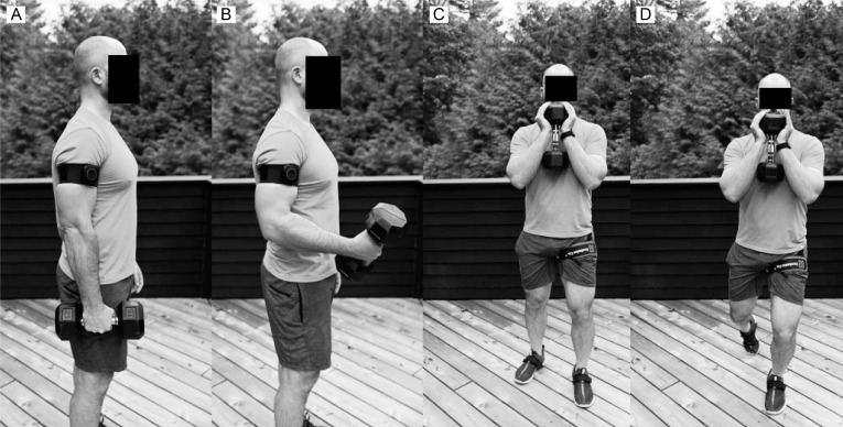
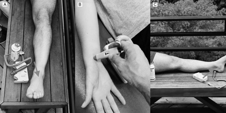
Muscle weakness and atrophy are common impairments after musculoskeletal injury. Blood flow restriction (BFR) training offers the ability to mitigate weakness and atrophy without overloading healing tissues. It appears to be a safe and effective approach to therapeutic exercise in sports medicine environments. This approach requires consideration of a wide range of factors, and the purpose of our article is to provide insights into proposed mechanisms of effectiveness, safety considerations, application guidelines, and clinical recommendations for BFR training after musculoskeletal injury. Whereas training with higher loads produces the most substantial increases in strength and hypertrophy, BFR training appears to be a reasonable option for bridging earlier phases of rehabilitation when higher loads may not be tolerated by the patient and later stages that are consistent with return to sport.

Keywords: clinical rehabilitation; hypertrophy; occlusion training; resistance training.


Figures

Figure 1
Figure 1
Cuff placement: A and B, upper extremity. C and D, lower extremity. The cuff should be placed proximally on the limb, which allows occlusion to occur in the majority of the muscle without interfering with movement.
Figure 2
Figure 2
Determining arterial occlusion pressure using a pulse oximeter or A, a handheld Doppler ultrasound. A pulse oximeter can be placed on a finger. The Doppler ultrasound is placed on a distal artery: B, upper extremity, radial artery; C, lower extremity, dorsalis pedis. Once the pulse has been identified, the cuff is slowly inflated (eg, start at 50 mm Hg and increase by 10-mm Hg increments every 10 seconds) to the point at which full occlusion occurs (ie, pulse is absent).

References

    1. Lisee C, Lepley AS, Birchmeier T, O'Hagan K, Kuenze C. Quadriceps strength and volitional activation after anterior cruciate ligament reconstruction: a systematic review and meta-analysis. Sports Health. 2019;11(2):163–179. doi: 10.1177/1941738118822739. - DOI - PMC - PubMed
    1. Ratamess NA, Alvar BA, Evetoch TK, et al. Progression models in resistance training for healthy adults. Med Sci Sports Exerc. 2009;41(3):687–708. doi: 10.1249/MSS.0b013e3181915670. - DOI - PubMed
    1. Schoenfeld BJ, Grgic J, Ogborn D, Krieger JW. Strength and hypertrophy adaptations between low- vs. high-load resistance training: a systematic review and meta-analysis. J Strength Cond Res. 2017;31(12):3508–3523. doi: 10.1519/JSC.0000000000002200. - DOI - PubMed
    1. Barber-Westin S, Noyes FR. Blood flow-restricted training for lower extremity muscle weakness due to knee pathology: a systematic review. Sports Health. 2019;11(1):69–83. doi: 10.1177/1941738118811337. - DOI - PMC - PubMed
    1. Hughes L, Paton B, Rosenblatt B, Gissane C, Patterson SD. Blood flow restriction training in clinical musculoskeletal rehabilitation: a systematic review and meta-analysis. Br J Sports Med. 2017;51(13):1003–1011. doi: 10.1136/bjsports-2016-097071. - DOI - PubMed