. 2021 Sep 16;14(1):183.
doi: 10.1186/s13068-021-02025-6.

Engineering the substrate binding site of the hyperthermostable archaeal endo-β-1,4-galactanase from Ignisphaera aggregans

Affiliations

Engineering the substrate binding site of the hyperthermostable archaeal endo-β-1,4-galactanase from Ignisphaera aggregans

Sebastian J Muderspach et al. Biotechnol Biofuels. .

Abstract

Background: Endo-β-1,4-galactanases are glycoside hydrolases (GH) from the GH53 family belonging to the largest clan of GHs, clan GH-A. GHs are ubiquitous and involved in a myriad of biological functions as well as being widely used industrially. Endo-β-1,4-galactanases, in particular hydrolyse galactan and arabinogalactan in pectin, a major component of the primary plant cell wall, with important functions in plant defence and application in the food and other industries. Here, we explore the family's biological diversity by characterizing the first archaeal and hyperthermophilic GH53 galactanase, and utilize it as a scaffold for engineering enzymes with different product lengths.

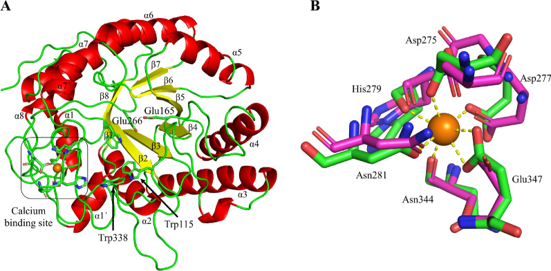
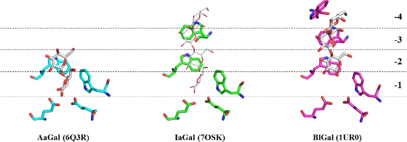
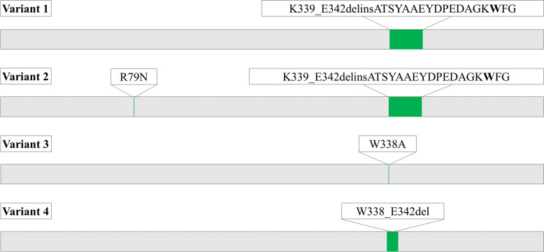
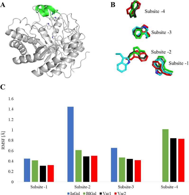
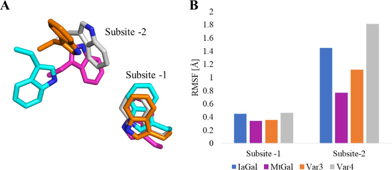
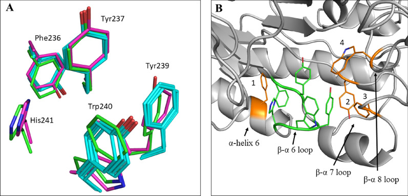
Results: A galactanase gene was identified in the genome of the anaerobic hyperthermophilic archaeon Ignisphaera aggregans, and the isolated catalytic domain expressed and characterized (IaGal). IaGal presents the typical (βα)8 barrel structure of clan GH-A enzymes, with catalytic carboxylates at the end of the 4th and 7th barrel strands. Its activity optimum of at least 95 °C and melting point over 100 °C indicate extreme thermostability, a very advantageous property for industrial applications. If enzyme depletion is reduced, so is the need for re-addition, and thus costs. The main stabilizing features of IaGal compared to other structurally characterized members are π-π and cation-π interactions. The length of the substrate binding site-and thus produced oligosaccharide products-is intermediate compared to previously characterized galactanases. Variants inspired by the structural diversity in the GH53 family were rationally designed to shorten or extend the substrate binding groove, in order to modulate product length. Subsite-deleted variants produced shorter products than IaGal, as do the fungal galactanases inspiring the design. IaGal variants engineered with a longer binding site produced a less expected degradation pattern, though still different from that of wild-type IaGal. All variants remained extremely stable.

Conclusions: We have characterized in detail the most thermophilic endo-β-1,4-galactanase known to date and successfully engineered it to modify the degradation profile, while maintaining much of its desirable thermostability. This is an important achievement as oligosaccharide products length is an important property for industrial and natural GHs alike.

Keywords: Archaea; Biomass degradation; Crystal structure; Degradation profiles; Extreme thermophile; Galactan; Glycoside hydrolase; High-performance anion-exchange chromatography; Ignisphaera aggregans; Rational design.


Conflict of interest statement

The authors declare the following conflicts of interest: KBRMK and KJ work for Novozymes, a major manufacturer of industrial enzymes.

Figures

Fig. 1
Fig. 1
Activity optima for native endo-β-1,4-galactanases. A Normalized temperature activity optimum of BlGal, HiGal and IaGal at pH 5.0. B Normalized pH activity profile of BlGal, HiGal and IaGal at 40 °C. All experiments were done in triplicates and the error bars represent the standard deviations of the normalized activity
Fig. 2
Fig. 2
Degradation profiles for native endo-β-1,4-galactanases. The degradation products of IaGal (A), BlGal (B), MtGal (C) and HiGal (D) hydrolysing lupin galactan are shown. The concentrations of G1 (galactose), G2 (galactobiose), G3 (galactotriose), G4 (galactotetraose), G5 (galactopentaose) and G > 5 (larger than galactopentaose) are reported in mM for each given DoH (shown on the x-axis). The %DoH is defined as the amount of reducing ends divided by the theoretical maximum amount of reducing ends, i.e. a full degradation to galactose
Fig. 3
Fig. 3
Overview of the IaGal structure. A The overall structure of the IaGal catalytic domain. Secondary structural elements are coloured (α-helices red, and β-sheets yellow). The catalytic acid/base (Glu165) and catalytic nucleophile (Glu266) are shown in sticks. Trp115 and Trp338 correspond to the − 2 and − 3 subsites, respectively. The calcium binding site is also highlighted. B The calcium binding site of IaGal (PDB ID: 7OSK, green) compared to that from the bacterial enzyme BlGal (PDB ID: 1UR0, magenta)
Fig. 4
Fig. 4
Substrate binding sites of AaGal, IaGal and BlGal. Substrate subsites at the non-reducing end increase incrementally from fungal enzymes (AaGal PDB ID 6Q3R) to archaeal enzymes (IaGal PDB ID 7OSK) to the bacterial BlGal enzyme (PDB ID 1UR0). The galactobiose and galactotriose ligands are shown as white sticks in the AaGal and BlGal structures, respectively. The same ligands are shown as white lines after superposition onto the IaGal structure
Fig. 5
Fig. 5
IaGal variant overview. In subsite-extended variant 1 and 2 the residues 339–342 were replaced by the sequence ATSYAAEYDPEDAGKWFG with variant 2 having an additional point mutation (R79N). The tryptophan intended as the aromatic platform for binding subsite − 4 is highlighted in bold. Subsite-deleted variant 3 is a point mutation (W388A) while in variant 4 the residue sequence 338–342 has been deleted. The variant nomenclature in the figure is adopted from [32]
Fig. 6
Fig. 6
Molecular dynamic simulations of subsite-extended variants. A Overview of variant 1 after 90 ns of simulation. The inserted sequence is shown in green and the platforms for the non-reducing end binding subsites are shown as sticks. B Comparison of the non-reducing end binding subsites for variant 1 (black), variant 2 (red) and IaGal (blue) after 90 ns simulation and BlGal (PDB ID 1R8L, green). C Comparison of the RMSF for the non-reducing end binding subsites using the same colour scheme
Fig. 7
Fig. 7
Molecular dynamic simulations of subsite-deleted variants. IaGal is shown in blue, MtGal is magenta, variant 3 is orange and variant 4 is grey. A Comparison of the − 1 and − 2 binding subsites for variant 3, variant 4, IaGal and MtGal after 90 ns of simulation. B Comparison of the RMSF value for the binding subsites
Fig. 8
Fig. 8
Degradation profiles of IaGal variants. The degradation products of variant 1 (A), variant 2 (B), variant 3 (C) and variant 4 (D) hydrolysing lupin galactan are shown. The concentrations of G1 (galactose), G2 (galactobiose), G3 (galactotriose), G4 (galactotetraose), G5 (galactopentaose) and G > 5 (larger than galactopentaose) are reported in mM for each given DoH. The %DoH is defined as the amount of reducing ends divided by the theoretical maximum amount of reducing ends, i.e. a full degradation to galactose
Fig. 9
Fig. 9
Highly conserved area containing π–π interactions in GH53 galactanases. A Superposition of the conserved aromatic residues localized in the 6th β/α-loop. The residues from IaGal are labelled and shown in green, BlGal is shown in magenta and all the fungal galactanases are shown in cyan. Unique for BlGal and IaGal is the extension of the aromatic cluster with an additional histidine residue. B An overview of the π–π cluster in IaGal. The residues located in the 6th β/α-loop are shown as greens sticks in the same orientation as in A. Adjacent aromatic residues which interact with these conserved residues are shown as orange sticks (1: Phe247, 2: Tyr269, 3: Phe283, 4: Trp346)

Similar articles

Cited by

References

    1. Lau JM, McNeil M, Darvill AG, Albersheim P. Structure of the backbone of rhamnogalacturonan I, a pectic polysaccharide in the primary cell walls of plants. Carbohydr Res. 1985;137 C:111–125. doi: 10.1016/0008-6215(85)85153-3. - DOI
    1. Leclere L, Van CP, Michiels C. Anti-cancer activities of pH- or heat-modified pectin. Front Pharmacol. 2013;4:1–8. doi: 10.3389/fphar.2013.00128. - DOI - PMC - PubMed
    1. Voragen AGJ, Coenen G-J, Verhoef RP, Schols HA. Pectin, a versatile polysaccharide present in plant cell walls. Struct Chem. 2009;20:263–275. doi: 10.1007/s11224-009-9442-z. - DOI
    1. Hahn M, Darvill A, Albersheim P. Host-pathogen interactions. Plant Physiol. 1981;68:1161. doi: 10.1104/pp.68.5.1161. - DOI - PMC - PubMed
    1. Lara-Espinoza C, Carvajal-Millán E, Balandrán-Quintana R, López-Franco Y, Rascón-Chu A. Pectin and pectin-based composite materials: beyond food texture. Molecules. 2018;23:942. doi: 10.3390/molecules23040942. - DOI - PMC - PubMed