Observational Study
. 2021 Nov 2;97(18):e1809-e1822.
doi: 10.1212/WNL.0000000000012778. Epub 2021 Sep 16.

Spontaneous ARIA-like Events in Cerebral Amyloid Angiopathy-Related Inflammation: A Multicenter Prospective Longitudinal Cohort Study

Affiliations
Observational Study

Spontaneous ARIA-like Events in Cerebral Amyloid Angiopathy-Related Inflammation: A Multicenter Prospective Longitudinal Cohort Study

Laura Antolini et al. Neurology. .

Abstract

Background and objectives: The goal of this work was to investigate the natural history and outcomes after treatment for spontaneous amyloid-related imaging abnormalities (ARIA)-like in cerebral amyloid angiopathy-related inflammation (CAA-ri).

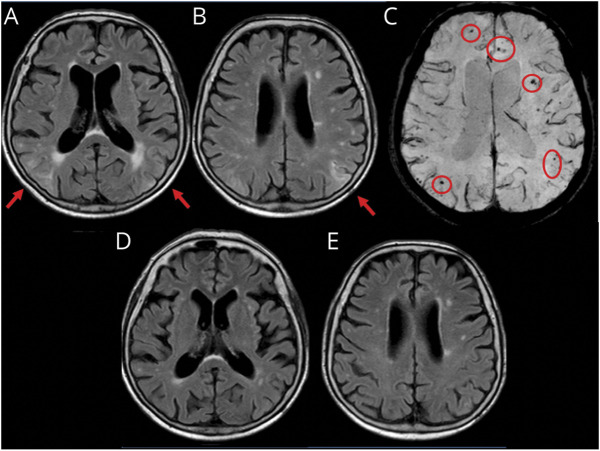
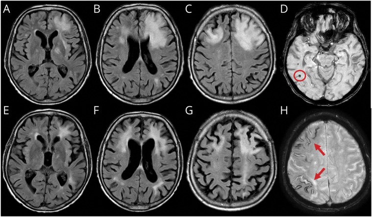
Methods: This was a multicenter, hospital-based, longitudinal, prospective observational study of inpatients meeting CAA-ri diagnostic criteria recruited through the Inflammatory Cerebral Amyloid Angiopathy and Alzheimer's Disease βiomarkers International Network from January 2013 to March 2017. A protocol for systematic data collection at first-ever presentation and at subsequent in-person visits, including T1-weighted, gradient recalled echo-T2*, fluid-suppressed T2-weighted (fluid-attenuated inversion recovery), and T1 postgadolinium contrast-enhanced images acquired on 1.5T MRI, was used at the 3-, 6-, 12-, and 24-month follow-up. Centralized reads of MRIs were performed by investigators blinded to clinical, therapeutic, and time-point information. Main outcomes were survival, clinical and radiologic recovery, intracerebral hemorrhage (ICH), and recurrence of CAA-ri.

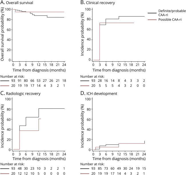
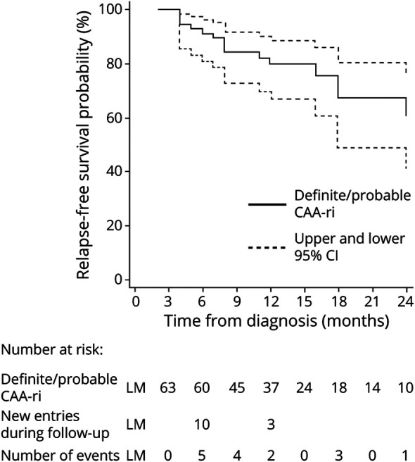
Results: The study enrolled 113 participants (10.6% definite, 71.7% probable, and 17.7% possible CAA-ri). Their mean age was 72.9 years; 43.4% were female; 37.1% were APOEε4 carriers; 36.3% had a history of Alzheimer disease; and 33.6% had a history of ICH. A history of ICH and the occurrence of new ICH at follow-up were more common in patients with cortical superficial siderosis at baseline (52.6% vs 14.3%, p < 0.0001 and 19.3% vs 3.6%, p < 0.009, respectively). After the first-ever presentation of CAA-ri, 70.3% (95% confidence interval [CI] 61.6%-78.5%) and 84.1% (95% CI 76.2%-90.6%) clinically recovered within 3 and 12 months, followed by radiologic recovery in 45.1% (95% CI 36.4%-54.8%) and 77.4% (95% CI 67.7%-85.9%), respectively. After clinicoradiologic resolution of the first-ever episode, 38.3% (95% CI 22.9%-59.2%) had at least 1 recurrence within the following 24 months. Recurrence was more likely if IV high-dose corticosteroid pulse therapy was suddenly stopped compared to slow oral tapering off (hazard ratio 4.68, 95% CI 1.57-13.93; p = 0.006).

Discussion: These results from the largest longitudinal cohort registry of patients with CAA-ri support the transient and potentially relapsing inflammatory nature of the clinical-radiologic acute manifestations of the disease and the effectiveness of slow oral tapering off after IV corticosteroid pulse therapy in preventing recurrences. Our results highlight the importance of differential diagnosis for spontaneous ARIA-like events in β-amyloid-driven diseases, including treatment-related ARIA in patients with Alzheimer disease exposed to immunotherapy drugs.


Figures

Figure 1
Figure 1. Spontaneous ARIA-like Imaging Findings at Presentation and After Radiologic Remission at 3-Month Follow-up of a Patient With Probable CAA-ri
(A and B) Fluid-attenuated inversion recovery (FLAIR) images (axial sequences) at presentation show cortico-subcortical regions of hyperintensity suggestive of vasogenic edema and sulcal effacement on both hemispheres (red arrows). (C) Susceptibility-weighted images show microbleeds (red circles). (D and E) MRI 3 months after corticosteroid pulse therapy (5 IV boluses of 1 g/d methylprednisolone followed by 1 mg/kg oral prednisone daily and slow tapering off over several months) showing the disappearance of the acute inflammatory white matter hyperintensity lesions in the corresponding planes of FLAIR sequences indexed at presentation. ARIA = amyloid-related imaging abnormalities; CAA-ri = cerebral amyloid angiopathy–related inflammation.
Figure 2
Figure 2. Spontaneous ARIA-like Imaging Findings of a Patient With Probable CAA-ri at Presentation and After 3 Months of Follow-up With No Radiologic Remission
(A–C) Fluid-attenuated inversion recovery (FLAIR) axial sequences at presentation show cortico-subcortical hyperintense areas on both frontal lobes with edema and mass effect on the anterior horn of the left lateral ventricle. (E–G) FLAIR sequences 3 months after corticosteroid pulse therapy (5 IV boluses of 1 g/d methylprednisolone followed by 1 mg/kg oral prednisone daily and slow tapering off over several months) treatment showing substantial reduction of the signal abnormalities on the right frontal lobe and persistent hyperintense signal abnormality on the left frontal lobe without mass effect and with atrophy of the corresponding cortico-subcortical region. (D and H) Susceptibility-weighted imaging sequences at presentation show microbleeds (red circle) and diffuse cortical superficial siderosis (red arrows). ARIA = amyloid-related imaging abnormalities; CAA-ri = cerebral amyloid angiopathy–related inflammation.
Figure 3
Figure 3. Kaplan-Meier Estimates of Probability of Survival, Clinical Recovery, Radiologic Recovery, and ICH Development of the iCAβ International Network Cohort Registry25 of 113 Patients With First-Ever Diagnosis of CAA-ri
(A) Overall survival, (B) clinical recovery, (C) radiologic recovery, and (D) intracerebral hemorrhage (ICH) development. Data are expressed as incidence probability (percentage). Diagnostic category (definite and probable vs possible) diagnosed by clinical presentation, radiologic criteria, or pathologic findings. Follow-up time defined as the time elapsed from the date of cerebral amyloid angiopathy–related inflammation (CAA-ri) diagnosis to the date of each in-person visit at 3, 6, 12, and 24 months or last follow-up available. Overall survival defined as the time elapsed from the date of CAA-ri diagnosis to the date of death or last follow-up visit. Any statistically significant effect emerged between the 2 diagnostic category groups (p = 0.40). Clinical recovery defined by stable recovery of the acute neurologic signs or symptoms of CAA-ri indexed at presentation. For the patients who did not show any recovery, survival time was set equal to the maximum follow-up time available. Any statistically significant effect emerged between the 2 diagnostic category groups (p = 0.49). Radiologic recovery defined as complete resolution (disappearance) or the almost unperceivable visualization of the acute inflammatory white matter hyperintensity lesions indexed at presentation based on the blind-to-clinical-features centralized evaluation. For the patients who did not show any recovery, survival time was set equal to the maximum follow-up time available. Any statistically significant effect emerged between the 2 diagnostic category groups (p = 0.08). ICH development defined as the time elapsed from diagnosis to the radiologic evidence of new ICH at follow-up. For the patients who did not develop any ICH, survival time was set equal to the maximum follow-up time available. Any statistically significant effect emerged between the 2 diagnostic category groups (p = 0.84). Continuous blue line indicates 93 total patients with diagnosis of definitive (n = 12) and probable (n = 81) CAA-ri. Dashed blue lines indicate upper and lower 95% confidence interval (CI) of relapses in definite/probable CAA-ri. Continuous red line indicates 20 patients with diagnosis of possible CAA-ri.
Figure 4
Figure 4. Kaplan-Meier Estimates of Probability of Relapse-Free Survival of the iCAβ International Network Cohort Registry25 of 113 Patients With First-Ever Diagnosis of CAA-ri.
Data are expressed as incidence probability (percentage). Definite and probable cerebral amyloid angiopathy–related inflammation (CAA-ri) (diagnostic category) diagnosed by clinical presentation, radiologic criteria, or pathologic findings. Follow-up time defined as the time elapsed from the date of CAA-ri diagnosis to the date of each in-person visit or last follow-up available. Relapse-free survival defined as the time elapsed from the date of ascertained clinical and radiologic recovery to the date of first relapse, death, or last follow-up, whichever occurred first. Relapse-free survival estimated starting from a 3-month landmark (LM) by the Simon-Makuch method to include the delayed entry of patients with clinical recovery over the entire follow-up. Analysis included all the 90 recovered patients (n = 76 with definite/probable CAA-ri; n = 14 with possible CAA-ri with first-ever diagnosis received at baseline). Relapses occurred only in definite/probable CAA-ri (continuous blue line). Dashed blue lines indicate upper and lower 95% confidence interval (CI) of relapses in definite/probable CAA-ri. iCAβ = Inflammatory Cerebral Amyloid Angiopathy and Alzheimer's Disease βiomarkers;

Comment in

References

    1. Piazza F, Greenberg SM, Savoiardo M, et al. . Anti-amyloid beta autoantibodies in cerebral amyloid angiopathy-related inflammation: implications for amyloid-modifying therapies. Ann Neurol. 2013;73(4):449-458. - PubMed
    1. Auriel E, Charidimou A, Gurol ME, et al. . Validation of clinicoradiological criteria for the diagnosis of cerebral amyloid angiopathy-related inflammation. JAMA Neurol. 2016;73(2):197-202. - PubMed
    1. Castro Caldas A, Silva C, Albuquerque L, Pimentel J, Silva V, Ferro JM. Cerebral amyloid angiopathy associated with inflammation: report of 3 cases and systematic review. J Stroke Cerebrovasc Dis. 2015;24(9):2039-2048. - PubMed
    1. Corovic A, Kelly S, Markus HS. Cerebral amyloid angiopathy associated with inflammation: a systematic review of clinical and imaging features and outcome. Int J Stroke. 2018;13(3):257-267. - PubMed
    1. Kimura A, Sakurai T, Yoshikura N, et al. . Corticosteroid therapy in a patient with cerebral amyloid angiopathy-related inflammation. J Neuroinflammation 2013;10:39. - PMC - PubMed

Publication types