. 2021 Sep 21;118(38):e2108242118.
doi: 10.1073/pnas.2108242118.

Activation loop phosphorylation of a non-RD receptor kinase initiates plant innate immune signaling

Affiliations

Activation loop phosphorylation of a non-RD receptor kinase initiates plant innate immune signaling

Kyle W Bender et al. Proc Natl Acad Sci U S A. .

Abstract

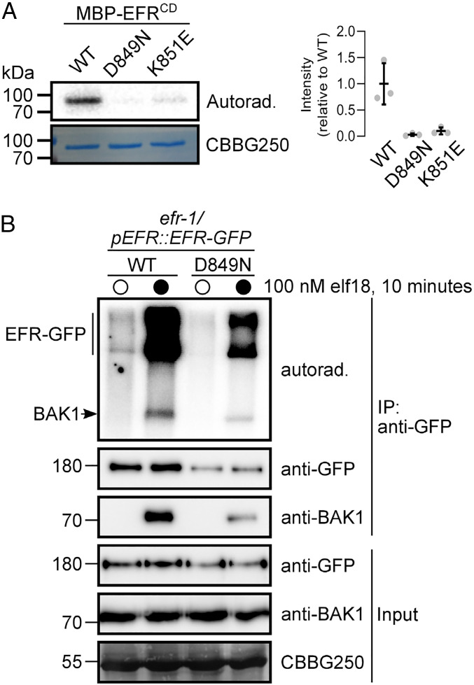
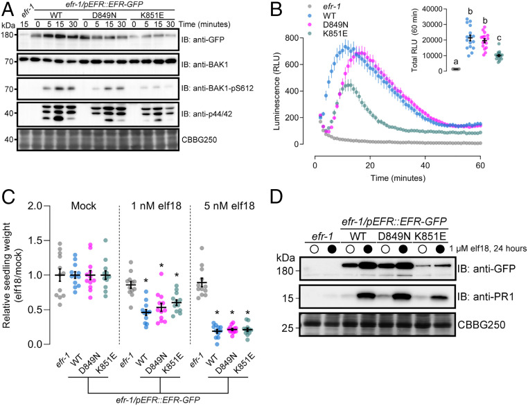
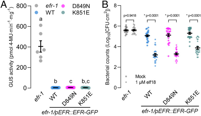
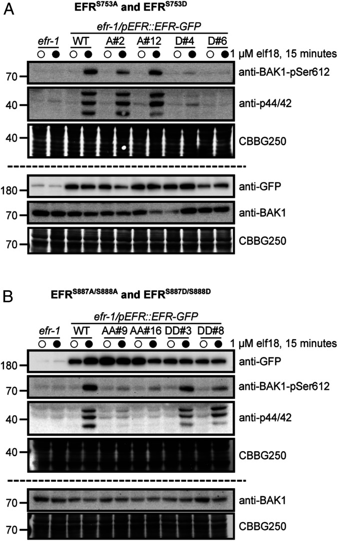
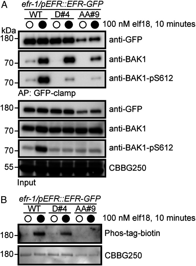
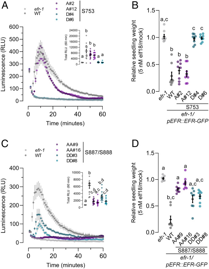
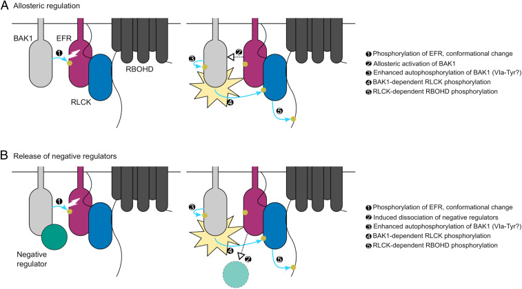
Receptor kinases (RKs) are fundamental for extracellular sensing and regulate development and stress responses across kingdoms. In plants, leucine-rich repeat receptor kinases (LRR-RKs) are primarily peptide receptors that regulate responses to myriad internal and external stimuli. Phosphorylation of LRR-RK cytoplasmic domains is among the earliest responses following ligand perception, and reciprocal transphosphorylation between a receptor and its coreceptor is thought to activate the receptor complex. Originally proposed based on characterization of the brassinosteroid receptor, the prevalence of complex activation via reciprocal transphosphorylation across the plant RK family has not been tested. Using the LRR-RK ELONGATION FACTOR TU RECEPTOR (EFR) as a model, we set out to understand the steps critical for activating RK complexes. While the EFR cytoplasmic domain is an active protein kinase in vitro and is phosphorylated in a ligand-dependent manner in vivo, catalytically deficient EFR variants are functional in antibacterial immunity. These results reveal a noncatalytic role for EFR in triggering immune signaling and indicate that reciprocal transphoshorylation is not a ubiquitous requirement for LRR-RK complex activation. Rather, our analysis of EFR along with a detailed survey of the literature suggests a distinction between LRR-RKs with RD- versus non-RD protein kinase domains. Based on newly identified phosphorylation sites that regulate the activation state of the EFR complex in vivo, we propose that LRR-RK complexes containing a non-RD protein kinase may be regulated by phosphorylation-dependent conformational changes of the ligand-binding receptor, which could initiate signaling either allosterically or through driving the dissociation of negative regulators of the complex.

Keywords: phosphorylation; receptor kinase; signaling.


Conflict of interest statement

The authors declare no competing interest.

Figures

Fig. 1.
Fig. 1.
EFR is an active protein kinase but its activity is not required for phosphorylation in an isolated receptor complex. (A) In vitro protein kinase activity of recombinant MBP-tagged EFRCD (WT) and catalytic site mutants (D849N and K851E). Recombinant proteins were incubated with 1 µCi γ[32P]ATP for 10 min and 32P incorporation was assessed by autoradiography. Relative quantification of 32P incorporation from three independent assays is shown. (B) On-bead kinase activity assay of immunopurified EFR-GFP (mock treatment, open circles) and EFR-GFP/BAK1 (elf18-treated, closed circles) complexes purified with GFP-Trap beads. Bead-bound receptor complexes were incubated with 5 µCi γ[32P]ATP for 30 min and 32P incorporation was assessed by autoradiography. On-bead kinase activity assays were performed three times with similar results each time. In A and B, Coomassie stain is shown as loading control (CBBG250).
Fig. 2.
Fig. 2.
Catalytically inactive EFR variants are competent for elf18-induced PTI signaling. (A) Immunoblot analysis of elf18-induced phosphorylation of BAK1 (anti-BAK1-pS612) and MAPKs (anti-p44/42) in 12-d-old seedlings treated with 1 µM elf18 for the indicated time. Anti-GFP shows protein accumulation of EFR and the site-directed mutants. Anti-BAK1 shows similar abundance of the coreceptor across all samples. Coomassie stain is shown as loading control (CBBG250). Blotting experiments were performed three times with similar results. (B) Time course of the oxidative burst in leaf discs from transgenic Arabidopsis expressing EFR-GFP (WT) or kinase-dead variants (D849N or K851E) in the efr-1 knockout background induced by treatment with 100 nM elf18. Points are mean with SEM. Inset shows mean with SEM of total luminescence over 60 min with individual data points. Means with like letter designations are not statistically different (Kruskal–Wallis ANOVA, n = 16 leaf discs, P < 0.000001, Dunn’s multiple comparisons test). The experiment was repeated three times with similar results. (C) Relative weight of seedlings grown in liquid media for 10 d with (1 or 5 nM) or without (Mock) the addition of elf18 peptide. Mean with SEM and individual values are shown. Asterisk indicates statistical difference from efr-1 within a given treatment (two-way ANOVA, n = 12 seedlings, P < 0.0001, Dunnett’s multiple comparison test). The experiment was repeated three times with similar results. (D) Accumulation of PR1 protein assessed by immunoblotting with anti-PR1 antibodies 24 h after infiltration of leaves from 3-wk-old plants with mock (open circles) or 1 µM elf18 (closed circles). Coomassie stain is shown as loading control (CBBG250). PR1 accumulation was assessed in three independent experiments with similar results each time.
Fig. 3.
Fig. 3.
Loss of EFR kinase activity does not compromise immune responses. (A) Fluorometric measurement of β-GUS activity in leaves of 3-wk-old plants 5 d after infiltration of leaves with Agrobacterium containing the pBIN19g:GUS plasmid. Mean with SEM and individual data points are shown. Means with like letter designations are not statistically different (Brown–Forsythe ANOVA, P = 0.000338, n = 5 or 6 plants, Dunnett’s multiple comparisons). The experiment was repeated three times with similar results. (B) Growth of Pto DC3000 2 d after infiltration in leaves pretreated with either mock or 1 µM elf18 for 24 h. Mean with SEM and individual data points (n = 23 or 24 plants) from three pooled independent experiments are shown. P values are derived from the comparison between elf18 pretreatment and mock, separately for each genotype as described in Experimental Procedures. Asterisk indicates a statistical difference between mock and elf18-treated leaves within each genotype.
Fig. 4.
Fig. 4.
EFR phosphorylation site mutants fail to trigger ligand-induced phosphorylation events. Immunoblot analysis of elf18-induced phosphorylation of BAK1 (anti-BAK1-pS612) and MAP kinases (anti-p44/42) in 12-d-old seedlings expressing WT EFR and (A) EFRS753A (A#2, A#12) or EFRS753D (D#4, D#6), or (B) EFRS887A/S888A (AA#9, AA#16) or EFRS887D/S888D (DD#3, DD#8) mutants. Seedlings were treated with mock (open circles) or 1 µM elf18 (closed circles) for 15 min. Anti-GFP shows protein accumulation of WT EFR-GFP and the site-directed mutants. Panels above and below the dashed line represent immunoblots derived from replicate SDS/PAGE gels. Coomassie stained blots are shown as loading control (CBBG250). Experiments were repeated three times with similar results.
Fig. 5.
Fig. 5.
EFR phosphorylation site mutants form a ligand-induced complex with BAK1. (A) Immunoblot analysis of elf18-induced receptor complex formation in 12-d-old seedlings expressing either WT EFR or phosphorylation site mutants (S753D, D#4; S887A/S888A, AA#9). Seedlings were treated with either mock (open circles) or 100 nM elf18 (closed circles) for 10 min, followed by coimmunoprecipitation with GFP-clamp beads and blotting with antibodies as indicated. (B) Analysis of in vivo phosphorylation of WT EFR or phosphorylation site mutants. Seedlings were treated with either mock (open circles) or 100 nM elf18 (closed circles) for 10 min, followed by immunoprecipitation of GFP-tagged receptors with GFP-Trap beads. Phospho-proteins were detected using a Zn2+-Phos-tag::biotin-Streptavidin::HRP complex. In both panels, Coomassie stain is shown as a loading control (CBBG250). Experiments in A and B were repeated four times with similar results.
Fig. 6.
Fig. 6.
Analysis of PTI responses in EFR phosphorylation site mutants. (A and C) Oxidative burst in leaf discs from the indicated genotype after treatment with 100 nM elf18. Points represent mean with SEM. Inset shows mean with SEM of total luminescence over 60 min. Means with like letter designations are not statistically different (Kruskal–Wallis ANOVA, n = 16 leaf discs, P < 0.000001, Dunn’s multiple comparisons test). (B and D) Seedling growth of the indicated genotypes in the presence of 5 nM elf18. Data are shown relative to mock treated seedlings for each genotype. Individual data points with mean and SEM are shown. Means with like letter designations are not statistically different (B, Kruskal–Wallis ANOVA, P = 0.00001, n = 8 seedlings, Dunn’s multiple comparison test; D, Kruskal–Wallis ANOVA, P < 0.000001, n = 8 seedlings, Dunn’s multiple comparison test). All experiments presented were repeated three times with similar results.
Fig. 7.
Fig. 7.
Potential mechanisms for phosphorylation-mediated activation of plant non-RD LRR-RK complexes. Ligand-triggered dimerization promotes phosphorylation of the EFR (purple) activation loop by BAK1 (light gray), inducing a conformational change of the EFR cytoplasmic domain. This conformational rearrangement feeds forward on BAK1 to enhance its catalytic activity either: (A) by direct allosteric activation of BAK1 or (B) by triggering the release of negative regulators (teal) of BAK1 activation. Either scenario permits full phosphorylation of the complex including on the VIa-Tyr residues. After full activation, BAK1 can phosphorylate the executor RLCKs (blue) to initiate downstream signaling, for example the RBOHD (dark gray)-dependent apoplastic oxidative burst. Yellow circles and blue arrows represent simplified requirements for activation of RBOHD-dependent ROS production by phosphorylation.

Similar articles

Cited by

References

    1. Shiu S. H., Bleecker A. B., Receptor-like kinases from Arabidopsis form a monophyletic gene family related to animal receptor kinases. Proc. Natl. Acad. Sci. U.S.A. 98, 10763–10768 (2001). - PMC - PubMed
    1. Shiu S. H., Bleecker A. B., Expansion of the receptor-like kinase/Pelle gene family and receptor-like proteins in Arabidopsis. Plant Physiol. 132, 530–543 (2003). - PMC - PubMed
    1. Hohmann U., Lau K., Hothorn M., The structural basis of ligand perception and signal activation by receptor kinases. Annu. Rev. Plant Biol. 68, 109–137 (2017). - PubMed
    1. Dufayard J.-F., et al. ., New insights on leucine-rich repeats receptor-like kinase orthologous relationships in angiosperms. Front. Plant Sci. 8, 381 (2017). - PMC - PubMed
    1. Dievart A., Gottin C., Périn C., Ranwez V., Chantret N., Origin and diversity of plant receptor-like kinases. Annu. Rev. Plant Biol. 71, 131–156 (2020). - PubMed

Publication types

MeSH terms