

Automated bone mineral density prediction and fracture risk assessment using plain radiographs via deep learning
- PMID: 34531406
- PMCID: PMC8446034
- DOI: 10.1038/s41467-021-25779-x
Automated bone mineral density prediction and fracture risk assessment using plain radiographs via deep learning
Abstract
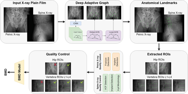
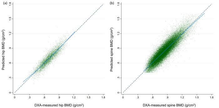
Dual-energy X-ray absorptiometry (DXA) is underutilized to measure bone mineral density (BMD) and evaluate fracture risk. We present an automated tool to identify fractures, predict BMD, and evaluate fracture risk using plain radiographs. The tool performance is evaluated on 5164 and 18175 patients with pelvis/lumbar spine radiographs and Hologic DXA. The model is well calibrated with minimal bias in the hip (slope = 0.982, calibration-in-the-large = -0.003) and the lumbar spine BMD (slope = 0.978, calibration-in-the-large = 0.003). The area under the precision-recall curve and accuracy are 0.89 and 91.7% for hip osteoporosis, 0.89 and 86.2% for spine osteoporosis, 0.83 and 95.0% for high 10-year major fracture risk, and 0.96 and 90.0% for high hip fracture risk. The tool classifies 5206 (84.8%) patients with 95% positive or negative predictive value for osteoporosis, compared to 3008 DXA conducted at the same study period. This automated tool may help identify high-risk patients for osteoporosis.
© 2021. The Author(s).
Conflict of interest statement
The authors declare no competing interests.
Figures
Similar articles
-
Predicting osteoporosis from kidney-ureter-bladder radiographs utilizing deep convolutional neural networks.Bone. 2024 Jul;184:117107. doi: 10.1016/j.bone.2024.117107. Epub 2024 Apr 25. Bone. 2024. PMID: 38677502
-
The utility of dual-energy X-ray absorptiometry, calcaneal quantitative ultrasound, and fracture risk indices (FRAX® and Osteoporosis Risk Assessment Instrument) for the identification of women with distal forearm or hip fractures: A pilot study.Endocr Res. 2016 Aug;41(3):248-60. doi: 10.3109/07435800.2015.1120744. Epub 2016 Feb 11. Endocr Res. 2016. PMID: 26864472
-
Evaluation of Singh Index and Osteoporosis Self-Assessment Tool for Asians as risk assessment tools of hip fracture in patients with type 2 diabetes mellitus.J Orthop Surg Res. 2017 Mar 3;12(1):37. doi: 10.1186/s13018-017-0539-6. J Orthop Surg Res. 2017. PMID: 28253896 Free PMC article.
-
Use of Trabecular Bone Score (TBS) as a Complementary Approach to Dual-energy X-ray Absorptiometry (DXA) for Fracture Risk Assessment in Clinical Practice.J Clin Densitom. 2017 Jul-Sep;20(3):334-345. doi: 10.1016/j.jocd.2017.06.019. Epub 2017 Jul 19. J Clin Densitom. 2017. PMID: 28734710 Review.
-
The role of DXA bone density scans in the diagnosis and treatment of osteoporosis.Postgrad Med J. 2007 Aug;83(982):509-17. doi: 10.1136/pgmj.2007.057505. Postgrad Med J. 2007. PMID: 17675543 Free PMC article. Review.
Cited by
-
Proximal humeral bone density assessment and prediction analysis using machine learning techniques: An innovative approach in medical research.Heliyon. 2024 Jul 31;10(15):e35451. doi: 10.1016/j.heliyon.2024.e35451. eCollection 2024 Aug 15. Heliyon. 2024. PMID: 39166094 Free PMC article.
-
A new osteogenic protein isolated from Dioscorea opposita Thunb accelerates bone defect healing through the mTOR signaling axis.Bioact Mater. 2023 Apr 23;27:429-446. doi: 10.1016/j.bioactmat.2023.04.018. eCollection 2023 Sep. Bioact Mater. 2023. PMID: 37152710 Free PMC article.
-
A Neural Network Model for Intelligent Classification of Distal Radius Fractures Using Statistical Shape Model Extraction Features.Orthop Surg. 2025 May;17(5):1513-1524. doi: 10.1111/os.70034. Epub 2025 Apr 3. Orthop Surg. 2025. PMID: 40180705 Free PMC article.
-
Establish and validate the reliability of predictive models in bone mineral density by deep learning as examination tool for women.Osteoporos Int. 2024 Jan;35(1):129-141. doi: 10.1007/s00198-023-06913-5. Epub 2023 Sep 20. Osteoporos Int. 2024. PMID: 37728768
-
Assessing the utility of osteoporosis self-assessment tool for Asians in patients undergoing hip surgery.Osteoporos Sarcopenia. 2024 Mar;10(1):16-21. doi: 10.1016/j.afos.2024.01.003. Epub 2024 Mar 2. Osteoporos Sarcopenia. 2024. PMID: 38690542 Free PMC article.
References
Publication types
MeSH terms
LinkOut - more resources
Full Text Sources
Other Literature Sources
Medical
