. 2021 Sep 16;4(1):1090.
doi: 10.1038/s42003-021-02615-y.

Venous puncture wound hemostasis results in a vaulted thrombus structured by locally nucleated platelet aggregates

Affiliations

Venous puncture wound hemostasis results in a vaulted thrombus structured by locally nucleated platelet aggregates

Sung W Rhee et al. Commun Biol. .

Abstract

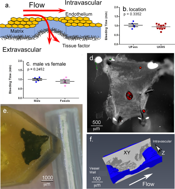
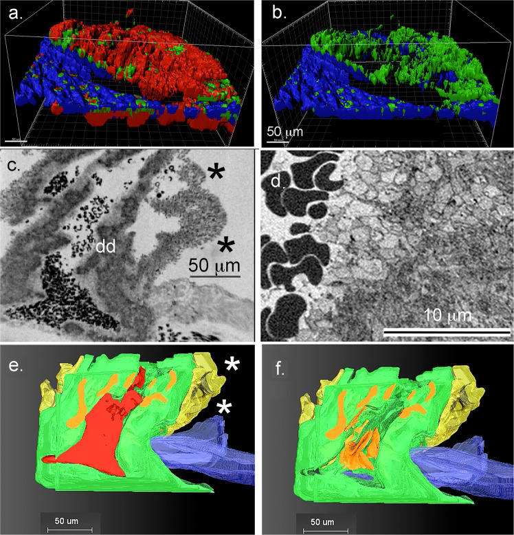
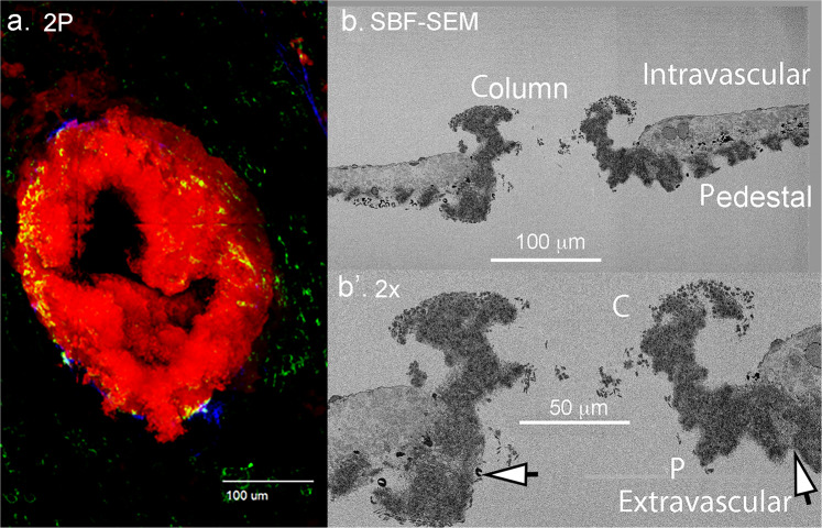
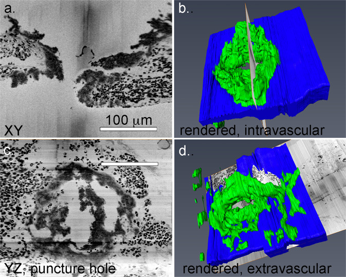
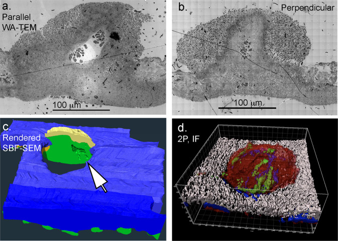
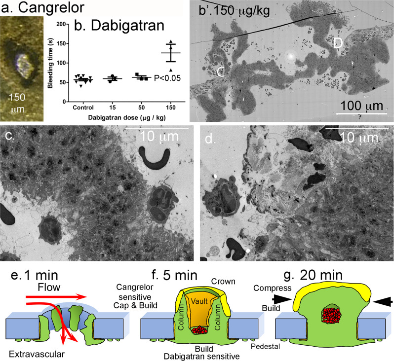
Primary hemostasis results in a platelet-rich thrombus that has long been assumed to form a solid plug. Unexpectedly, our 3-dimensional (3D) electron microscopy of mouse jugular vein puncture wounds revealed that the resulting thrombi were structured about localized, nucleated platelet aggregates, pedestals and columns, that produced a vaulted thrombus capped by extravascular platelet adherence. Pedestal and column surfaces were lined by procoagulant platelets. Furthermore, early steps in thrombus assembly were sensitive to P2Y12 inhibition and late steps to thrombin inhibition. Based on these results, we propose a Cap and Build, puncture wound paradigm that should have translational implications for bleeding control and hemostasis.


Conflict of interest statement

The authors declare no competing interests.

Figures

Fig. 1
Fig. 1. Experimental system.
a Schematic of flow and tissue factor exposure in a jugular vein puncture wound. Tissue factor exposure leads to coagulation factor activation, e.g., thrombin generation. b, c Bleeding time cessation times at the two different locations where punctures were performed and the effect of mouse sex on bleeding times, mean plus and minus standard deviation. Comparative positioning of the thrombus (red fluorescence in (d)). Upon opening the jugular vein (d) and embedding in plastic (e, f). Schematic of repeated, SBF-SEM imaging across the puncture wound hole. The resulting image slices are positioned so that flow is from left to right and the direction of progressive block face exposure into the plane of the page.
Fig. 2
Fig. 2. Bleeding cessation is through extravascular capping the vaulted 5 min post-puncture thrombus.
a/b Rendered images from 2P, light microscopy of a 5 min thrombus. a Three-color image: Red, CD 41 staining, a general platelet stain; Green, p-selectin antibody staining, an indicator of platelet α-granule secretion, Blue, fibrin. b Two-color: p-selectin (green) and fibrin (blue). Much of this staining is extravsascular, c Raw image, SBF-SEM, 100 nm XY pixel size in a raw captured image, mid-thrombus, showing an extravascular platelet layer that caps the puncture hole from the extravascular side. Asterisks, loosely adherent platelets. dd, approximate area shown in (d). d Raw image, SBF-SEM, 20 nm XY pixel size in a raw captured image of an area approximating dd in frame (c). Note that morphologically the platelets are most activated, i.e., degranulated on the surface of the column and least at the center. Furthermore, the trapped RBCs are distorted in shape, tending to polyhedral morphology. e/f 3D rendering of the thrombus with and without red blood cells (RBCs). RBCs, red; tightly adherent platelets, green, degranulated platelets, orange; loosely adherent platelets, yellow; vessel wall, blue.
Fig. 3
Fig. 3. Overall 3D structure reveals limited loosely adherent platelet sheathing of 5 min post-puncture wound thrombi.
a/b Example of single segmented image slice from a 5 min post-puncture jugular vein thrombus showing loosely adherent platelets (yellow), tightly adherent platelets (green), vessel wall (blue), RBCs (red) trapped intravascularly with a vault, and a small RBC patch on the extravascular presumably entrapped by extravascular fibrin. Flow from left to right. cf Full 3D rendering showing various features of the thrombus when viewed from different angles. Loosely adherent platelet sheathing of the intravascular thrombus crown is limited to small patches. RBC (red), tightly adherent platelets (green), degranulated platelets (orange), loosely adherent platelets (yellow), vessel wall (blue).
Fig. 4
Fig. 4. Early, nucleated platelet accumulation as revealed by 2P and SBF-SEM imaging of 1 min post-puncture thrombi.
a 2P, light microscope imaging of the thrombus, maximum intensity projection. Red, CD41 antibody staining, a marker for platelet accumulation; green: p-selectin antibody staining, a marker for α-granule fusion with the platelet plasma membrane; blue, fibrin antibody staining, a marker for what is principally extravascular accumulation of fibrin (see). b/b’ SBF-SEM visualization, 20 nm raw XY pixel size, one of a series of images taken every 20 μm across the forming thrombus. The intravascular lumen of the mouse jugular vein is to the top and the extravascular side of the vessel wall is to the bottom. The vein is lined by a layer of endothelial cells and collagen-rich adventitia below in these images. C, columnar platelet aggregate; P, pedestal (apparent nucleated platelet accumulation anchored to vessel wall adventitial layer. Arrows point to lightly staining platelet areas indicative of platelet degranulation. These presumably correspond to mobilized/exposed p-selectin staining in the 2P image. Electron micrograph slice is parallel to flow.
Fig. 5
Fig. 5. Morphological evidence for pedestal extension as the starting point for intravascular growth and puncture wound hole sealing.
XY cross-section slice through a 1 min post puncture wound hole (a) and its placement within the thrombus (b). YZ cross-section within the same puncture hole seen as imaged (c) and placed within a rendering (d). Raw images are 100 nm pixel size, 200 nm spacing in Z-dimension. Viewed from either orientation, the extension of the pedestals through further platelet aggregation appears to be a starting point for further thrombus growth. See also Supplementary Movies 1 and 4.
Fig. 6
Fig. 6. Thrombus remodeling leads to the retention of vaulting in 20 min thrombi.
The central portion of the thrombi appears to contain a “compressed” zone of tightly adherent platelets. a/b wide area TEM images of 20 min post wounding jugular thrombi taken either parallel to flow or perpendicular to flow. Note that the parallel image appears shaped by flow while the perpendicular to flow image appears to be nearly symmetric with respect to left and right. c A split open 3D rendering from SBF-SEM imaging of a 20 min post puncture thrombus (representative example, n = 2, SBF-SEM image sets) showing that the limited vaulting in the thrombus is enclosed by tightly adherent platelets. Tightly adherent platelets (green), loosely adherent platelets (yellow), vessel wall (blue). Note that extravascular platelet accumulation is found in hundreds of microns from the ~200 μm diameter puncture hole. d 2P, immunofluorescence rendering of protein distributions within a representative 20 min thrombus. Platelets (red, CD41), mobilized/exposed p-selectin (green), fibrin (blue), vessel wall (white). Note that fibrin staining “bands” within the intravascular interior of the thrombus appear to be in proximity to parallel bands of mobilized/exposed p-selectin staining. WA-TEM, wide area transmission electron microscopy. SBF-SEM, serial block face scanning electron microscopy. If, immunofluorescence.
Fig. 7
Fig. 7. Drug sensitivity of thrombus formation.
a Cangrelor treatment has a major effect on 5 min thrombus formation with osmium black platelets accumulating only around the rim of the puncture hole. b Dabigatran dose-response bleeding curve. Mean plus or minus standard deviation. b’ WA-TEM (wide-area transmission microscope) cross-section across the full puncture, dabigatran at 150 μg/Kg. WA-TEM, 3 nm raw pixel size. c/d Blowups of areas noted with C and D in (b’). Light staining, degranulated platelets are at most a very thin layer on the column surfaces with dabigatran treatment. Note that apparent PMN (polymorphonucleocyte) attachment, although rare, is more obvious in dabigatran treated cells with PMNs appearing to make attachments to degranulated, procoagulant platelets through pseudopods. eg A working model, Cap and Build, green: tightly adherent platelets, orange: areas of degranulated platelets obvious by SBF-SEM, yellow: loosely adherent platelets., red: trapped RBCs, RBCs are seen in variable amounts in 5 min thrombi as all preparations are in situ saline washed and intravascular continuities to vessel lumen are variable in amount.

References

    1. Falati S, Gross P, Merrill-Skoloff G, Furie BC, Furie B. Real-time in vivo imaging of platelets, tissue factor, and fibrin during arterial thrombus formation in the mouse. Nat. Med. 2002;8:1175–1181. doi: 10.1038/nm782. - DOI - PubMed
    1. Furie B, Furie BC. Thrombus formation in vivo. J. Clin. Invest. 2005;115:3355–3362. doi: 10.1172/JCI26987. - DOI - PMC - PubMed
    1. Getz TM, et al. Novel mouse hemostasis model for real-time determination of bleeeding time and hemostatic plug composition. J. Thromb. Haemost. 2015;13:417–425. doi: 10.1111/jth.12802. - DOI - PMC - PubMed
    1. Lee RH, et al. Impaired hemostatic activity of healthy transfused platelets in inherited and acquired platelet disorders: mechanisms and implications. Sci. Transl. Med. 2019;11:eaay0203. doi: 10.1126/scitranslmed.aay0203. - DOI - PMC - PubMed
    1. Chauhan AK, et al. Systemic antithrombotic effects of ADAMTS13. J. Exp. Med. 2006;203:767–778. doi: 10.1084/jem.20051732. - DOI - PMC - PubMed

Publication types