. 2021 Aug 31:12:654998.
doi: 10.3389/fimmu.2021.654998. eCollection 2021.

HCV Core Protein Induces Chemokine CCL2 and CXCL10 Expression Through NF-κB Signaling Pathway in Macrophages

Affiliations

HCV Core Protein Induces Chemokine CCL2 and CXCL10 Expression Through NF-κB Signaling Pathway in Macrophages

Xiaotian Song et al. Front Immunol. .

Abstract

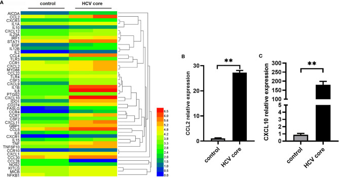
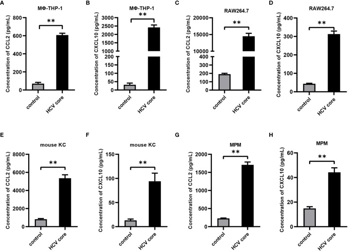
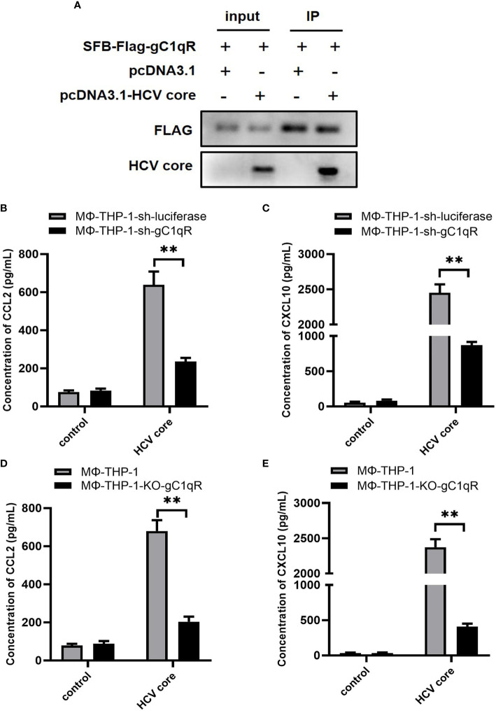
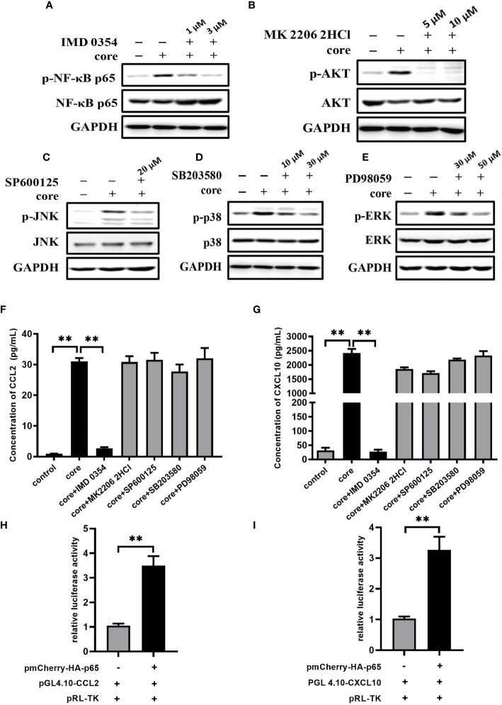
HCV core protein is the first structural protein synthesized during hepatitis C virus (HCV) infection and replication. It is released from virus infected liver cells and mediates multiple functions to affect host cell response. The innate immune response is the first line of defense against viral infection. After HCV infection, Kupffer cells (KCs) which are liver macrophages play an important role in host innate immune response. Kupffer cells act as phagocytes and release different cytokines and chemokines to counter viral infection and regulate inflammation and fibrosis in liver. Earlier, we have demonstrated that HCV core protein interacts with gC1qR and activates MAPK, NF-κB and PI3K/AKT pathways in macrophages. In this study, we explored the effect of HCV core protein on CCL2 and CXCL10 expression in macrophages and the signaling pathways involved. Upon silencing of gC1qR, we observed a significant decrease expression of CCL2 and CXCL10 in macrophages in the presence of HCV core protein. Inhibiting NF-κB pathway, but not P38, JNK, ERK and AKT pathways greatly reduced the expression of CCL2 and CXCL10. Therefore, our results indicate that interaction of HCV core protein with gC1qR could induce CCL2 and CXCL10 secretion in macrophages via NF-κB signaling pathway. These findings may shed light on the understanding of how leukocytes migrate into the liver and exaggerate host-derived immune responses and may provide novel therapeutic targets in HCV chronic inflammation.

Keywords: CCL2; CXCL10; HCV core protein; chemokines; macrophages.


Conflict of interest statement

The authors declare that the research was conducted in the absence of any commercial or financial relationships that could be construed as a potential conflict of interest.

Figures

Figure 1
Figure 1
Identification of differentially expressed genes in macrophages treated with HCV core protein. (A) Heat map showing the gene expression profile data in macrophages (MΦ-THP-1) treated or untreated with HCV core protein (10 μg/mL) for 8 h based on PCR array analysis. Red: up-regulation; Blue: down-regulation. (B, C) MΦ-THP-1 were treated or untreated with HCV core protein (10 μg/mL) for 8 h. Relative expressions of CCL2 and CXCL10 were determined by quantitative real-time PCR (qPCR). Data are shown as mean ± SD for three independent experiments. **p < 0.01.
Figure 2
Figure 2
HCV core protein up-regulates CCL2 and CXCL10 expression in macrophages. Human macrophages cell line (MΦ-THP-1) (A, B), mouse macrophage cell line RAW 264.7 (C, D), mouse Kupffer cells (KC) (E, F) and mouse peritoneal macrophages (MPM) (G, H) were treated or untreated with HCV core protein (10 μg/mL) for 24 h. Cell culture supernatants were collected and analyzed for CCL2 and CXCL10 production using ELISA kits. Data are shown as mean ± SD for three independent experiments. **p < 0.01.
Figure 3
Figure 3
gC1qR is involved in the expression of CCL2 and CXCL10 in macrophages. (A) HEK 293T cells were co-transfected with SFB-Flag-gC1qR and pcDNA3.1-HCV core plasmid (co-transfected SFB-Flag-gC1qR and pcDNA3.1 plasmid as control group) for 48 h. Cells were lysed with NTEN buffer and analyzed by immunoprecipitation and immunoblotting using antibodies of FLAG and HCV Core protein. (B, C) gC1qR knock-down cell line (MΦ-THP-1-sh-gC1qR) and control cell line (MΦ-THP-1-sh-luciferase) were either treated or untreated with HCV core protein (10 μg/mL) for 24 h. Cell culture supernatants were collected and analyzed for CCL2 and CXCL10 production using ELISA kits. (D, E) gC1qR knock-out cell line (MΦ-THP-1-KO-gC1qR) and control cell line (MΦ-THP-1) were either treated or untreated with HCV core protein (10 μg/mL) for 24 h. Cell culture supernatants were collected and analyzed for CCL2 and CXCL10 production using ELISA kits. Data are shown as mean ± SD for three independent experiments. **p < 0.01.
Figure 4
Figure 4
NF-κB signaling pathway plays pivotal role in CCL2 and CXCL10 expression in macrophages induced by HCV core protein. (A–E) MΦ-THP-1 were pretreated with specific inhibitor in different doses for 30 min and then treated with HCV core protein for 30 min. Cells were lysed and analyzed by immunoblotting with anti-p-NF-κB p65, anti-NF-κB p65, anti-p-AKT, anti-AKT, anti-p-JNK, anti-JNK, anti-p-p38, anti-p38, anti-p-ERK, anti-ERK and anti-GAPDH antibodies to determine the optimum concentration of inhibitors: IKKβ inhibitor IMD 0354 was 3 μM, Akt inhibitor MK 2206 2HCl was 5 μM, JNK inhibitor SP600125 was 20 μM, P38 inhibitor SB203580 was 30 μM, ERK inhibitor PD98059 was 50 μM. (F, G) MΦ-THP-1 were pretreated with optimal concentration of specific inhibitor and then treated with HCV core protein for 24 h. The supernatants were analyzed by the ELISA to detect CCL2 and CXCL10 production. (H, I) Luciferase activities were detected in HEK 293T cells co-transfected with NF-κB p65expressing plasmid, CCL2 or CXCL10 reporter plasmid and pRL-TK plasmid as described in Materials and Methods (co-transfected with promoter reporter plasmid and pRL-TK plasmid as control group). Data are presented as the relative ratio of Firefly luciferase activity to Renilla luciferase activity. Data are shown as mean ± SD for three independent experiments. **p < 0.01.
Figure 5
Figure 5
CCL2 and CXCL10 expression is significantly increased in liver biopsy samples of HCV patients. (A, B) Six liver biopsy samples from patients infected with HCV and three control samples from healthy liver tissues were collected. The samples were handled in BSL-2 laboratory using standard precautions. Relative expressions of CCL2 and CXCL10 were determined by quantitative real-time PCR (qPCR). Data are shown as mean ± SD for three independent experiments. **p < 0.01.

References

    1. Lavanchy D. Evolving Epidemiology of Hepatitis C Virus. Clin Microbiol Infect (2011) 172:107–15. 10.1111/j.1469-0691.2010.03432.x - DOI - PubMed
    1. Shepard CW, Finelli L, Alter MJ. Global Epidemiology of Hepatitis C Virus Infection. Lancet Infect Dis (2005) 59:558–67. 10.1016/s1473-3099(05)70216-4 - DOI - PubMed
    1. Brown RS. Hepatitis C And Liver Transplantation. Nature (2005) 4367053:973–8. 10.1038/nature04083 - DOI - PubMed
    1. Pickett BE, Striker R, Lefkowitz EJ. Evidence for Separation of HCV Subtype 1a Into Two Distinct Clades. J Viral Hepat (2011) 189:608–18. 10.1111/j.1365-2893.2010.01342.x - DOI - PMC - PubMed
    1. Poynard T, Yuen MF, Ratziu V, Lai CL. Viral Hepatitis C. Lancet (2003) 3629401:2095–100. 10.1016/s0140-6736(03)15109-4 - DOI - PubMed

Publication types

MeSH terms