

Expression, clinical significance and correlation of RUNX3 and HER2 in colorectal cancer
- PMID: 34532112
- PMCID: PMC8421900
- DOI: 10.21037/jgo-21-403
Expression, clinical significance and correlation of RUNX3 and HER2 in colorectal cancer
Abstract
Background: The incidence of colorectal cancer is high and on the rise. The genetic and protein expressions of RUNT-associated transcription factor 3 (RUNX3) and human epidermal growth factor receptor 2 (HER2) in colorectal cancer (CRC) and adjacent normal tissues were detected to preliminarily explore their correlation and clinical significance.
Methods: CRC specimens excised during general surgery were selected for localization and quantitative analysis of protein and gene expression by SP (Streptavidin-peroxidase conjugated method) immunohistochemical staining, reverse transcription-polymerase chain reaction (RT-qPCR) and western blot. Combined with the patients' data, the relationship between the expression of the two genes and tumor characteristics was analyzed. Log-rank test was used to analyze the correlation between the two proteins and survival prognosis of CRC patients. The expression of RUNX3 in RKO and HCT-116 was knocked down, and the relative expression of HER2 in the two cell lines was detected.
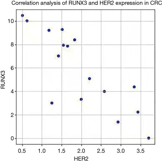
Results: Immunohistochemical, RT-qPCR and Western blot results showed that the positive expression rate of RUNX3 in CRC was lower than in the normal group, and HER2 in CRC was higher than in the normal group. The positive expression of the two proteins correlated with the pT and pN stages of CRC. A significant negative correlation between the expression of the two genes in CRC. Follow-up results showed that the number of Runx3-positive patients was higher than negative ones, while HER2 positive were fewer than negative ones. In vitro experiments showed that RUNX3 protein and gene expression decreased, HER2 protein and gene expression increased in RUNX3 knockdown RKO and HCT-116 cells, respectively (P<0.05).
Conclusions: The expression of RUNX3 and HER2 in CRC is related to the occurrence and development of CRC, and the two genes have a negative regulating effect.
Keywords: Colorectal cancer (CRC); HER2; RUNX3; molecular markers.
2021 Journal of Gastrointestinal Oncology. All rights reserved.
Conflict of interest statement
Conflicts of Interest: All authors have completed the ICMJE uniform disclosure form (available at https://dx.doi.org/10.21037/jgo-21-403). The authors have no conflicts of interest to declare.
Figures


References
-
- National Health Commission of the People's Republic of China . Chinese Protocol of Diagnosis and Treatment of Colorectal Cancer (2020 edition). Zhonghua Wai Ke Za Zhi 2020;58:561-85. - PubMed
-
- Ren Y, Cai J, Chen J. The significance of anti-oncogene Runx3 expression in colorectal cancer. Journal of Modern Oncology 2018;1:72-6.
-
- Xue J, Wu X, Wang L, et al. Expression of RUNX3 in colorectal cancer, adenoma and normal colorectal tissues and its clinical significance. Clin Exp Pathol 2014;30:605-9.
-
- Wu L, Li Y, Liu H, et al. The role of RUNX3, β-catenin and C-myc in carcinogenesis of colorectal adenoma Chinese Journal of Difficult and Complicated Cases 2018;17:176-179.
LinkOut - more resources
Full Text Sources
Research Materials
Miscellaneous