Anatomical Increased/Decreased Changes in the Brain Area Following Individuals with Chronic Traumatic Complete Thoracic Spinal Cord Injury
- PMID: 34532212
- PMCID: PMC8419593
- DOI: 10.1298/ptr.E10076
Anatomical Increased/Decreased Changes in the Brain Area Following Individuals with Chronic Traumatic Complete Thoracic Spinal Cord Injury
Abstract
Objectives: This study aimed to investigate anatomical changes in the brain following chronic complete traumatic thoracic spinal cord injury (ThSCI) using voxel-based morphometry (VBM). That is, it attempted to examine dynamic physical change following thoracic injury and the presence or absence of regions with decreased and increased changes in whole brain volume associated with change in the manner of how activities of daily living are performed.
Methods: Twelve individuals with chronic traumatic complete ThSCI (age; 21-63 years, American Spinal Injury Association Impairment Scale; grade C-D) participated in this study. VBM was used to investigate the regions with increased volume and decreased volume in the brain in comparison with healthy control individuals.
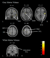
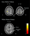
Results: Decreases in volume were noted in areas associated with motor and somatosensory functions, including the right paracentral lobule (PCL)-the primary motor sensory area for lower limbs, left dorsal premotor cortex, and left superior parietal lobule (SPL). Furthermore, increased gray matter volume was noted in the primary sensorimotor area for fingers and arms, as well as in higher sensory areas.
Conclusions: Following SCI both regions with increased volume and regions with decreased volume were present in the brain in accordance with changes in physical function. Using longitudinal observation, anatomical changes in the brain may be used to determine the rehabilitation effect by comparing present cases with cases with cervical SCI or cases with incomplete palsy.
Keywords: Anatomical change; Brain; Rehabilitation; Spinal cord injury; Voxel-based morphometry.
2021, JAPANESE PHYSICAL THERAPY ASSOCIATION.
Conflict of interest statement
The authors declare no conflict of interest.
Figures


Similar articles
-
Reorganization of the somatosensory pathway after subacute incomplete cervical cord injury.Neuroimage Clin. 2019;21:101674. doi: 10.1016/j.nicl.2019.101674. Epub 2019 Jan 9. Neuroimage Clin. 2019. PMID: 30642754 Free PMC article.
-
Specific Brain Morphometric Changes in Spinal Cord Injury: A Voxel-Based Meta-Analysis of White and Gray Matter Volume.J Neurotrauma. 2019 Aug 1;36(15):2348-2357. doi: 10.1089/neu.2018.6205. Epub 2019 Apr 23. J Neurotrauma. 2019. PMID: 30794041
-
Brain sensorimotor system atrophy during the early stage of spinal cord injury in humans.Neuroscience. 2014 Apr 25;266:208-15. doi: 10.1016/j.neuroscience.2014.02.013. Epub 2014 Feb 20. Neuroscience. 2014. PMID: 24561217
-
Non-concomitant cortical structural and functional alterations in sensorimotor areas following incomplete spinal cord injury.Neural Regen Res. 2017 Dec;12(12):2059-2066. doi: 10.4103/1673-5374.221165. Neural Regen Res. 2017. PMID: 29323046 Free PMC article.
-
Anatomical changes in human motor cortex and motor pathways following complete thoracic spinal cord injury.Cereb Cortex. 2009 Jan;19(1):224-32. doi: 10.1093/cercor/bhn072. Epub 2008 May 14. Cereb Cortex. 2009. PMID: 18483004
Cited by
-
Brain morphology changes after spinal cord injury: A voxel-based meta-analysis.Front Neurol. 2022 Sep 1;13:999375. doi: 10.3389/fneur.2022.999375. eCollection 2022. Front Neurol. 2022. PMID: 36119697 Free PMC article.
References
-
- Dietz V and Curt A: Neurological aspects of spinal-cord repair: promises and challenges. Lancet Neurol. 2006; 5: 688-694. - PubMed
-
- Tetzlaff W, Kobayashi NR, et al.: Response of rubrospinal and corticospinal neurons to injury and neurotrophins. Prog Brain Res. 1994; 103: 271-286. - PubMed
-
- Florence SL, Taub HB, et al.: Large-scale sprouting of cortical connections after peripheral injury in adult macaque monkeys. Science. 1998; 282: 1117-1121. - PubMed
-
- Hains BC, Black JA, et al.: Primary cortical motor neurons undergo apoptosis after axotomizing spinal cord injury. J Comp Neurol. 2003; 462: 328-341. - PubMed
-
- Lee BH, Lee KH, et al.: Injury in the spinal cord may produce cell death in the brain. Brain Res. 2004; 1020: 37-44. - PubMed

