

Animal models of diabetic retinopathy
- PMID: 34532409
- PMCID: PMC8421981
- DOI: 10.21037/atm-20-6737
Animal models of diabetic retinopathy
Abstract
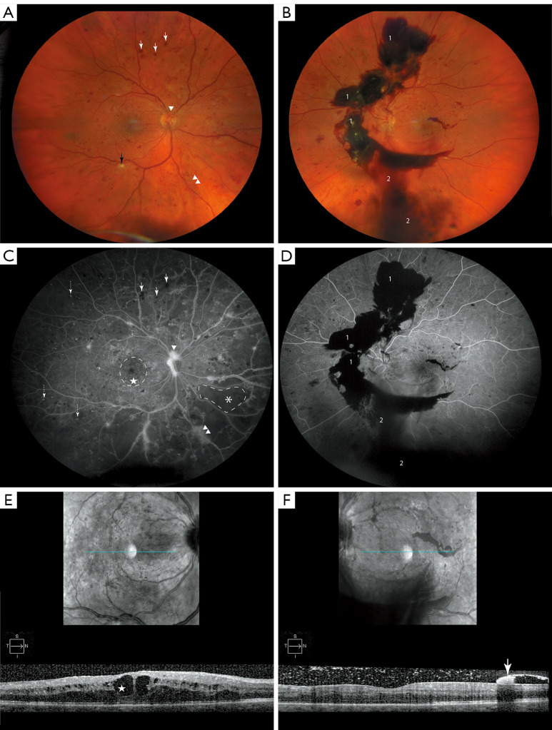
The retina is the posterior neuro-integrated layer of the eye that conducts impulses induced by light to the optic nerve for human vision. Diseases of the retina often leads to diminished vision and in some cases blindness. Diabetes mellitus (DM) is a worldwide public health issue and globally, there is an estimated 463 million people that are affected by DM and its consequences. Diabetic retinopathy (DR) is a blinding complication of chronic uncontrolled DM and is the most common cause of blindness in the United States between the ages 24-75. It is estimated that the global prevalence of DR will increase to 191.0 million by 2030, of those 56.3 million possessing vision-threatening diabetic retinopathy (VTDR). For the most part, current treatment modalities control the complications of DR without addressing the underlying pathophysiology of the disease. Therefore, there is an unmet need for new therapeutics that not only repair the damaged retinal tissue, but also reverse the course of DR. The key element in developing these treatments is expanding our basic knowledge by studying DR pathogenesis in animal models of proliferative and non-proliferative DR (PDR and NPDR). There are numerous models available for the research of both PDR and NPDR with substantial overlap. Animal models available include those with genetic backgrounds prone to hyperglycemic states, immunologic etiologies, or environmentally induced disease. In this review we aimed to comprehensively summarize the available animal models for DR while also providing insight to each model's ocular therapeutic potential for drug discovery.
Keywords: Diabetes mellitus (DM); animal model; diabetic retinopathy (DR).
2021 Annals of Translational Medicine. All rights reserved.
Conflict of interest statement
Conflicts of Interest: Both authors have completed the ICMJE uniform disclosure form (available at https://dx.doi.org/10.21037/atm-20-6737). The series “Novel Tools and Therapies for Ocular Regeneration” was commissioned by the editorial office without any funding or sponsorship. The authors have no other conflicts of interest to declare.
Figures
References
Publication types
Grants and funding
LinkOut - more resources
Full Text Sources