Review
. 2021 Sep 21;10(18):e021987.
doi: 10.1161/JAHA.121.021987. Epub 2021 Sep 17.

Evolving Diagnostic Criteria for Arrhythmogenic Cardiomyopathy

Affiliations
Review

Evolving Diagnostic Criteria for Arrhythmogenic Cardiomyopathy

Domenico Corrado et al. J Am Heart Assoc. .

Abstract

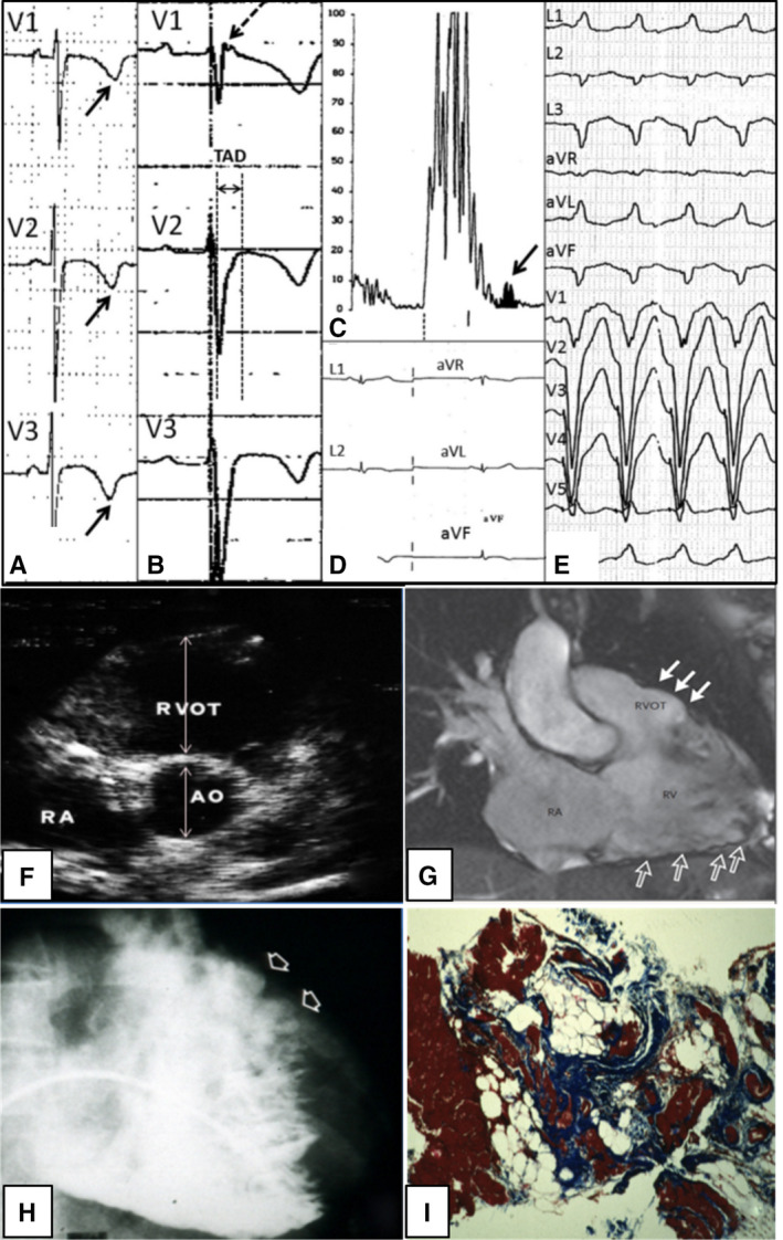
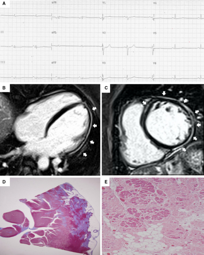
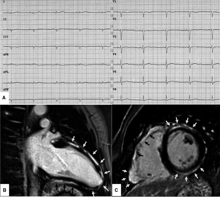
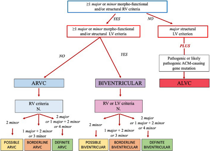
Criteria for diagnosis of arrhythmogenic cardiomyopathy (ACM) were first proposed in 1994 and revised in 2010 by a Task Force. Although the Task Force criteria demonstrated a good accuracy for diagnosis of the original right ventricular phenotype (arrhythmogenic right ventricular cardiomyopathy), they lacked sensitivity for identification of the expanding phenotypic spectrum of ACM, which includes left-sided variants and did not incorporate late-gadolinium enhancement findings by cardiac magnetic resonance. The 2020 International criteria ("Padua criteria") have been developed by International experts with the aim to improve the diagnosis of ACM by providing new criteria for the diagnosis of left ventricular phenotypic features. The key upgrade was the incorporation of tissue characterization findings by cardiac magnetic resonance for noninvasive detection of late-gadolinium enhancement/myocardial fibrosis that are determinants for characterization of arrhythmogenic biventricular and left ventricular cardiomyopathy. The 2020 International criteria are heavily dependent on cardiac magnetic resonance, which has become mandatory to characterize the ACM phenotype and to exclude other diagnoses. New criteria regarding left ventricular depolarization and repolarization ECG abnormalities and ventricular arrhythmias of left ventricular origin were also provided. This article reviews the evolving approach to diagnosis of ACM, going back to the 1994 and 2010 International Task Force criteria and then grapple with the modern 2020 International criteria.

Keywords: cardiac magnetic resonance; cardiomyopathy; diagnosis; sudden death; ventricular arrhythmia.


Conflict of interest statement

None.

Figures

Figure 1
Figure 1. Clinical features of arrhythmogenic right ventricular cardiomyopathy.
Right precordial negative T waves in leads V1 to V3 and prolongation of QRS complex because of delayed S‐wave upstroke leading to a significant terminal activation delay (A), epsilon waves (arrow) (B), late potentials on signal‐averaged ECG (arrow) (C), low QRS voltages (<0.5 mV) in the limb leads (D), and ventricular tachycardia with a left bundle branch block (E). Two‐dimensional echocardiogram (parasternal short‐axis view), showing dilatation of the RVOT (parasternal long axis‐RVOT=37 mm) (F). Cardiac magnetic resonance imaging scan (systolic frame of right ventricular 2‐chamber long‐axis view on cine sequences) evidencing an aneurysm (with dyskinesia, not shown) of the RVOT (solid arrows) and multiple sacculations of the inferior and apical regions (open arrows) (G). Angiography showing RV dilatation with a bulging of the RVOT (arrows) (H). Endomyocardial biopsy revealing myocyte loss with fibrofatty replacement (I). AO indicates aorta; RA, right atrium; RV, right ventricle; and RVOT, right ventricular outflow tract. Adapted from Corrado et al.
Figure 2
Figure 2. Clinical and histopathologic features of arrhythmogenic left ventricular cardiomyopathy.
ECG, and CMR findings of a patient with ALVC related to a DSP gene defect. Basal ECG showing low voltages in limb leads and flattened T‐waves in the inferolateral leads (A). Postcontrast CMR images in long‐axis (B) and short‐axis (C) views showing normal LV cavity size and subepicardial LGE (white arrows) involving the whole LV free wall and the anterior septum (“ring‐like” pattern), from basal to apical regions. Histology of the LV inferolateral region showing fibrofatty myocardial replacement affecting the subepicardial layer (Heidenhain trichrome stain) (D); close‐up detailing residual myocytes embedded within fibrous and fatty tissue (hematoxylin and eosin stain) (E). ALVC indicates arrhythmogenic left ventricular cardiomyopathy; CMR, cardiac magnetic resonance; DSP, desmoplakin gene; LGE, late gadolinium enhancement; and LV, left ventricle. Adapted from Cipriani et al.
Figure 3
Figure 3. Clinical features of biventricular arrhythmogenic cardiomyopathy.
ECG and CMR findings in a 40‐year‐old patient with biventricular ACM caused by a pathogenic DSG‐2 gene mutation. Basal ECG showing low QRS voltages (<0.5 mV, peak to peak) in the limb leads, in the absence of other repolarization and depolarization ECG abnormalities. Premature ventricular beats on Holter monitoring <500/24 hours; no sustained or nonsustained ventricular tachycardia (not shown) (A). Postcontrast cardiovascular magnetic resonance images—end‐diastolic frame on long‐axis view (B) and short‐axis view (C)—showing normal cavity size of both RV and LV and LGE of the myocardium of the basal anterolateral right ventricular wall and anterior and inferior LV wall (arrows). On cine sequences (not shown) the RV shows regional akinesia with a mild reduction of the ejection fraction (ie, 50%) and the LV an inferolateral ipokinesia with a preserved systolic function. While this phenotypic variant of ACM does not fulfill the 2010 TF criteria, it is diagnosed according to the 2020 International criteria as definite biventricular ACM based on the low QRS voltages, the regional akinesia of the RV, the regional hypokinesia of the LV, and the biventricular LGE other than the pathogenic gene mutation. ACM indicates arrhythmogenic cardiomyopathy; CMR, cardiac magnetic resonance; DSG‐2, desmoglein‐2 gene; LGE, late gadolinium enhancement; LV, left ventricle; RV, right ventricle; and TF, task force.
Figure 4
Figure 4. Diagnostic flow‐chart for ACM phenotypic variants.
According to the 2020 International criteria, any diagnosis of ACM requires that at least 1 criterion either major or minor from category I (ie, morpho‐functional abnormalities) or II (ie, structural abnormalities) be fulfilled. For diagnosis of possible, borderline, or definite biventricular ACM, besides the need for ≥1 morpho‐functional and/or structural criteria from both the RV and LV, the remaining criteria are from either the RV or the LV (see text for details). ACM indicates arrhythmogenic left ventricular cardiomyopathy; ALVC, arrhythmogenic left ventricular cardiomyopathy; ARVC, arrhythmogenic right ventricular cardiomyopathy; LV, left ventricle; and RV, right ventricle.

References

    1. Marcus FI, Fontaine GH, Guiraudon G, Frank R, Laurenceau JL, Malergue C, Grosgogeat Y. Right ventricular dysplasia: a report of 24 adult cases. Circulation. 1982;65:384–398. DOI: 10.1161/01.CIR.65.2.384. - DOI - PubMed
    1. Thiene G, Nava A, Corrado D, Rossi L, Pennelli N. Right ventricular cardiomyopathy and sudden death in young people. N Engl J Med. 1988;318:129–133. DOI: 10.1056/NEJM198801213180301. - DOI - PubMed
    1. Corrado D, Link MS, Calkins H. Arrhythmogenic right ventricular cardiomyopathy. N Engl J Med. 2017;376:61–72. DOI: 10.1056/NEJMra1509267. - DOI - PubMed
    1. Corrado D, Basso C, Judge DP. Arrhythmogenic cardiomyopathy. Circ Res. 2017;121:784–802. DOI: 10.1161/CIRCRESAHA.117.309345. - DOI - PubMed
    1. Corrado D, van Tintelen PJ, McKenna WJ, Hauer RNW, Anastastakis A, Asimaki A, Basso C, Bauce B, Brunckhorst C, Bucciarelli‐Ducci C, International Experts , et al. Arrhythmogenic right ventricular cardiomyopathy: evaluation of the current diagnostic criteria and differential diagnosis. Eur Heart J. 2020;41:1414–1429. DOI: 10.1093/eurheartj/ehz669. - DOI - PMC - PubMed

MeSH terms