

Role of CT angiography in detecting acute pulmonary embolism associated with COVID-19 pneumonia
- PMID: 34533699
- PMCID: PMC8446165
- DOI: 10.1007/s11547-021-01415-y
Role of CT angiography in detecting acute pulmonary embolism associated with COVID-19 pneumonia
Abstract
Purpose: Recently coronavirus disease (COVID-19) caused a global pandemic, characterized by acute respiratory distress syndrome (ARDS). The aim of our study was to detect pulmonary embolism (PE) in patients with severe form of COVID-19 infection using pulmonary CT angiography, and its associations with clinical and laboratory parameters.
Methods: From March to December 2020, we performed a prospective monocentric study collecting data from 374 consecutive patients with confirmed SARS-CoV-2 infection, using real-time reverse-transcriptase polymerase-chain-reaction (rRT-PCR) assay of nasopharyngeal swab specimens. We subsequently selected patients with at least two of the following inclusion criteria: (1) severe acute respiratory symptoms (such as dyspnea, persistent cough, fever > 37.5 °C, fatigue, etc.); (2) arterial oxygen saturation ≤ 93% at rest; (3) elevated D-dimer (≥ 500 ng/mL) and C-reactive protein levels (≥ 0.50 mg/dL); and (4) presence of comorbidities. A total of 63/374 (17%) patients met the inclusion criteria and underwent CT angiography during intravenous injection of iodinated contrast agent (Iomeprol 400 mgI/mL). Statistical analysis was performed using Wilcoxon rank-sum and Chi-square tests.
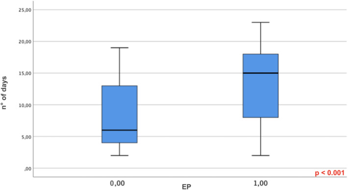
Results: About, 26/60 patients (40%) were found positive for PE at chest CT angiography. In these patients, D-dimer and CRP values were significantly higher, while a reduction in SaO2 < 93% was more common than in patients without PE (P < 0.001). Median time between illness onset and CT scan was significantly longer (15 days; P < 0.001) in patients with PE. These were more likely to be admitted to the Intensive Care Unit (19/26 vs. 11/34 patients; P < 0.001) and required mechanical ventilation more frequently than those without PE (15/26 patients vs. 9/34 patients; P < 0.001). Vascular enlargement was significantly more frequent in patients with PE than in those without (P = 0.041).
Conclusions: Our results pointed out that patients affected by severe clinical features of COVID-19 associated with comorbidities and significant increase of D-dimer levels developed acute mono- or bi-lateral pulmonary embolism in 40% of cases. Therefore, the use of CT angiography rather than non-contrast CT should be considered in these patients, allowing a better evaluation, that can help the management and improve the outcomes.
Keywords: ARDS; COVID-19; CT angiography; Pulmonary embolism.
© 2021. The Author(s).
Conflict of interest statement
The authors declare that they have no conflict of interest.
Figures


References
MeSH terms
LinkOut - more resources
Full Text Sources
Medical
Research Materials
Miscellaneous
