. 2021 Sep 17;17(9):e1009929.
doi: 10.1371/journal.ppat.1009929. eCollection 2021 Sep.

In vitro selection of Remdesivir resistance suggests evolutionary predictability of SARS-CoV-2

Affiliations

In vitro selection of Remdesivir resistance suggests evolutionary predictability of SARS-CoV-2

Agnieszka M Szemiel et al. PLoS Pathog. .

Abstract

Remdesivir (RDV), a broadly acting nucleoside analogue, is the only FDA approved small molecule antiviral for the treatment of COVID-19 patients. To date, there are no reports identifying SARS-CoV-2 RDV resistance in patients, animal models or in vitro. Here, we selected drug-resistant viral populations by serially passaging SARS-CoV-2 in vitro in the presence of RDV. Using high throughput sequencing, we identified a single mutation in RNA-dependent RNA polymerase (NSP12) at a residue conserved among all coronaviruses in two independently evolved populations displaying decreased RDV sensitivity. Introduction of the NSP12 E802D mutation into our SARS-CoV-2 reverse genetics backbone confirmed its role in decreasing RDV sensitivity in vitro. Substitution of E802 did not affect viral replication or activity of an alternate nucleoside analogue (EIDD2801) but did affect virus fitness in a competition assay. Analysis of the globally circulating SARS-CoV-2 variants (>800,000 sequences) showed no evidence of widespread transmission of RDV-resistant mutants. Surprisingly, we observed an excess of substitutions in spike at corresponding sites identified in the emerging SARS-CoV-2 variants of concern (i.e., H69, E484, N501, H655) indicating that they can arise in vitro in the absence of immune selection. The identification and characterisation of a drug resistant signature within the SARS-CoV-2 genome has implications for clinical management and virus surveillance.


Conflict of interest statement

The authors have declared that no competing interests exist.

Figures

Fig 1
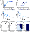
Fig 1. Continuous passage of SARS-CoV-2Engl2 in RDV selects for partial resistant populations.
(A) Schematic of the experimental layout to select for RDV resistant viruses. The passage (p) number of the input virus SARS-CoV-2Engl2 is given. We consider p1 as the stock supplied by Public Health England, it was passaged twice in VeroE6 prior to selection in RDV. SARS-CoV-2Engl2 p3 is the input stock that was sequenced and used for analysis. Each condition is given a different colour and the amount of virus used at the start of the experiment is indicated. Populations that failed to amplify (no CPE or/and virus titre) are indicated by the empty circles. All lineages sequenced are indicated. (B) Virus titers (pfu/ml) at p1, p4, p7 and p10. 6 lineages per condition and two different virus inputs; 1000 pfu (solid circle) and 2000 pfu (open circle). Median for each is shown. (C) Virus growth kinetics in VeroE6 in the presence (dashed line) or absence (solid line) of 7.5μM RDV for 3 different virus populations. Data is from 2 independent experiments with 3 replicates. Error bars represent SEM. Unpaired t-tests (Holm-Šídák method; *,P< 0.05; **,P< 0.01; ***,P< 0.001. ****, P<0.0001). (D) EIDD2801 dose dependency curve. EIDD2801 treated VeroE6-ACE2-TMPRSS2 infected with 8400 pfu/ml of each virus. (E) RDV dose dependency curves determined in A549NPro-ACE2 infected with 8400 pfu/ml of each virus. (F) Bar graph of RDV EC50 for different viruses in A549NPro-ACE2 with 8400 pfu/well. For all panels, error bars represent SEM.
Fig 2
Fig 2. Common mutations in partial RDV resistance populations.
(A) Location of E802 within structure of SARS-CoV-2 NSP12 in association with NSP7 and NSP8 (PDB ID 6YYT). Three focused panels are WT (upper) and two potential confirmations of E802D. H-bonds are indicated by light blue line. (B) Conservation of E802 amino acid across coronaviruses. Accession numbers for the coronavirus sequences are in the materials and methods. (C) NSP6 I168 amino acid is not conserved across coronaviruses.
Fig 3
Fig 3. NSP12 E802 mutation recapitulates change in RDV susceptibility.
All viruses were derived by reverse genetics and have a SARS-CoV-2Wu1 backbone with specific point mutations as indicated. (A) Virus replication kinetics of rescued viruses with single mutation in either NSP12, NSP6 or both in Calu-3. Data is from 3 independent experiments with 3 replicates, there was no significant difference between growth of the mutants versus the wild-type rSARS-CoV-2. (B) RDV dose-dependent inhibition for each mutant virus. Post-treatment with RDV, VeroE6-ACE2-TMPRSS2 were infected with 500 pfu/ml of each virus. Mutations in NSP12 decrease the sensitivity to RDV. (C) RDV dose effect on virus titers at 24 h (left) and 48 h (right). Calu-3 were treated with decreasing doses of RDV and infected with an MOI~0.01. Data from 2 independent virus stocks with 2 replicates except for rSARS-CoV-2 and rNSP12-E802A. All error bars are SEM. (D) Co-infection competition assay virus titres for the input (p0) and each subsequent passage (p1, p2 and p3) expressed as genome copies/ml. VeroE6-ACE2-TMPRSS2 were infected with different ratios of rNSP12-E802D to rSARS-CoV-2. The assay does not discriminate between rSARS-CoV-2 and rNSP12-E802D. Data from 3 independent infections. (E) Co-infection competition assay with different input ratios (1:9 and 9:1) of rSARS-CoV-2 WT and rNSP12E802D in VeroE6-ACE2-TMPRSS2. The percentage of each virus population over successive passages is shown (Data from 3 independent infections).
Fig 4
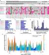
Fig 4. Sequence analysis of continuous in vitro passaged SARS-CoV-2Engl2.
(A) Alignment of serially passaged viruses and SARS-CoV-2Engl2 to SARS-CoV-2Wu1. Non-synonymous (pink) and synonymous (green) changes from Wuhan-1 are highlighted. Light pink are sites fixed at 50% in SARS-CoV-2Engl2 and black box is a deletion mutation in serially passaged virus. Positions of mutations are indicated, and mutations only found in RDV selected populations are in bold. (B) Synonymous vs non-synonymous changes observed in continually passaged virus populations compare to input SARS-CoV-2Engl2. (C) Transversion vs transitional changes observed in continually passaged virus populations compare to input SARS-CoV-2Engl2. (D) Number of in vitro passaged viruses with non-synonymous changes in Spike in comparison to SARS-CoV-2Engl2. Mutation fixed in the consensus genomes (dark blue) are compared to the total number of viruses with evidence of the mutation at sub-consensus levels (light blue). Amino acid residues in common with the emerging variants of concern (Alpha (UK, B.1.1.7), Gamma (Brazil, P.1); and Beta (South Africa, B.1.351) are highlighted by a star. (E) Worldwide diversity of Spike protein sites of circulating SARS-CoV-2 variants. The average number of different substitutions at each codon is calculated along a 20 amino acid residues wide sliding windows. Data was calculated using only substitutions observed in a minimum of 5 sequences from the publicly available SARS-CoV-2 genomes (n = 1384). The position of the amino acid substitutions in the in vitro passaged viruses are indicated at the bottom, residues are red are shared with variants of concerns, black are specific to in vitro virus, residues in italics were synonymous. Mutations in amino acid residues that are also mutated in the variants of concern Alpha (UK, B.1.1.7), Gamma (Brazil, P.1), & Beta (South Africa, B.1.351) are shown in purple triangles.

Comment in

References

    1. WHO Coronavirus (COVID-19) Dashboard. [cited 21 Jul 2021]. Available: https://covid19.who.int
    1. Kaye AD, Okeagu CN, Pham AD, Silva RA, Hurley JJ, Arron BL, et al. Economic impact of COVID-19 pandemic on healthcare facilities and systems: International perspectives. Best Pract Res Clin Anaesthesiol. 2020. [cited 29 Dec 2020]. doi: 10.1016/j.bpa.2020.11.009 - DOI - PMC - PubMed
    1. Gordon CJ, Tchesnokov EP, Woolner E, Perry JK, Feng JY, Porter DP, et al. Remdesivir is a direct-acting antiviral that inhibits RNA-dependent RNA polymerase from severe acute respiratory syndrome coronavirus 2 with high potency. J Biol Chem. 2020;295: 6785–6797. doi: 10.1074/jbc.RA120.013679 - DOI - PMC - PubMed
    1. Pruijssers AJ, George AS, Schäfer A, Leist SR, Gralinksi LE, Dinnon KH, et al. Remdesivir Inhibits SARS-CoV-2 in Human Lung Cells and Chimeric SARS-CoV Expressing the SARS-CoV-2 RNA Polymerase in Mice. Cell Rep. 2020;32: 107940. doi: 10.1016/j.celrep.2020.107940 - DOI - PMC - PubMed
    1. Wit E de, Feldmann F, Cronin J, Jordan R, Okumura A, Thomas T, et al. Prophylactic and therapeutic remdesivir (GS-5734) treatment in the rhesus macaque model of MERS-CoV infection. Proc Natl Acad Sci. 2020;117: 6771–6776. doi: 10.1073/pnas.1922083117 - DOI - PMC - PubMed

Publication types

MeSH terms