Review
. 2021 Dec 7;42(46):4717-4730.
doi: 10.1093/eurheartj/ehab649.

Application of artificial intelligence to the electrocardiogram

Affiliations
Review

Application of artificial intelligence to the electrocardiogram

Zachi I Attia et al. Eur Heart J. .

Abstract

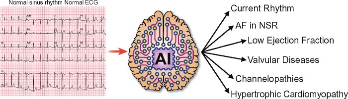
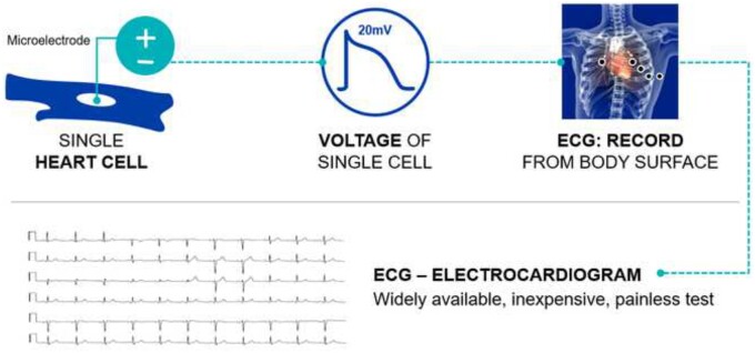
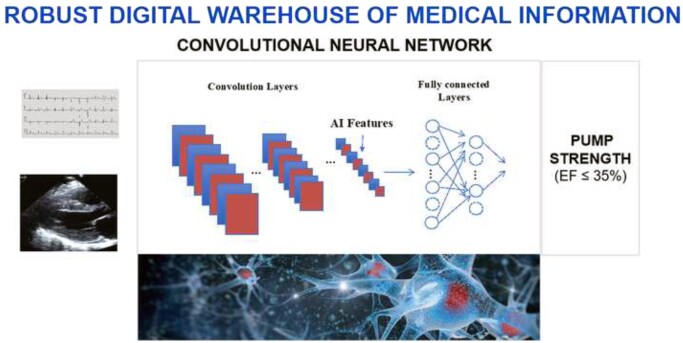
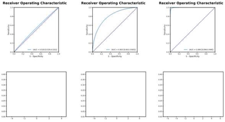
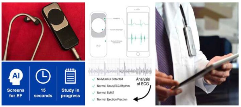
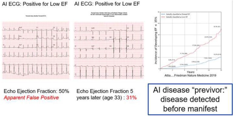
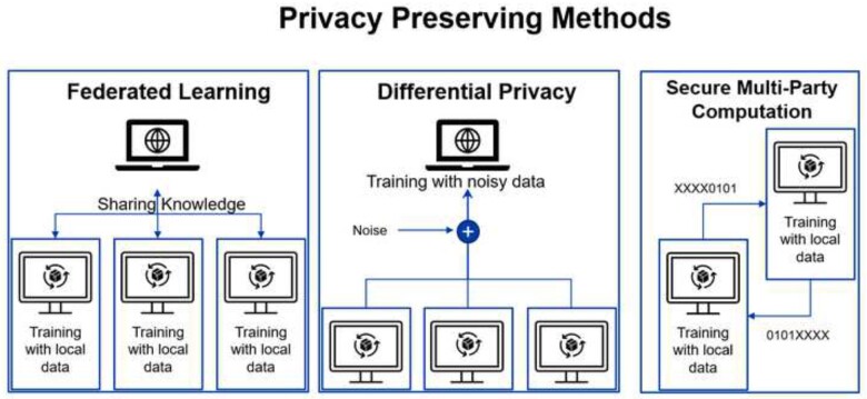
Artificial intelligence (AI) has given the electrocardiogram (ECG) and clinicians reading them super-human diagnostic abilities. Trained without hard-coded rules by finding often subclinical patterns in huge datasets, AI transforms the ECG, a ubiquitous, non-invasive cardiac test that is integrated into practice workflows, into a screening tool and predictor of cardiac and non-cardiac diseases, often in asymptomatic individuals. This review describes the mathematical background behind supervised AI algorithms, and discusses selected AI ECG cardiac screening algorithms including those for the detection of left ventricular dysfunction, episodic atrial fibrillation from a tracing recorded during normal sinus rhythm, and other structural and valvular diseases. The ability to learn from big data sets, without the need to understand the biological mechanism, has created opportunities for detecting non-cardiac diseases as COVID-19 and introduced challenges with regards to data privacy. Like all medical tests, the AI ECG must be carefully vetted and validated in real-world clinical environments. Finally, with mobile form factors that allow acquisition of medical-grade ECGs from smartphones and wearables, the use of AI may enable massive scalability to democratize healthcare.

Keywords: Artificial intelligence; Digital health; Electrocardiograms; Machine learning.


Figures

Graphical Abstract
Graphical Abstract
The application of artificial intelligence to the standard electrocardiogram enables it to diagnose conditions not previously identifiable by an electrocardiogram, or to do so with a greater performance than previously possible. This includes identification of the current rhythm, identification of episodic atrial fibrillation from an ECG acquired during sinus rhythm, the presence of ventricular dysfunction (low ejection fraction), the presence of valvular heart disease, channelopathies (even when electrocardiographically ‘concealed’), and the presence of hypertrophic cardiomyopathy.
Figure 1
Figure 1
Microelectrodes in a single myocyte (top left) record an action potential (depicted middle panel). Ionic currents and their propagation are sensitive to cardiac and non-cardiac conditions and structural changes. When the aggregated action potentials are recorded at the body surface (top right), the insuring tracing is the electrocardiogram (bottom). ECG, electrocardiogram.
Figure 2
Figure 2
A convolutional neural network is trained by feeding in labelled data (in this case voltage time waveforms), and through repetition it identifies the patterns in the data that are associated with the data labels (in this example, heart pump strength, or ejection fraction). The network has two components, convolution layers that extract image components to create the artificial intelligence features, and the fully connected layers that comprise the model, that leads to the network output. While large data sets and robust computing are required to train networks, once trained, the computation requirements are substantially reduced, permitting smartphone application. AI, artificial intelligence; EF, ejection fraction.
Figure 3
Figure 3
The receiver operating characteristic curve and model performance. Left panel: A test with an area under the curve of 0.529 (top) results in very poor separation of the classes (bottom left). As the area under the curve increases (0.803 middle panel, top and 0.998 right panel, top) the separation of the classes and utility of the test improves (bottom panels). This results in improved sensitivity and specificity. See text for additional details.
Figure 4
Figure 4
Embedding of an artificial intelligence electrocardiogram into a stethoscope. Electrodes on the stethoscope acquire an electrocardiogram during normal auscultation, permitting the identification of ventricular dysfunction with 15 s of skin contact time. This workflow provides immediate notification to the healthcare provider via a smartphone connection. ECG, electrocardiogram; EF, ejection fraction.
Figure 5
Figure 5
Artificial intelligence disease previvor. Left panel—an apparently normal electrocardiogram is identified by artificial intelligence as being associated with a low ejection fraction. A contemporaneous echocardiogram depicts normal ventricular function (ejection fraction 50%). Middle panel—5 years later, at age 33, additional electrocardiograms changes are now visible to the human eye, and repeat echocardiography shows depressed ventricular function (ejection fraction 31%). Right panel—risk of developing ejection fraction <35% with a positive artificial intelligence electrocardiogram (red line) vs. with a negative artificial intelligence electrocardiogram for low ejection fraction (blue line). Further details in the text. AI, artificial intelligence; ECG, electrocardiogram; EF, ejection fraction.
Figure 6
Figure 6
Privacy preserving methods. Details in the text.

References

    1. Attia ZI, Friedman PA, Noseworthy PA, Lopez-Jimenez F, Ladewig DJ, Satam G, Pellikka PA, Munger TM, Asirvatham SJ, Scott CG, Carter RE, Kapa S. Age and sex estimation using artificial intelligence from standard 12-lead ECGs. Circ Arrhythm Electrophysiol 2019;12:e007284. - PMC - PubMed
    1. Attia ZI, Kapa S, Lopez-Jimenez F, McKie PM, Ladewig DJ, Satam G, Pellikka PA, Enriquez-Sarano M, Noseworthy PA, Munger TM, Asirvatham SJ, Scott CG, Carter RE, Friedman PA. Screening for cardiac contractile dysfunction using an artificial intelligence-enabled electrocardiogram. Nat Med 2019;25:70–74. - PubMed
    1. Attia ZI, Noseworthy PA, Lopez-Jimenez F, Asirvatham SJ, Deshmukh AJ, Gersh BJ, Carter RE, Yao X, Rabinstein AA, Erickson BJ, Kapa S, Friedman PA. An artificial intelligence-enabled ECG algorithm for the identification of patients with atrial fibrillation during sinus rhythm: a retrospective analysis of outcome prediction. Lancet 2019;394:861–867. - PubMed
    1. Siontis KC, Noseworthy PA, Attia ZI, Friedman PA. Artificial intelligence-enhanced electrocardiography in cardiovascular disease management. Nat Rev Cardiol 2021;18:465–478. - PMC - PubMed
    1. Selvaraju RR, Cm Das A, Vedantam R, Parikh D, Batra D. Grad-CAM: visual explanations from deep networks via gradient-based localization. IEEE Int Conf Comput Vis (ICCV) 2017;618–626.