. 2021 Nov 7;42(42):4373-4385.
doi: 10.1093/eurheartj/ehab605.

Untargeted metabolomics identifies succinate as a biomarker and therapeutic target in aortic aneurysm and dissection

Affiliations

Untargeted metabolomics identifies succinate as a biomarker and therapeutic target in aortic aneurysm and dissection

Hongtu Cui et al. Eur Heart J. .

Erratum in

Abstract

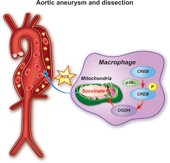
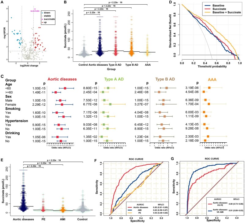
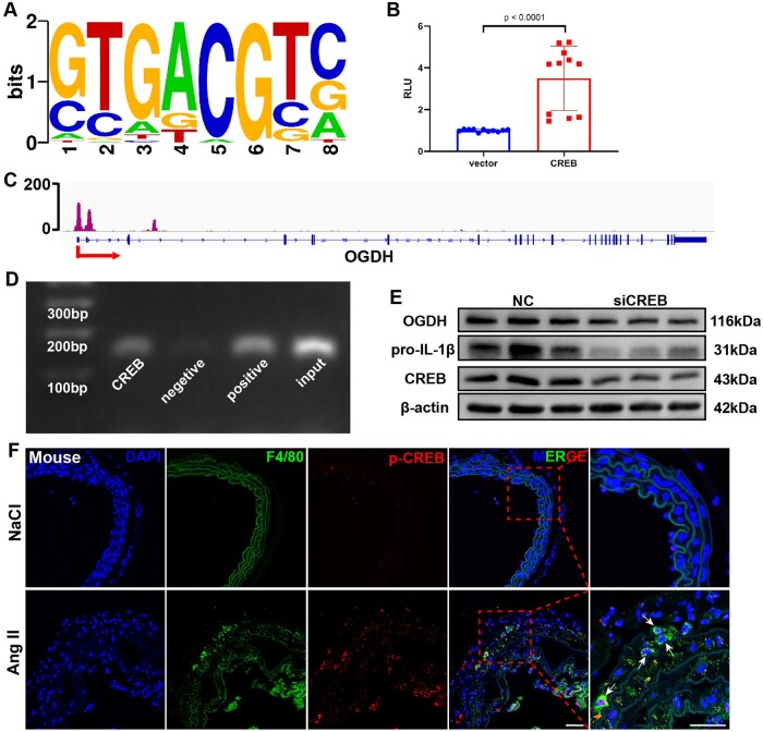
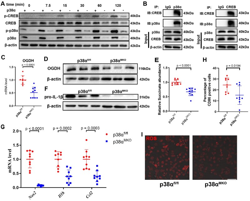
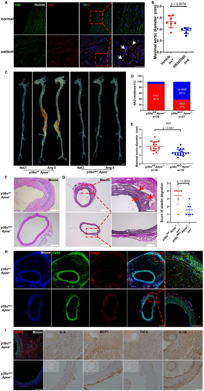
Aims: Aortic aneurysm and dissection (AAD) are high-risk cardiovascular diseases with no effective cure. Macrophages play an important role in the development of AAD. As succinate triggers inflammatory changes in macrophages, we investigated the significance of succinate in the pathogenesis of AAD and its clinical relevance.

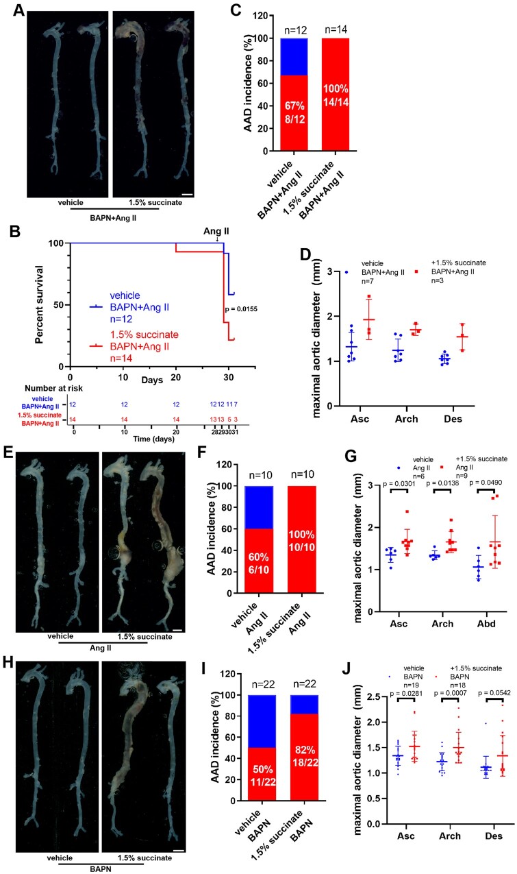
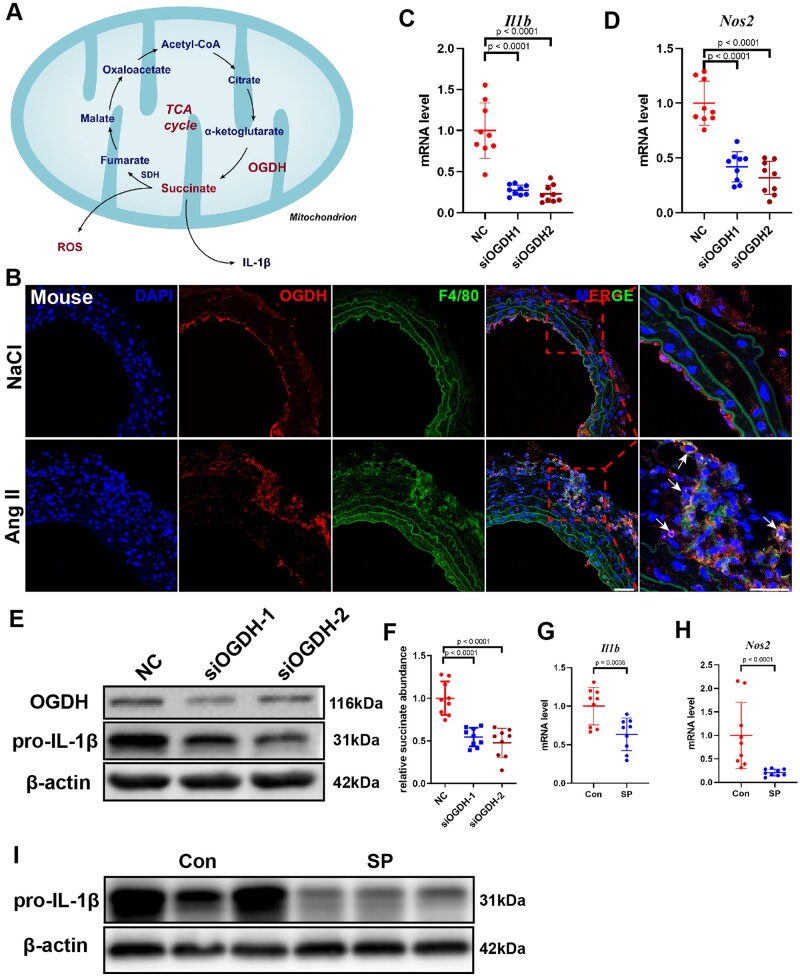
Methods and results: We used untargeted metabolomics and mass spectrometry to determine plasma succinate concentrations in 40 and 1665 individuals of the discovery and validation cohorts, respectively. Three different murine AAD models were used to determine the role of succinate in AAD development. We further examined the role of oxoglutarate dehydrogenase (OGDH) and its transcription factor cyclic adenosine monophosphate-responsive element-binding protein 1 (CREB) in the context of macrophage-mediated inflammation and established p38αMKOApoe-/- mice. Succinate was the most upregulated metabolite in the discovery cohort; this was confirmed in the validation cohort. Plasma succinate concentrations were higher in patients with AAD compared with those in healthy controls, patients with acute myocardial infarction (AMI), and patients with pulmonary embolism (PE). Moreover, succinate administration aggravated angiotensin II-induced AAD and vascular inflammation in mice. In contrast, knockdown of OGDH reduced the expression of inflammatory factors in macrophages. The conditional deletion of p38α decreased CREB phosphorylation, OGDH expression, and succinate concentrations. Conditional deletion of p38α in macrophages reduced angiotensin II-induced AAD.

Conclusion: Plasma succinate concentrations allow to distinguish patients with AAD from both healthy controls and patients with AMI or PE. Succinate concentrations are regulated by the p38α-CREB-OGDH axis in macrophages.

Keywords: Aortic aneurysm and dissection; Macrophage; Oxoglutarate dehydrogenase; Succinate.


Figures

None
Graphical abstract
Figure 1
Figure 1
High level of succinate could increase the diagnostic performance for aortic disease. (A) Volcano plot of plasma metabolites in all patients vs. healthy controls. (B) Distribution of succinate according to disease status in the validation cohort, before propensity score-matching. (C) Forest plots showing the adjusted per-standard deviation odds ratio (95% confidence interval) for plasma succinate levels associated with aortic diseases in subgroups using the integration of discovery and validation cohort data. Binary logistic regression was adjusted for age, sex, smoking and drinking status, history of hypertension, diabetes, or systolic blood pressure, and levels of glucose, total cholesterol, and triglycerides. (D) Decision curve analysis: aortic diseases vs. healthy controls for baseline, succinate, and the combination of baseline and succinate using the integration of discovery and validation cohort data. Baseline refers to age, sex, smoking and drinking status, and history of hypertension, diabetes, and systolic blood pressure. (E) Distribution of succinate plasma levels in patients with aortic diseases, acute myocardial infarction, and pulmonary embolism and in healthy controls. (F) Receiver operating characteristic curves: patients with aortic diseases, acute myocardial infarction, and pulmonary embolism vs. healthy controls for succinate. (G) Receiver operating characteristic curves: acute myocardial infarction and pulmonary embolism vs. aortic diseases for succinate.
Figure 2
Figure 2
Supplementation with succinate aggravates aortic aneurysm and dissection in vivo. (AD) Four-week-old male mice were administered with 0.25% BAPN (wt/vol) for 28 days with or without 1.5% sodium succinate (wt/vol) and then infused with saline or angiotensin II (1000 ng/kg/min) for 3 days: vehicle + BAPN/angiotensin II, n = 12; and 1.5% succinate + BAPN/angiotensin II, n = 14. (A) Representative morphology of the aortas for each group. Scale bar = 2 mm. (B) Survival curve and number at risk, log-rank test. (C) Incidence of aortic aneurysm and dissection for each group. (D) Measurement of the maximal aortic diameter ex vivo. n = 7 for mice administered BAPN/angiotensin II and n = 3 for 1.5% succinate + BAPN/angiotensin II (mice died of aortic rupture were not included). (EG) Nine-week-old Apoe–/– male mice were infused with saline or angiotensin II (1000 ng/kg/min) for 28 days with or without 1.5% sodium succinate (wt/vol): vehicle + angiotensin II, n = 10; and 1.5% succinate + angiotensin II, n = 10. (E) Representative morphology of the aortas from different groups of mice. Scale bar = 2 mm. (F) Incidence of aortic aneurysm and dissection in different groups of mice. (G) Measurement of the maximal aortic diameter ex vivo. n = 6 for mice administered angiotensin II and n = 9 for 1.5% succinate + angiotensin II (mice died of aortic rupture were not included). H–J, Three-week-old male mice were administered with 0.25% BAPN (wt/vol) for 28 days with or without 1.5% sodium succinate (wt/vol): vehicle + BAPN, n = 22; and 1.5% succinate + BAPN, n = 22. (H) Representative morphology of the aortas from the different groups of mice. Scale bar = 2 mm. (I) Incidence of aortic aneurysm and dissection from the different groups of mice. (J) Measurements of the maximal aortic diameter ex vivo. n = 19 for mice administered BAPN and n = 18 for 1.5% succinate + BAPN (mice died of aortic rupture were not included). Apoe–/–, apolipoprotein E-deficient; Asc, ascending aorta; Des, descending aorta.
Figure 3
Figure 3
Inhibition of oxoglutarate dehydrogenase (OGDH) reduces the expression of inflammatory factors in macrophages. (A) Schematic diagram of the tricarboxylic acid cycle. B, Representative immunofluorescence staining for F4/80 and OGDH in the suprarenal abdominal aortas from NaCl- and angiotensin II-infused mice. Scale bar = 50 μm. Arrows indicate macrophages expressing OGDH. (C and D) Differentially expressed genes were validated by qRT-PCR in bone marrow-derived macrophages stimulated with lipopolysaccharide after transfection with negative control (NC) or siOGDH. Data are pooled from three independent experiments. n =9 per group. (E) Representative western blot of pro-IL-1β in lipopolysaccharide-stimulated bone marrow-derived macrophages after transfection with NC or siOGDH. (F) Relative succinate abundance in lipopolysaccharide-stimulated bone marrow-derived macrophages after transfection with NC or siOGDH. Data are pooled from three independent experiments. n =9 per group. (G and H) Differentially expressed genes were validated by qRT-PCR in bone marrow-derived macrophages stimulated with lipopolysaccharide with or without the addition of succinyl phosphonate. Data are pooled from three independent experiments. n =9 per group (G and H). (I) Representative western blot of pro-IL-1β in bone marrow-derived macrophages stimulated with lipopolysaccharide with or without the addition of succinyl phosphonate.
Figure 4
Figure 4
Cyclic AMP-responsive element-binding protein 1 (CREB) regulates oxoglutarate dehydrogenase OGDH expression via direct promoter binding. (A) CREB-binding motifs. (B) Luciferase reporter analysis in 293T cells. Results are presented as the mean ± standard deviation. Data are pooled from three independent experiments. n =11 per group. (C) Chromatin immunoprecipitation-seq assay of the occupancy of OGDH gene promoters by CREB (data from GSM2663863 dataset). (D) Chromatin immunoprecipitation assay: binding of CREB to the OGDH gene promoter in bone marrow-derived macrophages. Three independent experiments were performed. (E) Representative western blot of OGDH in bone marrow-derived macrophages stimulated with lipopolysaccharide for 24 h with or without addition of siCREB. Three independent experiments were performed. (F) Representative immunofluorescence staining for F4/80 and phosphorylated CREB in the suprarenal abdominal aortas from control and aortic aneurysm and dissection mice. Scale bar = 50 μm.
Figure 5
Figure 5
p38α affects macrophage inflammatory phenotype by regulating the expression of OGDH and succinate via CREB phosphorylation. (A) Representative western blot of phosphorylated p38α (p-p38α), total p38α, p-CREB, and total CREB in p38αfl/fl and p38αMKO bone marrow-derived macrophages stimulated with lipopolysaccharide for 0, 7.5, 15, 30, 60, or 120 min. (B) The co-immunoprecipitation assay of CREB and p38α in bone marrow-derived macrophages. (C) Oxoglutarate dehydrogenase (Ogdh) mRNA level was measured by qRT-PCR in p38αfl/fl and p38αMKO bone marrow-derived macrophages stimulated with lipopolysaccharide. Data are pooled from three independent experiments. n =9 per group. (D) Representative western blot of OGDH in p38αfl/fl and p38αMKO bone marrow-derived macrophages stimulated with lipopolysaccharide. (E) Relative succinate abundance in p38αfl/fl and p38αMKO bone marrow-derived macrophages stimulated with lipopolysaccharide. Data are pooled from three independent experiments. n =9 per group. (F) Representative western blot of pro-IL-1β in p38αfl/fl and p38αMKO bone marrow-derived macrophages stimulated with lipopolysaccharide. (G) Expression levels of Nos2, Il1b, and Ccl2 determined by qRT-PCR in p38αfl/fl and p38αMKO bone marrow-derived macrophages stimulated with lipopolysaccharide. (H) Percentage of CD86+ cells determined by flow cytometry in p38αfl/fl and p38αMKO bone marrow-derived macrophages stimulated with lipopolysaccharide. Data are pooled from three independent experiments. n =9 per group. (I) Dihydroethidium staining of reactive oxygen species in p38αfl/fl and p38αMKO bone marrow-derived macrophages stimulated with lipopolysaccharide. Scale bar = 50 μm.
Figure 6
Figure 6
Macrophage-specific p38α deficiency attenuates angiotensin II-induced aortic aneurysm and dissection. (A) Representative immunofluorescence staining for CD68 (marker of macrophages) and p38α in the aortas from healthy individuals and patients with aortic aneurysm and dissection. (B) Measurement of the maximal suprarenal aortic diameters ex vivo from elastase-treated male mice injected with either SB203580 or vehicle. n =7 for mice administered vehicle and n =6 for mice administered SB203580. (CI) 14-weeks-old p38αfl/fl  Apoe−/− and p38αMKO  Apoe−/− male mice were infused with either saline or angiotensin II (1000 ng/kg/min) for 28 d. p38αfl/fl  Apoe−/− + saline, n =4; p38αMKO  Apoe−/− + saline, n =4; p38αfl/fl  Apoe−/− + angiotensin II, n =26; and p38αMKO  Apoe−/− + angiotensin II, n =27. (C) Representative morphology of the abdominal aortas from the different groups of mice. Scale bar = 2 mm. (D) Incidence of aortic aneurysm and dissection in different groups of mice. (E) Measurement of the maximal suprarenal aortic diameters ex vivo in different groups of mice. p38αfl/fl  Apoe−/− + angiotensin II, n =16; p38αMKO  Apoe−/− + angiotensin II, n =19 (mice died of aortic rupture were not included). Representative images of haematoxylin and eosin (F; scale bar = 100 μm) and Elastic Van Gieson (G; scale bar = 200 and 50 μm, n =7 per group) staining in the suprarenal abdominal aortas of mice, and the grade of elastin degradation. (H) Representative immunofluorescence staining images for F4/80 and CD206 in the suprarenal abdominal aortas from the different groups of mice. Scale bar = 100 μm. (I) Representative immunohistochemical staining images for matrix metalloproteinase-9, IL-1β, IL-6, monocyte chemoattractant protein-1, and tumour necrosis factor-alpha in the suprarenal abdominal aortas from the different groups of mice Scale bar = 50 μm.

Comment in

References

    1. Miniño AM, Murphy SL, Xu J, Kochanek KD. Deaths: final data for 2008. Natl Vital Stat Rep 2011;59:1–126. - PubMed
    1. Wang SW, Huang YB, Huang JW, Chiu CC, Lai WT, Chen CY. Epidemiology, clinical features, and prescribing patterns of aortic aneurysm in Asian population from 2005 to 2011. Medicine (Baltimore) 2015;94:e1716. - PMC - PubMed
    1. Erbel R, Aboyans V, Boileau C, Bossone E, Bartolomeo RD, Eggebrecht H, Evangelista A, Falk V, Frank H, Gaemperli O, Grabenwöger M, Haverich A, Iung B, Manolis AJ, Meijboom F, Nienaber CA, Roffi M, Rousseau H, Sechtem U, Sirnes PA, Allmen RSv, Vrints CJM. 2014 ESC Guidelines on the diagnosis and treatment of aortic diseases: document covering acute and chronic aortic diseases of the thoracic and abdominal aorta of the adult. The Task Force for the Diagnosis and Treatment of Aortic Diseases of the European Society of Cardiology (ESC). Eur Heart J 2014;35:2873–2926. - PubMed
    1. Guo DC, Papke CL, He R, Milewicz DM. Pathogenesis of thoracic and abdominal aortic aneurysms. Ann N Y Acad Sci 2006;1085:339–352. - PubMed
    1. Lu H, Daugherty A. Aortic aneurysms. Arterioscler Thromb Vasc Biol 2017;37:e59–e65. - PMC - PubMed

Publication types