. 2021 Oct 12;16(10):2422-2431.
doi: 10.1016/j.stemcr.2021.08.009. Epub 2021 Sep 16.

Tetraspanin CD82 regulates S1PR1-mediated hematopoietic stem and progenitor cell mobilization

Affiliations

Tetraspanin CD82 regulates S1PR1-mediated hematopoietic stem and progenitor cell mobilization

Chelsea A Saito-Reis et al. Stem Cell Reports. .

Abstract

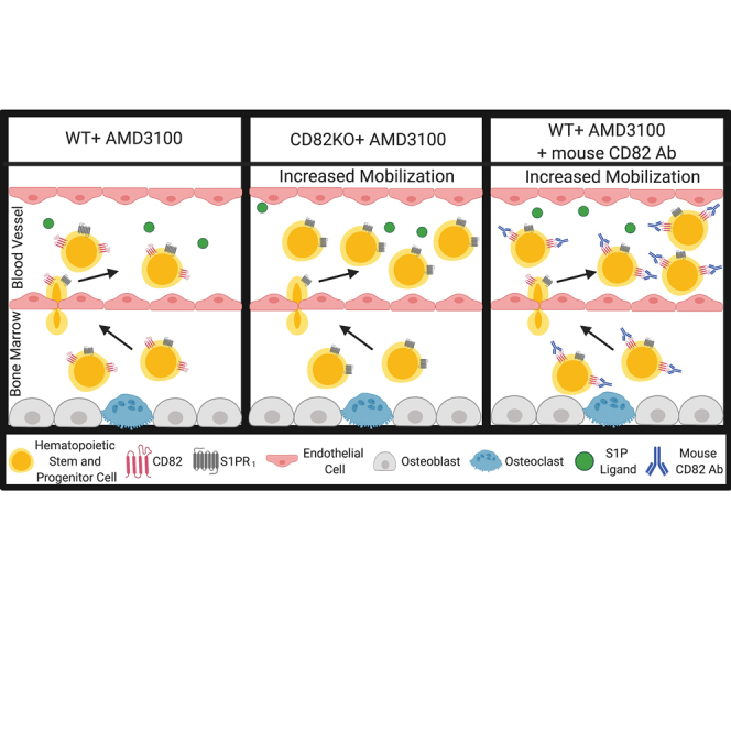
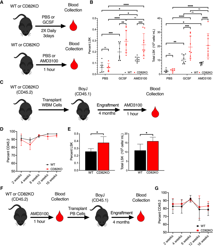
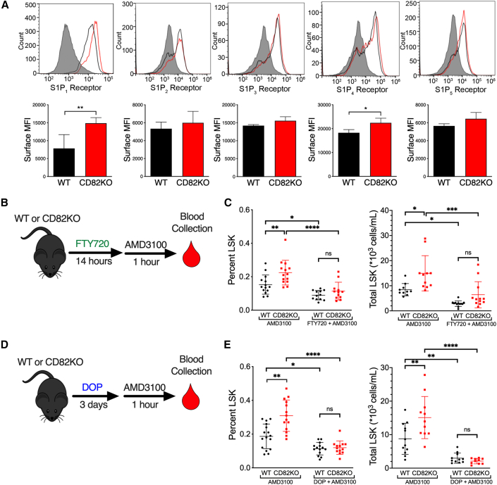
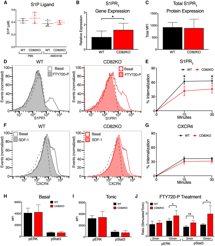
Hematopoietic stem and progenitor cell (HSPC) mobilization into the blood occurs under normal physiological conditions and is stimulated in the clinic to enable the isolation of HSPCs for transplantation therapies. In the present study, we identify the tetraspanin CD82 as a novel regulator of HSPC mobilization. Using a global CD82 knockout (CD82KO) mouse, we measure enhanced HSPC mobilization after granulocyte-colony stimulating factor (G-CSF) or AMD3100 treatment, which we find is promoted by increased surface expression of the sphingosine 1-phosphate receptor 1 (S1PR1) on CD82KO HSPCs. Additionally, we identify a disruption in S1PR1 internalization in CD82-deficient HSPCs, suggesting that CD82 plays a critical role in S1PR1 surface regulation. Finally, combining AMD3100 and anti-CD82 treatments, we detect enhanced mobilization of mouse HSPCs and human CD34+ cells in animal models. Together, these data provide evidence that CD82 is an important regulator of HSPC mobilization and suggests exploiting the CD82 scaffold as a therapeutic target to enhance stem cell isolation.

Keywords: CD82; hematopoietic stem and progenitor cells; mobilization; sphingosine-1-phosphate receptor; tetraspanins.


Figures

None
Graphical abstract
Figure 1
Figure 1
CD82KO HSPCs display enhanced mobilization (A) WT and CD82KO mice were treated with PBS, G-CSF, or AMD3100 prior to peripheral blood collection. (B) Flow cytometry analysis of %LSK and total LSK cells in peripheral blood collected from WT and CD82KO mice treated with PBS, AMD3100, or G-CSF (n = 5–9 mice/group, four independent experiments; ∗∗∗∗p < 0.0001, ∗∗∗p < 0.001, p < 0.05, two-way ANOVA). (C) BM cells from WT or CD82KO mice were transplanted into lethally irradiated BoyJ mice and engrafted for 4 months prior to AMD3100 mobilization. (D) Percentage of donor cells (CD45.2) repopulated in peripheral blood over 4 months measured by flow cytometry. (E) Flow cytometry analysis of %LSK and total LSK cells in peripheral blood collected after AMD3100-induced HSPC mobilization of WT and CD82KO transplanted BoyJ mice (n = 4–5 mice/group, three independent experiments,p < 0.05, unpaired t test). (F) Mobilized PBMCs from WT or CD82KO mice were transplanted into lethally irradiated BoyJ mice and engrafted for 4 months. (G) Percentage of donor cells (CD45.2) repopulated in peripheral blood over 4 months measured by flow cytometry (n = 4–5 mice/group, three independent experiments,p < 0.05, unpaired t test). Error bars, SD.
Figure 2
Figure 2
Increased S1PR1 expression on CD82KO HSPCs promotes enhanced mobilization (A) MFI of S1PR1-5 surface expression on WT and CD82KO LSK cells (n = 4 mice/group ∗∗p < 0.01 and p < 0.05, unpaired t test). (B) FTY720, which activates and internalizes S1PR1, was injected into WT or CD82KO mice 14 h prior to AMD3100 treatment followed by peripheral blood collection. (C) Flow cytometry analysis of %LSK and total LSK cells in peripheral blood collected from WT and CD82KO mice treated with AMD3100 or AMD3100/FTY720. (D) The S1P lyase inhibitor, DOP, increases S1P resulting in S1PR1 internalization. Drinking water for WT and CD82KO mice was supplemented with DOP for 3 days. AMD3100 treatment occurred 1 h prior to peripheral blood collection. (E) Flow cytometry analysis of %LSK and total LSK cells in peripheral blood collected from WT and CD82KO mice treated with AMD3100 or AMD3100/DOP. (B–E) n = 12–16 mice/group, four independent experiments, ∗∗∗∗p < 0.0001, ∗∗p < 0.01, p < 0.05, two-way ANOVA. Error bars, SD.
Figure 3
Figure 3
CD82 regulates S1PR1 internalization and signaling (A) S1P plasma levels of WT and CD82KO mice post PBS or AMD3100 treatment (n = 3–4 mice/group). (B) Quantitative PCR analysis of relative S1PR1 gene expression compared with GAPDH in WT and CD82KO LSK cells (n = 4 mice/group p < 0.05, unpaired t test). (C) MFI of total S1PR1 expression of fixed and permeabilized WT and CD82KO LSK cells (n = 4 mice/group p < 0.05, unpaired t test). (D) Representative histograms of S1PR1 surface MFI at 0 (basal) and 30 min post 10 μM FTY720-P treatment in WT and CD82KO LSK cells. Isotype indicated by dotted line. (E) Percentage internalization of S1PR1 at 15 and 30 min post 10 μM FTY720-P treatment (n = 3 independent experiments; p < 0.05, unpaired t test). (F) Representative histograms of CXCR4 surface MFI at 0 (basal) and 30 min post 100 ng/mL SDF-1 treatment in WT and CD82KO LSK cells. Isotype indicated by dotted line. (G–I) (G) Percentage internalization of CXCR4 at 15 and 30 min post 100 ng/mL SDF-1 treatment (n = 3 independent experiments). Phosphoflow cytometry analysis of (H) basal and (I) tonic conditions to assess MFI of pERK and pStat3 signaling in the LSK population (n = 3–4 mice/group; ∗∗∗p < 0.001, unpaired t test). (J) Phosphoflow cytometry analysis of LSK pERK and pSTAT3 signaling after 10 μM FTY720-P treatment at 2 and 10 min (n = 3 independent experiments; p < 0.05; ns, non-significant, unpaired t test). Error bars, SD.
Figure 4
Figure 4
CD82 Ab treatment enhances HSPC mobilization (A) MFI of CD82 surface expression on WT and CD82KO LSK cells (n = 4–5 mice/group; ∗∗∗p < 0.001, unpaired t test). (B) WT mice were injected with either IgG control or CD82 Ab for 2 h followed by PBS or AMD3100 treatment for 1 h prior to blood collection. (C) Flow cytometry analysis of %LSK and total LSK cells in peripheral blood collected from WT mice treated with IgG control or CD82 Ab (n = 4–5 mice/group, three independent experiments; ∗∗∗∗p < 0.0001, ∗∗∗p < 0.001, ∗∗p < 0.01, or p < 0.05, two-way ANOVA). (D) WT mice were injected with either IgG control or CD82 Ab once daily for 2 days, then injected with PBS or AMD3100 treatment the following day 1 h prior to blood collection. (E) Flow cytometry analysis of %LSK and total LSK cells in peripheral blood collected from WT mice treated with IgG control or CD82 Ab (n = 5 mice/group). (F) Lethally irradiated NSG mice were injected with human CD34+ cells and allowed to engraft for 6 weeks prior to IgG or human CD82 Ab injection and AMD3100 treatment. (G) Flow cytometry analysis of %CD45+ and total CD45+ cells in peripheral blood collected from NSG mice treated with IgG control or CD82 (n = 5–6 mice/group, three independent experiments; ∗∗p < 0.01, unpaired t test). Error bars, SD.

References

    1. Bendall L.J., Basnett J. Role of sphingosine 1-phosphate in trafficking and mobilization of hematopoietic stem cells. Curr. Opin. Hematol. 2013;20:281–288. - PubMed
    1. Broxmeyer H.E., Orschell C.M., Clapp D.W., Hangoc G., Cooper S., Plett P.A., Liles W.C., Li X., Graham-Evans B., Campbell T.B. Rapid mobilization of murine and human hematopoietic stem and progenitor cells with AMD3100, a CXCR4 antagonist. J. Exp. Med. 2005;201:1307–1318. - PMC - PubMed
    1. Craddock C.F., Nakamoto B., Andrews R.G., Priestley G.V., Papayannopoulou T. Antibodies to VLA4 integrin mobilize long-term repopulating cells and augment cytokine-induced mobilization in primates and mice. Blood. 1997;90:4779–4788. - PubMed
    1. Custer M.C., Risinger J.I., Hoover S., Simpson R.M., Patterson T., Barrett J.C. Characterization of an antibody that can detect the Kai1/CD82 murine metastasis suppressor. Prostate. 2006;66:567–577. - PubMed
    1. Danglot L., Chaineau M., Dahan M., Gendron M.C., Boggetto N., Perez F., Galli T. Role of TI-VAMP and CD82 in EGFR cell-surface dynamics and signaling. J. Cell Sci. 2010;123:723–735. - PubMed

Publication types