Review
. 2021 Sep 16;184(19):4857-4873.
doi: 10.1016/j.cell.2021.08.013.

The expanding amyloid family: Structure, stability, function, and pathogenesis

Affiliations
Review

The expanding amyloid family: Structure, stability, function, and pathogenesis

Michael R Sawaya et al. Cell. .

Abstract

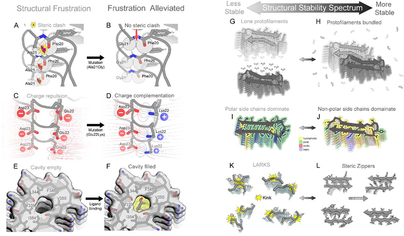
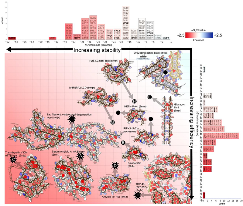
The hidden world of amyloid biology has suddenly snapped into atomic-level focus, revealing over 80 amyloid protein fibrils, both pathogenic and functional. Unlike globular proteins, amyloid proteins flatten and stack into unbranched fibrils. Stranger still, a single protein sequence can adopt wildly different two-dimensional conformations, yielding distinct fibril polymorphs. Thus, an amyloid protein may define distinct diseases depending on its conformation. At the heart of this conformational variability lies structural frustrations. In functional amyloids, evolution tunes frustration levels to achieve either stability or sensitivity according to the fibril's biological function, accounting for the vast versatility of the amyloid fibril scaffold.


Conflict of interest statement

Declaration of interests D.S.E. is SAB Chair and an equity holder in ADRx, Inc. J.A.R. is a founder and equity shareholder of Medstruc Inc.

Figures

Figure 1.
Figure 1.. A fundamental component of amyloid fibrils is the steric zipper motif.
Amyloid fibrils of α-synuclein are associated with Parkinson’s disease and other synucleinopathies. The crystal structure of residues 47–56 offered atomic resolution details of its assembly at 1.4 Å resolution (PDB ID 4ZNN)(Rodriguez et al., 2015). (A) Thousands of copies of this segment stack to form a pair of β-sheets (light and dark orange). (B) A view oblique to the fibril axis reveals the tight fit between side chains of the mated sheets. (C) A view perpendicular to the fibril axis (vertical line) reveals that this zipper-like mating of side chains extends along the entire length of the fibril. (D) The remaining orthogonal view shows how the β-strands stack into β-sheets via main chain hydrogen bonds (dotted lines), parallel and in-register. Amide C=O and N-H groups point up and down, nearly parallel to the fibril axis. (E,F) Steric zipper formation liberates protein-bound water molecules, contributing a hydrophobic effect to fibril stability. (G) As determined by cryoEM at 2.8 resolution (Ni et al., 2019), full-length α-synuclein molecules stack in hydrogen-bonded sheets like the segment, but with a slight twist of each molecule compared to the molecule 4.8 Å below (PDB ID 6osj). Two protofilaments (brown and gray) intertwine to form a fibril. The preNAC region, is colored orange. (H) A view oblique to the fibril axis reveals each molecule is confined to a nearly flat layer. Each chain adopts the same meandering path comprising a series of β-strands and turns which mate together side chains in steric zippers. The steric zipper motif bridging the two protofilaments (orange) is analogous to that of the segment in panel B. (I,J) Views perpendicular to the fibril axis reveal steric zipper interactions and hydrogen bonding patterns analogous to the segment in panels C and D. (K,L) Assembly of 7 steric zippers (orange and cyan shades) liberates water molecules, a process that contributes greatly to amyloid stability.
Figure 2
Figure 2. Folding patterns of amyloid proteins in compact, 2D layers.
(A) 2D layers of α-synuclein (upper left) stack into a fibril with a slight twist between layers. Thick lines trace the protein backbone. Brown and gray lines distinguish separate protofilaments. Sidechains are colored according to physical property. A view perpendicular to the fibril axis (right) reveals that identical residues stack in ladders along the fibril axis. Four types of amino acid ladders are depicted in detail (lower left), revealing stabilizing van der Waals contacts and/or hydrogen bonding and/or charge complementation. (B-H) Two dimensional layers of seven diverse amyloid fibrils reveal patterns of side chain association akin to those in globular proteins; hydrophobic residues (yellow) cluster together and tend to be buried. Polar residues also cluster together or reside on the surface. Charged residues associate in complementary pairs (black circle with +/− inscribed) or reside on the surface. β-arches (main chain U-turns) are ubiquitous. Steric zippers (extended β-strands running side-by-side) are evident in all but panel E and H.
Figure 3.
Figure 3.. A single protein sequence attains multiple amyloid polymorph structures.
At the center, the sequence of α-synuclein is represented in an unfolded state. At the outside of the circle are depicted seven different polymorphs of α-synuclein fibrils (A-G), each obtained from different conditions, ex vivo and in vitro as noted. The seven structures represent distinct groups obtained by structural similarity clustering analysis of 25 structures determined to date. The PDB codes for all members of a group are noted. Each polymorph is composed of a series of β-strands and turns, which are well conserved as evidenced by (H) secondary structure alignment. The turns are most often located near glycine (depicted by prominent yellow spheres centered on backbone), or near clusters of charged residues (pink and blue). Despite the conservation of secondary structure, the diversity of tertiary structure is striking. Some interfaces between strands are conserved between structural groups; however, not one interface is conserved among all seven groups. It appears all polymorphs are built from the same secondary structure building blocks but differ in the angles between them. This point is illustrated by the varied angles observed between strands comprising residues 51–58 and 60–66 (darkest brown color). A 90° angle between these two strands defines the “boot” polymorphs in the upper half of the circle. A 180° angle between these strands defines the “sandal” polymorphs in the lower half of the circle. The alternative paths adopted by the protein chains of distinct polymorphs represent alternative ways to pair hydrophobes together (yellow) and complement opposite charges (pink and blue). Paths are evidently determined by different chemicals in different growth conditions (unmodeled density).
Figure 4.
Figure 4.. Structural features that tune amyloid fibril stability.
(A) Torsion angle frustrations manifest as steric clashes. Here, strain in the phi angle of Ala21 produces steric clash between an oxygen atom of Phe20 and hydrogen atom of Ala21 (spheres) in Aβ, (2nao). (B) The Ala21Gly mutation eliminates some steric bulk, so Phe20 atoms do not clash with Gly21 atoms. (C) An example of charge frustration. Close proximity of negatively charged residues Glu22 and Asp23 of Aβ, (2nao) destabilizes the fibril. (D) The Glu22Lys mutation eliminates frustration by introducing a complementary positive charge. (E) A cavity is evident in tau filaments from CTE patients (6nwp) destabilizes the fibril. (F) A complementary ligand fills the cavity and stabilizes the filament. (G, H) A lone filament gains stability when it bundles with other filaments; formation of new interfaces liberates surface bound solvent molecules. (I, J) A fibril gains stability as its polar residues are replaced with hydrophobic residues. (K) Reversible amyloids contain LARKS motifs (Hughes et al., 2018),(Guenther et al., 2018b) which are kinked and exhibit small interfaces, making them less stable than steric zippers (L). Four examples of each are shown here.
Figure 5.
Figure 5.. Stabilization energy maps reveal structural features that influence reversibility.
Amyloid fibril structures are colored by energy: strongly stabilizing sidechains are red; destabilizing sidechains are blue. The standard free energies of 75 amyloid structures (indicated by PDB ID codes) are ranked in two dimensions: on a per molecule basis (horizontal histogram) which inform about stability of a molecule, and on a per residue basis (vertical histogram), which inform about energy efficiency (independent of molecule size). Select fibril structures are pictured within the graph. Energy estimates for pathological/irreversible fibrils are indicated with starburst icons. Energy estimates for presumably functional/reversible fibrils are indicated with black dots. Transthyretin V30M (lower left) is evaluated as the most stable structure (62.1 kcal/mol/molecule) and one of the most efficiently stable (−0.68 kcal/mol/residue). It features an abundance of buried hydrophobic residues (deep red). Notably, it is also a pathogenic fibril, extracted from the heart of a patient with hereditary transthyretin amyloidosis. At the opposite extreme, FUS (upper right) is relatively unstable (−12.2 kcal/mol/molecule), and inefficient (−0.20 kcal/mol/residue). It lacks a hydrophobic core. In contrast to transthyretin, FUS aggregation is functional and presumably reversible.

References

    1. Ader C, Frey S, Maas W, Schmidt HB, Görlich D, and Baldus M (2010). Amyloid-like interactions within nucleoporin FG hydrogels. Proc. Natl. Acad. Sci. U.S.A 107, 6281–6285. - PMC - PubMed
    1. Arakhamia T, Lee CE, Carlomagno Y, Duong DM, Kundinger SR, Wang K, Williams D, DeTure M, Dickson DW, Cook CN, et al. (2020). Posttranslational Modifications Mediate the Structural Diversity of Tauopathy Strains. Cell 180, 633–644.e12. - PMC - PubMed
    1. Astbury WT, Dickinson S, and Bailey K (1935). The X-ray interpretation of denaturation and the structure of the seed globulins. Biochem. J 29, 2351–2360.1. - PMC - PubMed
    1. Baba M, Nakajo S, Tu PH, Tomita T, Nakaya K, Lee VM, Trojanowski JQ, and Iwatsubo T (1998). Aggregation of alpha-synuclein in Lewy bodies of sporadic Parkinson’s disease and dementia with Lewy bodies. Am. J. Pathol 152, 879–884. - PMC - PubMed
    1. Balbirnie M, Grothe R, and Eisenberg DS (2001). An amyloid-forming peptide from the yeast prion Sup35 reveals a dehydrated beta-sheet structure for amyloid. Proc. Natl. Acad. Sci. U.S.A 98, 2375–2380. - PMC - PubMed

Publication types

Substances