

Shared and distinct mechanisms of skeletal muscle atrophy: A narrative review
- PMID: 34534682
- PMCID: PMC8524783
- DOI: 10.1016/j.arr.2021.101463
Shared and distinct mechanisms of skeletal muscle atrophy: A narrative review
Abstract
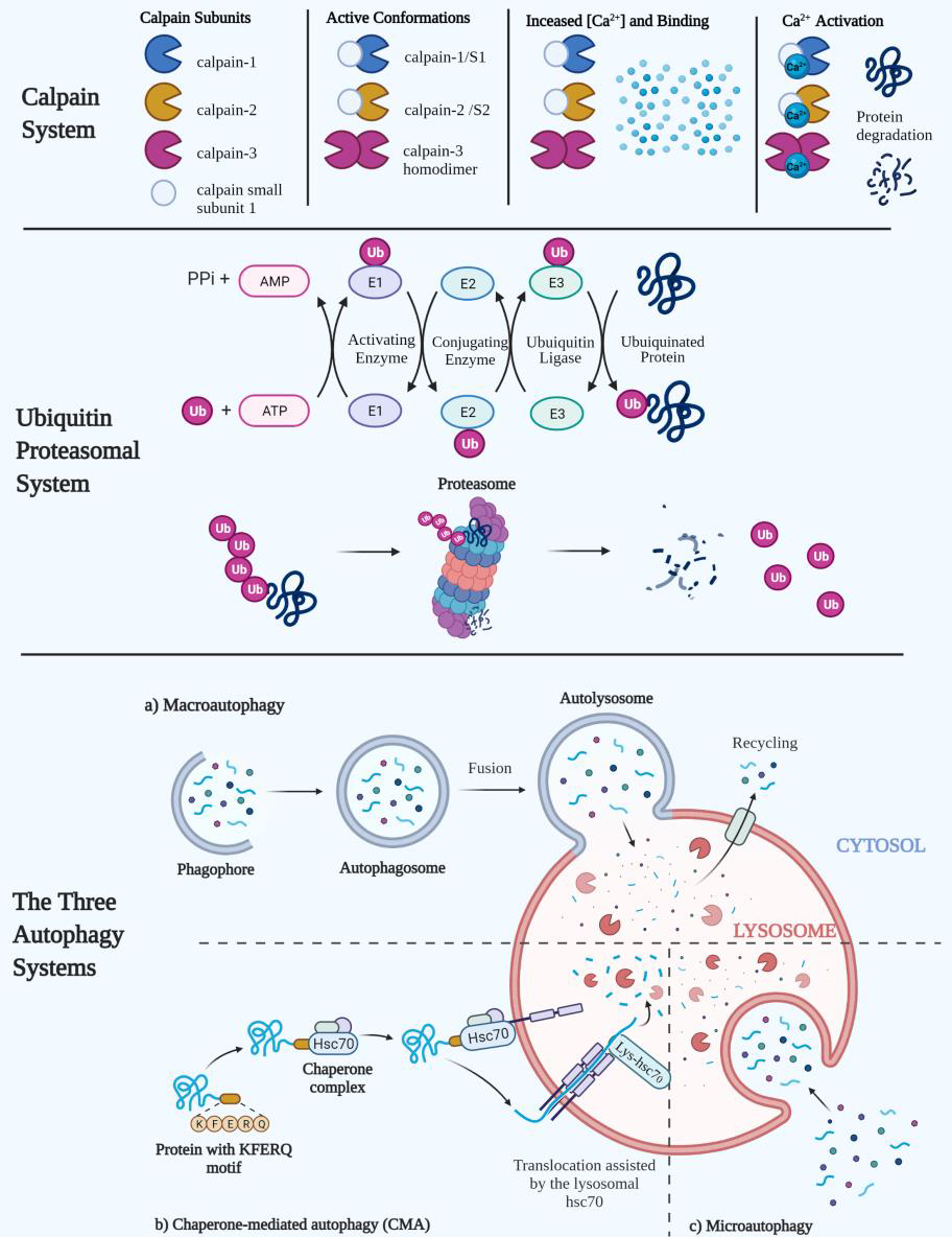
Maintenance of skeletal muscle mass and function is an incredibly nuanced balance of anabolism and catabolism that can become distorted within different pathological conditions. In this paper we intend to discuss the distinct intracellular signaling events that regulate muscle protein atrophy for a given clinical occurrence. Aside from the common outcome of muscle deterioration, several conditions have at least one or more distinct mechanisms that creates unique intracellular environments that facilitate muscle loss. The subtle individuality to each of these given pathologies can provide both researchers and clinicians with specific targets of interest to further identify and increase the efficacy of medical treatments and interventions.
Keywords: Atrophy; Cachexia; Catabolism; Muscle loss; Muscle protein degradation; Sarcopenia.
Copyright © 2021 Elsevier B.V. All rights reserved.
Conflict of interest statement
Figures
References
-
- Acharyya S, Villalta SA, Bakkar N, Bupha-Intr T, Janssen PML, Carathers M, Li Z-W, Beg AA, Ghosh S, Sahenk Z, Weinstein M, Gardner KL, Rafael-Fortney JA, Karin M, Tidball JG, Baldwin AS, Guttridge DC, 2007. Interplay of IKK/NF-kappaB signaling in macrophages and myofibers promotes muscle degeneration in Duchenne muscular dystrophy. J. Clin. Invest 117, 889–901. 10.1172/JCI30556 - DOI - PMC - PubMed
-
- Allen DL, Bandstra ER, Harrison BC, Thorng S, Stodieck LS, Kostenuik PJ, Morony S, Lacey DL, Hammond TG, Leinwand LL, Argraves WS, Bateman TA, Barth JL, 2009. Effects of spaceflight on murine skeletal muscle gene expression. J. Appl. Physiol. Bethesda Md 1985 106, 582–595. 10.1152/japplphysiol.90780.2008 - DOI - PMC - PubMed
Publication types
MeSH terms
Substances
Grants and funding
LinkOut - more resources
Full Text Sources
