Review
. 2021 Oct:50:173-182.
doi: 10.1016/j.coviro.2021.08.010. Epub 2021 Sep 8.

Structure of SARS-CoV-2 spike protein

Affiliations
Review

Structure of SARS-CoV-2 spike protein

Jun Zhang et al. Curr Opin Virol. 2021 Oct.

Abstract

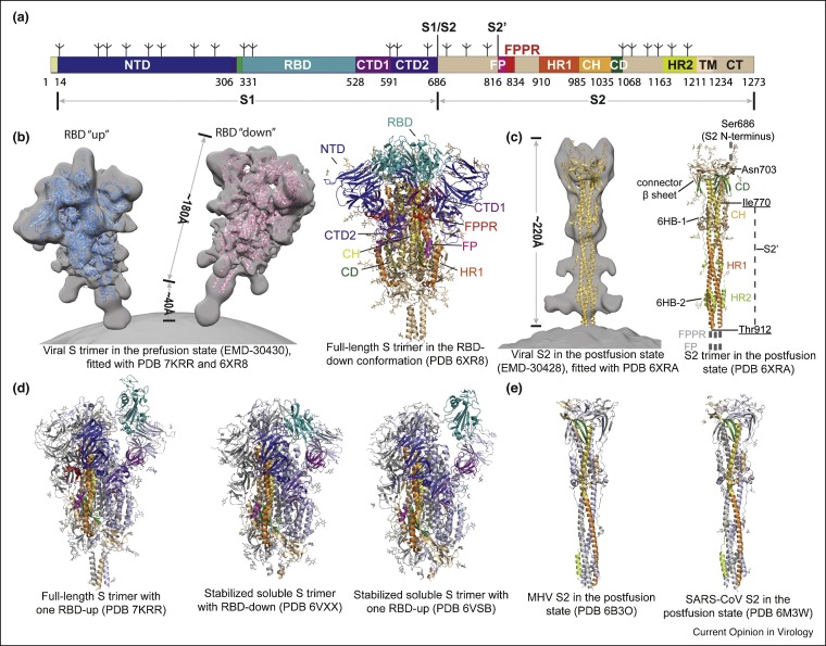
The COVID-19 (coronavirus disease 2019) pandemic, caused by severe acute respiratory syndrome coronavirus 2 (SARS-CoV-2), has led to loss of human life in millions and devastating socio-economic consequences worldwide. The disease has created urgent needs for intervention strategies to control the crisis and meeting these needs requires a deep understanding of the structure-function relationships of viral proteins and relevant host factors. The trimeric spike (S) protein of the virus decorates the viral surface and is an important target for development of diagnostics, therapeutics and vaccines. Rapid progress in the structural biology of SARS-CoV-2 S protein has been made since the early stage of the pandemic, advancing our knowledge on the viral entry process considerably. In this review, we summarize our latest understanding of the structure of the SARS-CoV-2 S protein and discuss the implications for vaccines and therapeutics.


Figures

Figure 1
Figure 1
Distinct conformational states of the SARS-CoV-2 spike protein. (a) Schematic representation of the SARS-CoV-2 spike protein organization. Segments of S1 and S2 include: NTD, N-terminal domain; RBD, receptor-binding domain; CTD1, C-terminal domain 1; CTD2, C-terminal domain 2; S1/S2, S1/S2 cleavage site; S2′, S2′ cleavage site; FP, fusion peptide; FPPR, fusion peptide proximal region; HR1, heptad repeat 1; CH, central helix region; CD, connector domain; HR2, heptad repeat 2; TM, transmembrane anchor; CT, cytoplasmic tail; and tree-like symbols for glycans. (b) Left: viral SARS-CoV-2 S trimer in the prefusion conformation (EMD-30430; Ref. [15]), fitted with the structures of purified proteins (PDB ID: 7KRR and 6XR8; Refs. [13••,50••]). Right: cryo-EM structure of the full-length S trimer in the RBD-down conformation (PDB ID: 6XR8). (c) Left: viral SARS-CoV-2 S2 trimer in the postfusion conformation (EMD-30428; Ref. [15]), fitted with the structure of the purified protein (PDB ID: 6XRA; Ref. [13••]). Right: cryo-EM structure of the full-length S2 trimer in the postfusion conformation (PDB ID: 6XRA). (d) Additional structures of coronavirus S proteins, including the full-length SARS-CoV-2 S trimer carrying G614 in the one RBD-up conformation (PDB ID: 7KRR), the stabilized soluble SARS-CoV-2 S trimer in the RBD-down conformation (PDB ID: 6VXX; Ref. [7]), the stabilized soluble SARS-CoV-2 S trimer in the one RBD-up conformation (PDB ID: 6VSB; Ref. [6••]). (e) MHV (mouse hepatitis virus) S2 in the postfusion state (PDB ID: 6B3O; Ref. [25]), and SARS-CoV S2 in the postfusion state (PDB ID: 6M3W; Ref. [24]).
Figure 2
Figure 2
Structures of NTD and its antibody complexes. (a) Cryo-EM structure of S1 fragment from the full-length SARS-CoV-2 S trimer (PDB ID: 6XR8), with the NTD highlighted in blue and the rest of S1 in gray. (b) Close-up view of the NTD in the SARS-CoV-2 S protein. (c) The NTD (in blue) from its complex with 4A8 is superposed with the domain from the full-length S trimer in gray, showing shifts of the five surface loops (N1–N5). (e) and (f) Close-up view of the binding interface for the NTD-4A8 and NTD-DH1205 complexes with contacting residues in the NTD highlighted in sticks. (d) Superposition of the structures of the NTD in complex with antibody 4A8 Fab (PDB ID: 7C2L; Ref. [28••]) and DH1052 Fab (PDB ID: 7LAB; Ref. [32]), as indicated. Heavy and light chains of 4A8 are colored in red and pink, respectively, and those of DH1052 are in green and cyan, respectively.
Figure 3
Figure 3
Structures of ACE2, ACE2-S complexes and RBD-antibody complexes. (a) Cryo-EM structure of the full-length ACE2 in complex with BoAT1 (PDB ID: 6M17; Ref. [10]), with the peptidase domain (PD) of ACE2 in pink, its neck domain in magenta, the transmembrane helix in yellow, and BoAT1 in gray. (b) The side view (left) and the top view (right) of cryo-EM structure of the soluble S trimer complexed with three ACE2s (PDB ID: 7KJ4; Ref. [37]). (c) Left, the crystal structure of SARS-CoV-2 RBD in complex with ACE2 (PDB ID: 6M0J; Ref. [8••]), with ACE2 in pink and the RBD in cyan. Middle and right, close-up views of the binding interface with contacting residues from the N-terminal helix of the ACE2 and the RBM of the RBD shown in sticks. (d) Left, cryo-EM structure of the RBD in complex with antibody REGN10933 (PDB ID: 6XDG; Ref. [43••]). Middle, cryo-EM structure of the RBD in complex with antibody REGN10987 (PDB ID: 6XDG; Ref. [43••]). Right, crystal structure of the RBD in complex with antibody CR3022 (PDB ID: 6YLA; Ref. [45]). The RBD is shown in cyan and heavy and light chains of the antibodies in various colors. The RBM is highlighted in dark blue.
Figure 4
Figure 4
Structures of CTDs. Structures of CTDs form the full-length S trimer (PDB ID: 7KRQ; Ref. [50••]) are shown, with CTD1 in magenta, CTD2 in purple, the 630-loop in yellow, and the β-strand in the CTD2 from S2 subunit in gray.
Figure 5
Figure 5
Structures and proposed conformational changes of SARS-CoV S2. (a) Close-up view of S2 in the prefusion (left) and postfusion (right) conformations from PDB ID: 6XR8 and 6XRA, with the fusion peptide (FP) highlighted in purple, the FPPR in red, central helix (CH) in gold, connector domain (CD) in green, HR1 in orange and HR2 in green. (b) Proposed structural transition of the HR1 from the prefusion to postfusion conformation. (c) Proposed conformational change of the HR2. (d). Six-helix bundle structures in the postfusion S2 with HR1 in orange and HR2 in green.

Similar articles

Cited by

References

    1. Zhou P., Yang X.L., Wang X.G., Hu B., Zhang L., Zhang W., Si H.R., Zhu Y., Li B., Huang C.L., et al. A pneumonia outbreak associated with a new coronavirus of probable bat origin. Nature. 2020;579:270–273. - PMC - PubMed
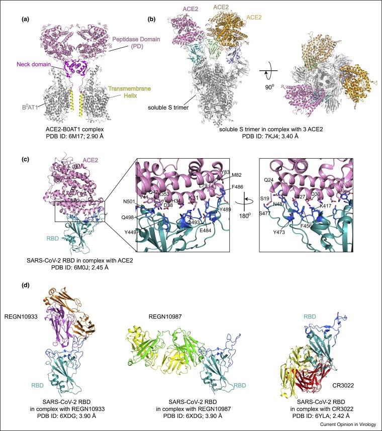
    2. This paper first reported the identification and characterization of SARS-CoV-2 (named 2019-nCoV at the time), which caused an epidemic of acute respiratory syndrome in humans in Wuhan, China. It shows that SARS-CoV-2 shares 79.6% sequence identity to SARS-CoV and 96% identity to a bat coronavirus. It has also confirmed that the new virus uses the same cell entry receptor—angiotensin converting enzyme II (ACE2)—as does SARS-CoV.

    1. Barcena M., Barnes C.O., Beck M., Bjorkman P.J., Canard B., Gao G.F., Gao Y., Hilgenfeld R., Hummer G., Patwardhan A., et al. Structural biology in the fight against COVID-19. Nat Struct Mol Biol. 2021;28:2–7. - PubMed
    1. Hoffmann M., Kleine-Weber H., Schroeder S., Kruger N., Herrler T., Erichsen S., Schiergens T.S., Herrler G., Wu N.H., Nitsche A., et al. SARS-CoV-2 cell entry depends on ACE2 and TMPRSS2 and is blocked by a clinically proven protease inhibitor. Cell. 2020;181:271–280. e278. - PMC - PubMed
    2. This paper has demonstrated that SARS-CoV-2 uses the SARS-CoV receptor ACE2 for entry and the serine protease TMPRSS2 for S protein priming.

    1. Zhao P., Praissman J.L., Grant O.C., Cai Y., Xiao T., Rosenbalm K.E., Aoki K., Kellman B.P., Bridger R., Barouch D.H., et al. Virus-receptor interactions of glycosylated SARS-CoV-2 spike and human ACE2 receptor. Cell Host Microbe. 2020;28:586–601. e586. - PMC - PubMed
    1. Watanabe Y., Allen J.D., Wrapp D., McLellan J.S., Crispin M. Site-specific glycan analysis of the SARS-CoV-2 spike. Science. 2020;369:330–333. - PMC - PubMed

Publication types

MeSH terms

Substances