. 2021 Sep 17;11(1):480.
doi: 10.1038/s41398-021-01578-2.

Proteomics of autism and Alzheimer's mouse models reveal common alterations in mTOR signaling pathway

Affiliations

Proteomics of autism and Alzheimer's mouse models reveal common alterations in mTOR signaling pathway

Shira Mencer et al. Transl Psychiatry. .

Abstract

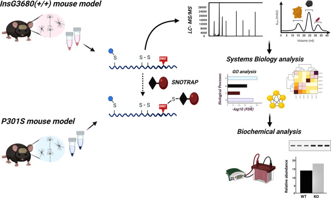
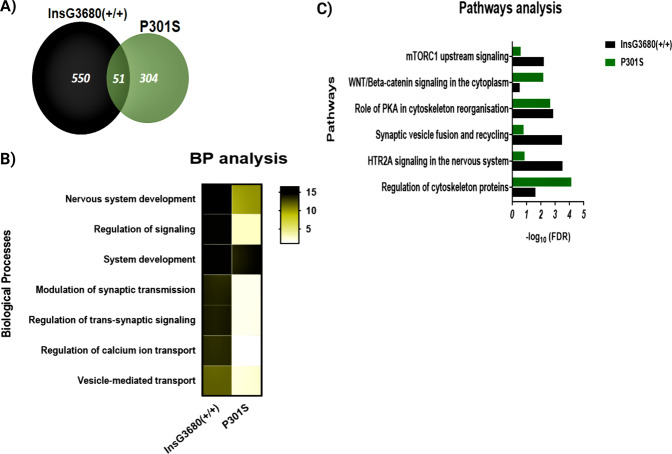
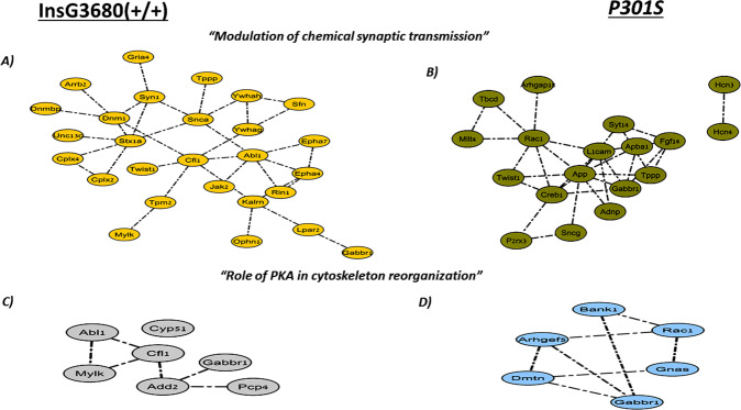
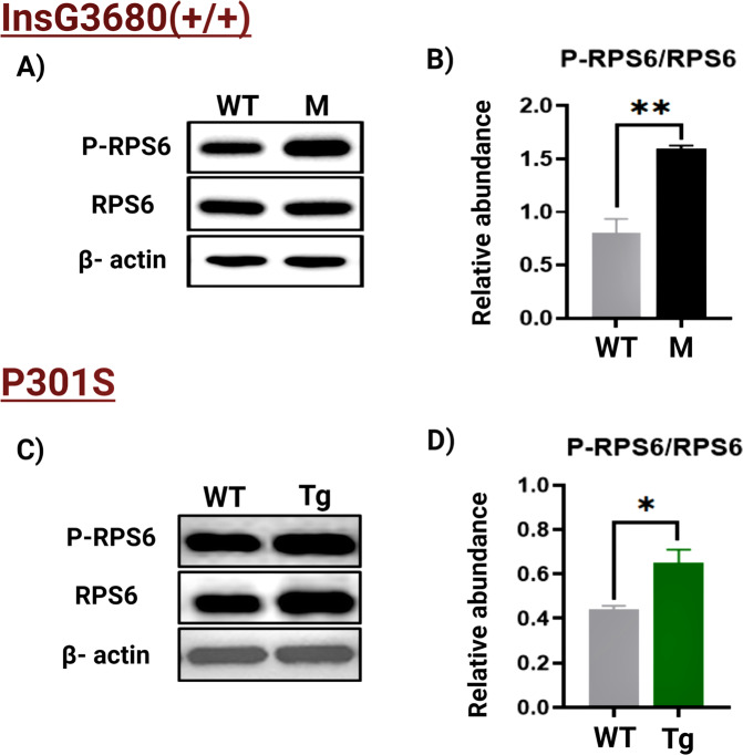
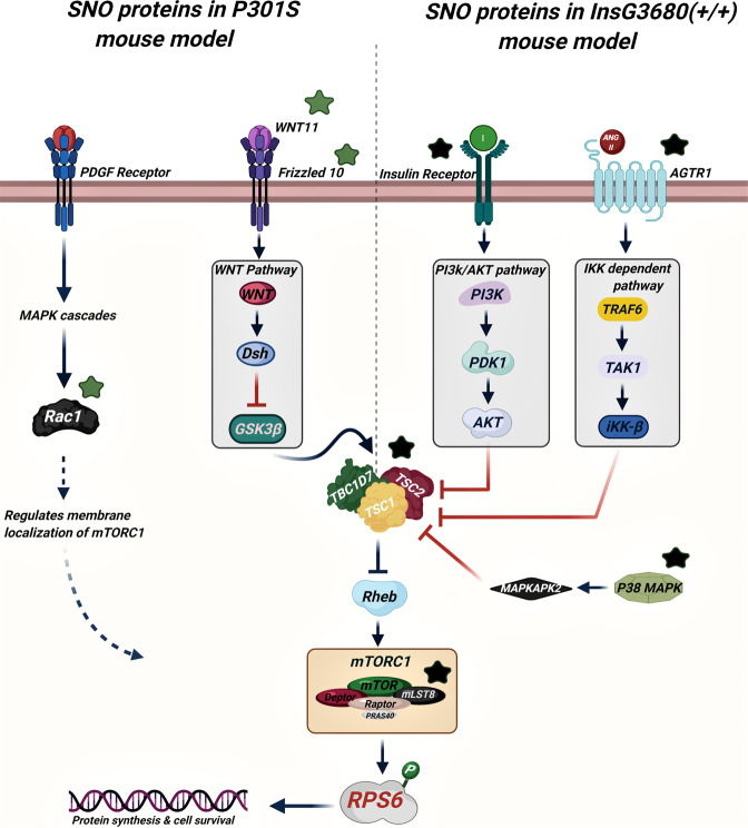
Autism spectrum disorder (ASD) and Alzheimer's disease (AD) are two different neurological disorders that share common clinical features, such as language impairment, executive functions, and motor problems. A genetic convergence has been proposed as well. However, the molecular mechanisms of these pathologies are still not well understood. Protein S-nitrosylation (SNO), the nitric oxide (NO)-mediated posttranslational modification, targets key proteins implicated in synaptic and neuronal functions. Previously, we have shown that NO and SNO are involved in the InsG3680(+/+) ASD and P301S AD mouse models. Here, we performed large-scale computational biology analysis of the SNO-proteome followed by biochemical validation to decipher the shared mechanisms between the pathologies. This analysis pointed to the mammalian target of rapamycin complex 1 (mTORC1) signaling pathway as one of the shared molecular mechanisms. Activation of mTOR in the cortex of both mouse models was confirmed by western blots that showed increased phosphorylation of RPS6, a major substrate of mTORC1. Other molecular alterations affected by SNO and shared between the two mouse models, such as synaptic-associated processes, PKA signaling, and cytoskeleton-related processes were also detected. This is the first study to decipher the SNO-related shared mechanisms between SHANK3 and MAPT mutations. Understanding the involvement of SNO in neurological disorders and its intersection between ASD and AD might help developing an effective novel therapy for both neuropathologies.


Conflict of interest statement

The authors declare no competing interests.

Figures

Fig. 1
Fig. 1. Schematic workflow of the study.
Schematic workflow of the SNOTRAP-based MS analysis of the ASD and AD cortex samples followed by large-scale systems analysis and biochemical validation.
Fig. 2
Fig. 2. Systems biology analysis of the SNO proteins of the ASD and AD cortex samples.
A Venn diagram of the SNO proteins. B Heat map representing the BP analysis conducted on the SNO proteins exclusive to ASD and AD models. *The scale represents the −log10 of the corrected false discovery rate (FDR). C Pathways analysis of the SNO proteins exclusive to ASD and AD models. *Bars represents the −log10 of the Benjamini corrected false discovery rate (FDR).
Fig. 3
Fig. 3. Clustering analysis of the SNOed proteins in ASD and AD mouse models.
“Modulation of chemical synaptic transmission” cluster was enriched in A ASD and B AD, and the “role of PKA in cytoskeleton reorganization” cluster was enriched in C ASD and D AD.
Fig. 4
Fig. 4. WB analysis.
A Representative WB of RPS6 and P-RPS6 prepared from the cortex tissues from the WT mice (n = 5) and ASD mouse model (abbreviated with M; n = 5). B The relative average WB intensity of P-RPS6, showing a significant increase in the phosphorylation levels of the RPS6 in mutant mice compared to the WT. The data are normalized to RPS6 and beta-actin presented as mean ± SEM. A two-tailed t-test was conducted. **P < 0.01. C Representative WB of RPS6 and P-RPS6 prepared from the cortex tissues from the WT mice (n = 5) and AD mouse model (abbreviated with Tg; n = 5). D The relative average WB intensity of P-RPS6, showing a significant increase in the phosphorylation levels of the RPS6 in Tg mice compared to their WT littermates. The data are normalized to RPS6 and beta-actin presented as mean ± SD. A two-tailed t-test was conducted. *P < 0.05.
Fig. 5
Fig. 5. The suggested scheme of the SNO-dependent mTOR activation in the ASD and AD mouse models.
Green stars are SNO proteins in AD. Black stars are SNO proteins in ASD.

References

    1. Sokol DK, Maloney B, Westmark CJ, Lahiri DK. Novel contribution of secreted smyloid-β precursor protein to white matter brain enlargement in autism spectrum disorder. Front Psychiatry. 2019;10:165–165. doi: 10.3389/fpsyt.2019.00165. - DOI - PMC - PubMed
    1. Besag FM. Epilepsy in patients with autism: links, risks and treatment challenges. Neuropsychiatr Dis Treat. 2017;14:1–10. doi: 10.2147/NDT.S120509. - DOI - PMC - PubMed
    1. Mullins C, Fishell G, Tsien RW. Unifying views of autism spectrum disorders: a consideration of autoregulatory feedback loops. Neuron. 2016;89:1131–56. doi: 10.1016/j.neuron.2016.02.017. - DOI - PMC - PubMed
    1. Thurm A, Swedo SE. The importance of autism research. Dialogues Clin Neurosci. 2012;14:219–22. doi: 10.31887/DCNS.2012.14.3/athurm. - DOI - PMC - PubMed
    1. Leigh JP, Du J. Brief Report: Forecasting the economic burden of autism in 2015 and 2025 in the United States. J Autism Dev Disord. 2015;45:4135–9. doi: 10.1007/s10803-015-2521-7. - DOI - PubMed

Publication types