. 2021 Sep 18;21(1):363.
doi: 10.1186/s12883-021-02352-z.

The role of hybrid FDG-PET/MRI on decision-making in presurgical evaluation of drug-resistant epilepsy

Affiliations

The role of hybrid FDG-PET/MRI on decision-making in presurgical evaluation of drug-resistant epilepsy

Márton Tóth et al. BMC Neurol. .

Abstract

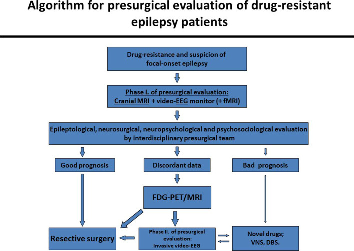
Background: When MRI fails to detect a potentially epileptogenic lesion, the chance of a favorable outcome after epilepsy surgery becomes significantly lower (from 60 to 90% to 20-65%). Hybrid FDG-PET/MRI may provide additional information for identifying the epileptogenic zone. We aimed to investigate the possible effect of the introduction of hybrid FDG-PET/MRI into the algorithm of the decision-making in both lesional and non-lesional drug-resistant epileptic patients.

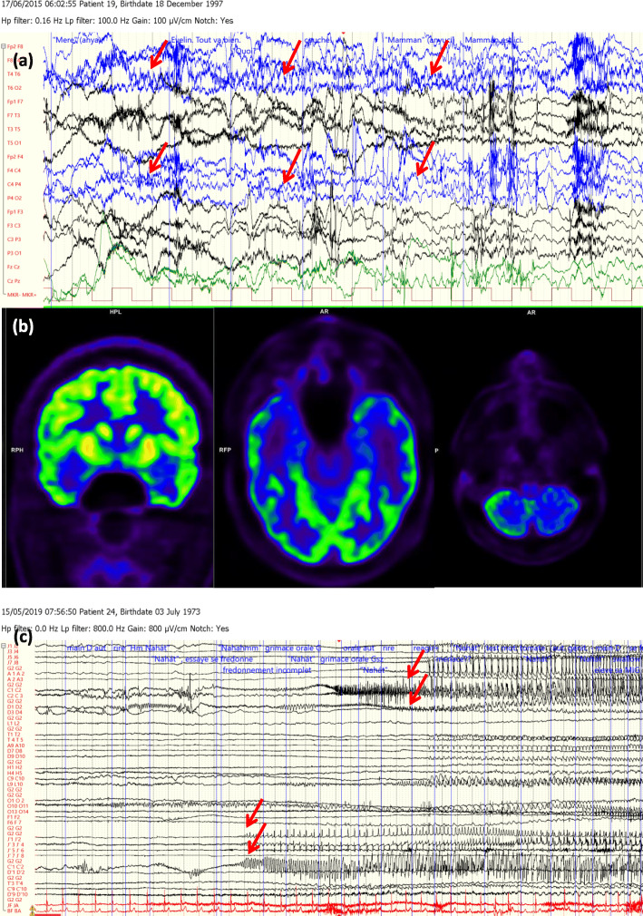
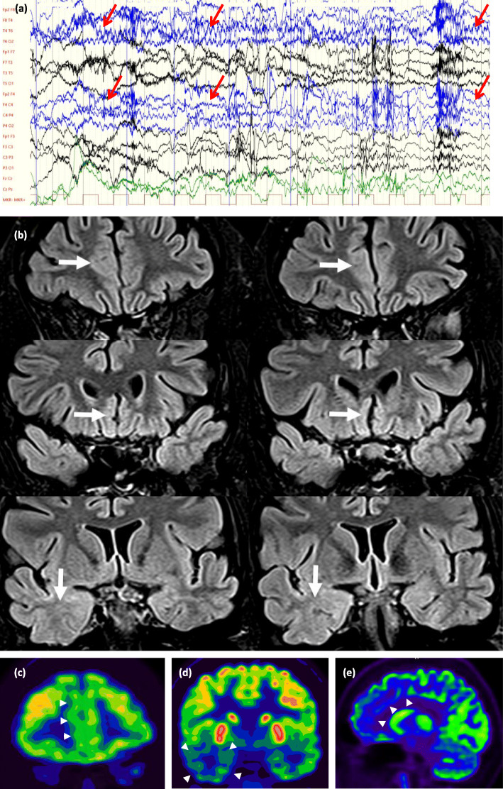
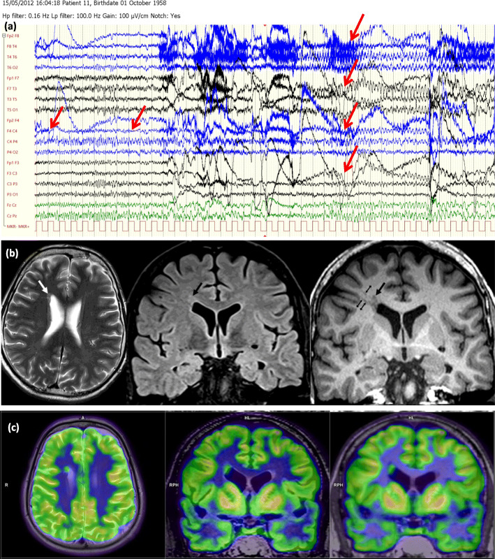
Methods: In a prospective study of patients suffering from drug-resistant focal epilepsy, 30 nonlesional and 30 lesional cases with discordant presurgical results were evaluated using hybrid FDG-PET/MRI.

Results: The hybrid imaging revealed morphological lesion in 18 patients and glucose hypometabolism in 29 patients within the nonlesional group. In the MRI positive group, 4 patients were found to be nonlesional, and in 9 patients at least one more epileptogenic lesion was discovered, while in another 17 cases the original lesion was confirmed by means of hybrid FDG-PET/MRI. As to the therapeutic decision-making, these results helped to indicate resective surgery instead of intracranial EEG (iEEG) monitoring in 2 cases, to avoid any further invasive diagnostic procedures in 7 patients, and to refer 21 patients for iEEG in the nonlesional group. Hybrid FDG-PET/MRI has also significantly changed the original therapeutic plans in the lesional group. Prior to the hybrid imaging, a resective surgery was considered in 3 patients, and iEEG was planned in 27 patients. However, 3 patients became eligible for resective surgery, 6 patients proved to be inoperable instead of iEEG, and 18 cases remained candidates for iEEG due to the hybrid FDG-PET/MRI. Two patients remained candidates for resective surgery and one patient became not eligible for any further invasive intervention.

Conclusions: The results of hybrid FDG-PET/MRI significantly altered the original plans in 19 of 60 cases. The introduction of hybrid FDG-PET/MRI into the presurgical evaluation process had a potential modifying effect on clinical decision-making.

Trial registration: Trial registry: Scientific Research Ethics Committee of the Medical Research Council of Hungary.

Trial registration number: 008899/2016/OTIG . Date of registration: 08 February 2016.

Keywords: Clinical decision-making; Drug-resistant epilepsy; Epilepsy surgery; Hybrid FDG-PET/MRI; Preoperative workflow.


Conflict of interest statement

Neither of the authors has any conflict of interest concerning the materials or methods used in this study or the findings specified in this paper to disclose.

Figures

Fig. 1
Fig. 1
An algorithm describing the steps of presurgical evaluation at our centers
Fig. 2
Fig. 2
(Case 28, Table 1, group nn, decision type 1.): A drug-resistant epileptic patient with the electroclinical features of humming epilepsy. a Video-EEG monitoring: during one of his habitual seizure, a right frontotemporal seizure activity was registered (red arrows). Originally, he was nonlesional and this MRI-status did not change yet after this study. b and c 18F-FDG PET and PET/MRI presented a bitemporal hypometabolism with a right predominance (red boxes) and d a left cerebellar hypometabolism (red box). c This patient remained as an iEEG candidate. iEEG monitor has been performed and showed a bitemporal seizure activity with a left side onset (red arrows, left side of the figure), a left-right propagation in between a 10-s interval (red arrows, right side of the figure), which was remote-controlled by a possible left orbitofrontal seizure onset zone. The patient did not allow neither a second iEEG intervention, nor VNS or DBS implantation
Fig. 3
Fig. 3
(Case 24, Table 1, group np, decision type 1.) A drug-resistant epileptic patient with the electroclinical features of a right frontotemporal epilepsy. a During video-EEG monitoring, her habitual hypermotor seizure with a right frontotemporal seizure activity was registered (red arrows). Originally, she was nonlesional and became lesional in this study. b Coronal FLAIR images: white arrows show possible focal cortical dysplasia in the right anterior cingulate cortex (upper row), in the medial cortex of the right straight gyrus (middle row), and the mildly increased signal intensity and blurred cortex-white matter interface in the right temporal lobe (lower row) can be seen. 18F-FDG PET and PET/MRI presented c and e a hypometabolism in the right mesiofrontal region (white arrowheads), d as well as in the right temporal lobe (white arroewheads). This patient remained as iEEG candidate; iEEG monitoring has not yet been realised
Fig. 4
Fig. 4
(Case 59, Table 2, group pp., decision type 2.): A drug-resistant epileptic patient with the electroclinical features of a right frontal epilepsy. a Video-EEG monitoring revealed his habitual seizure, a right frontocentral seizure activity was seen, which rapidly became bilateral (marked with red arrows). Concordantly, cranial MRI showed a nodular heterotopia in the right inferior frontal gyrus. b Axial T2 (left), coronal FLAIR (middle) and coronal T1 MPR (right) images. The white arrow on the T2 image and the large black arrows on the FLAIR and T1 images show focal nodular subependymal grey matter heterotopia. The small black arrows on the coronal T1 MPR image (right) show probable migrational bands. c Exceptionally compared to the other cases, during 18F-FDG PET and PET/MRI, a circumscribed FDG accumulation reaching the intensity of cortical tracer uptake (and highly exceeding white matter uptake) can be observed, identically to the right periventricular heterotopia. In this case, resective surgery became available instead of iEEG. Because the patient was left-handed, fMRI and also Wada-test were performed and they proved that in this case, active Broca region is localized in the right hemisphere. Thus, resective surgery was performed in awake state and finally, only a partial resection was possible. After the resective surgery, patient had much shorter (1–3 s long) seizures
Fig. 5
Fig. 5
(Case 43, Table 2, group pp.+, decision type 3.) A drug-resistant epileptic patient with the electroclinical features of a bitemporal lobe epilepsy. a Video-EEG monitoring. During her habitual seizure, a right frontotemporal seizure rapid activity was seen (marked with red arrows). Meanwhile, original cranial MRI (made before this study) showed an FCD along the left collateral sulcus. b Cranial MRI made in this study (coronal FLAIR images): horizontal arrows show the originally detected FCD along the left collateral sulcus while the oblique arrows show the newly observed FCD along the right collateral sulcus. The vertical arrows show the typical configuration of bilateral hippocampal malrotation, while c and d 18F-FDG PET and PET/MRI presented a hypometabolism in the right and left frontotemporal lobe, with a right predominance (red boxes). In summary, this patient was considered as not eligible for any further invasive procedures instead of iEEG

References

    1. Bell GS, Sander JW. The epidemiology of epilepsy: the size of the problem. Seizure. 2001;10(4):306–316. doi: 10.1053/seiz.2001.0584. - DOI - PubMed
    1. Ekman M, Forsgren L. Economic evidence in epilepsy: a review. Eur J Health Econ. 2004;5(SUPPL. 1):36–42. doi: 10.1007/s10198-005-0287-0. - DOI - PubMed
    1. Banerjee PN, Filippi D, Allen HW. The descriptive epidemiology of epilepsy-a review. Epilepsy Res. 2009;85(1):31–45. doi: 10.1016/j.eplepsyres.2009.03.003. - DOI - PMC - PubMed
    1. Marson A, Jacoby A, Johnson A, Kim L, Gamble C, Chadwick D. Immediate versus deferred antiepileptic drug treatment for early epilepsy and single seizures: a randomised controlled trial. Lancet. 2005;365(9476):2007–2013. doi: 10.1016/S0140-6736(05)66694-9. - DOI - PubMed
    1. Mula M, Cock HR. More than seizures: improving the lives of people with refractory epilepsy. Eur J Neurol. 2015;22(1):24–30. doi: 10.1111/ene.12603. - DOI - PubMed

Substances