Review
. 2021 Oct;42(5):463-473.
doi: 10.1053/j.sult.2021.07.004. Epub 2021 Aug 1.

Current Clinical Applications of Intracranial Vessel Wall MR Imaging

Affiliations
Review

Current Clinical Applications of Intracranial Vessel Wall MR Imaging

Raghav R Mattay et al. Semin Ultrasound CT MR. 2021 Oct.

Abstract

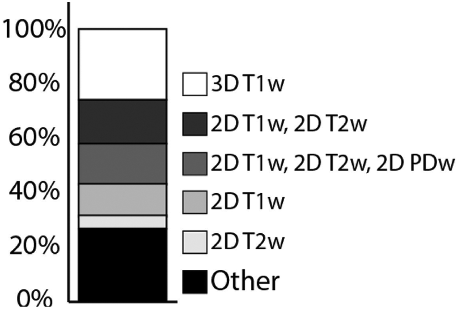
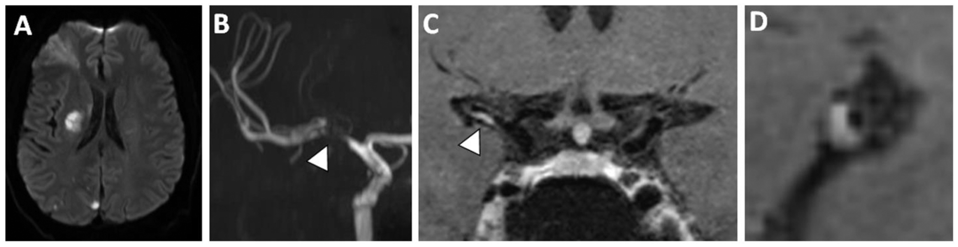
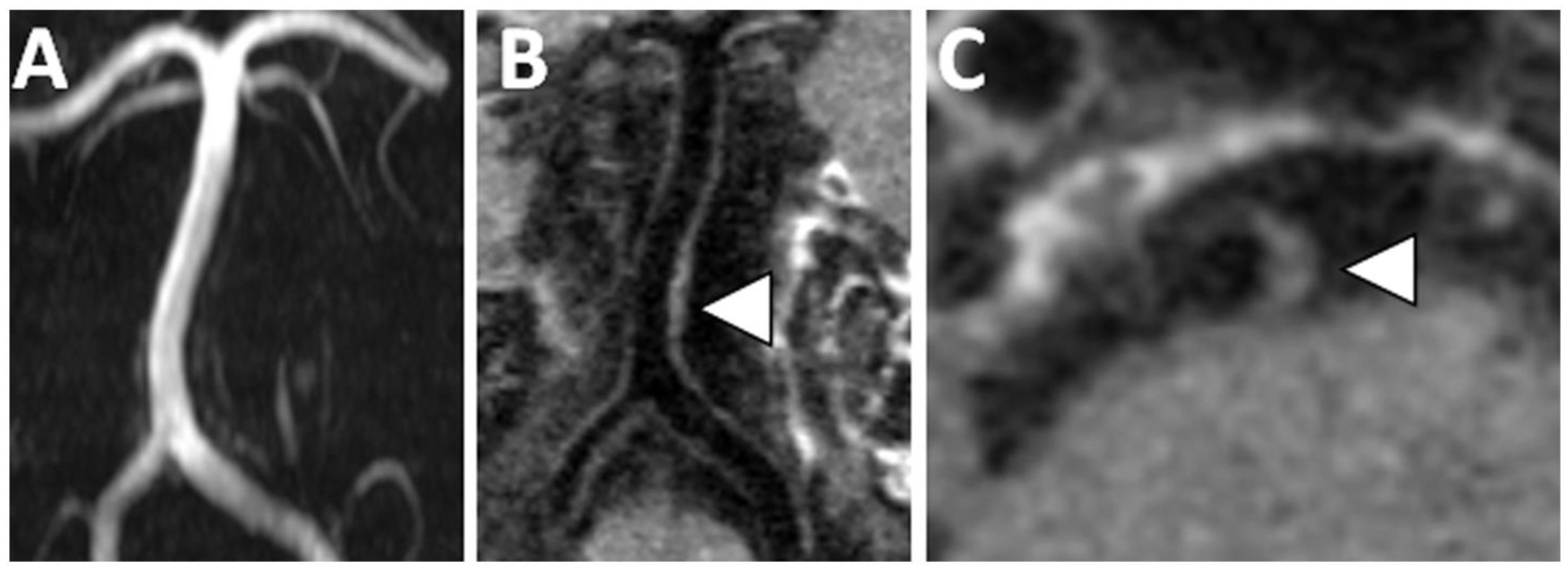
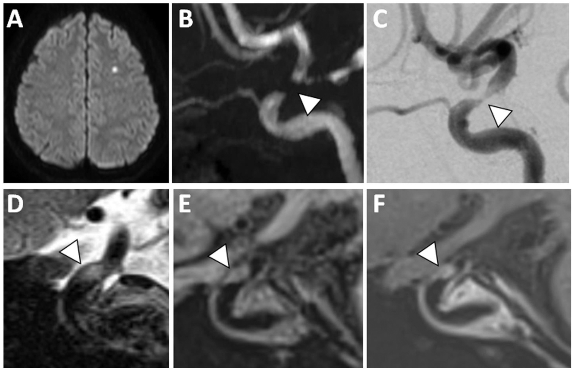
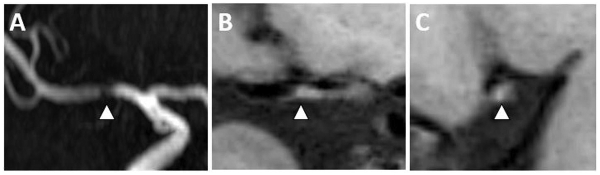
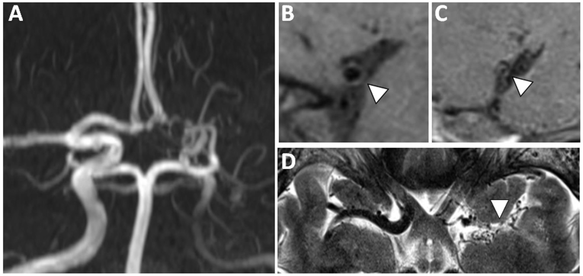
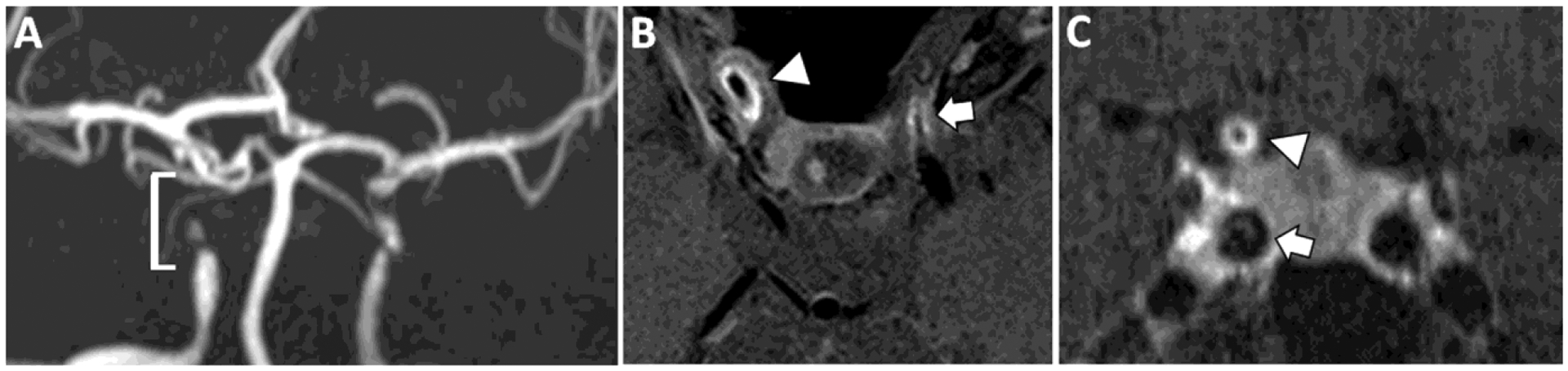
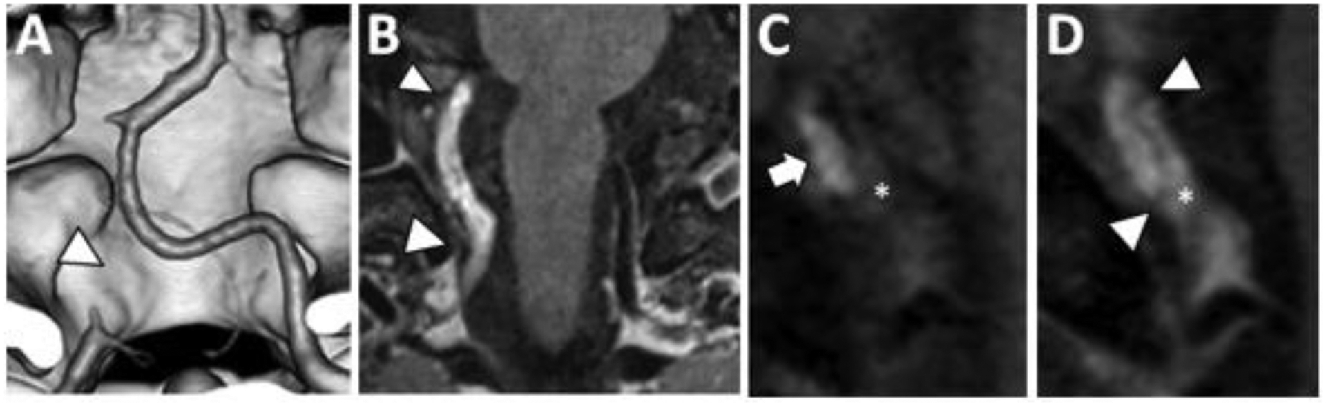
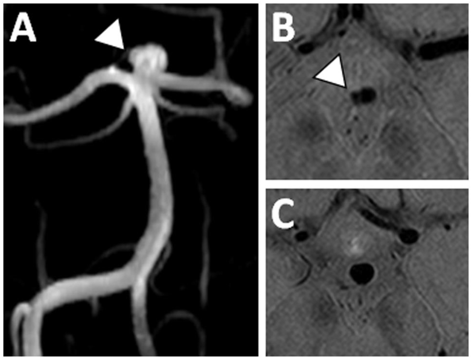
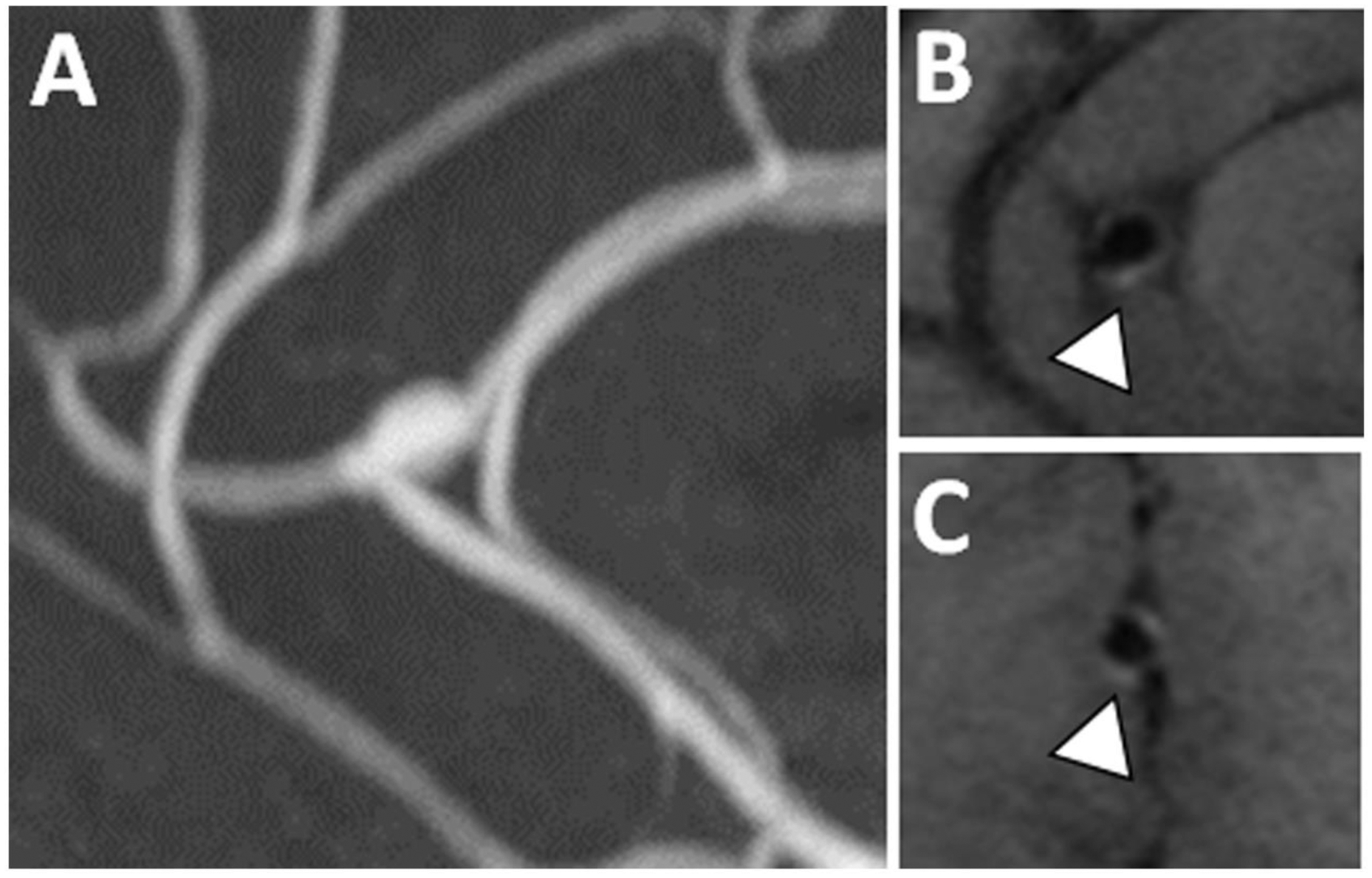
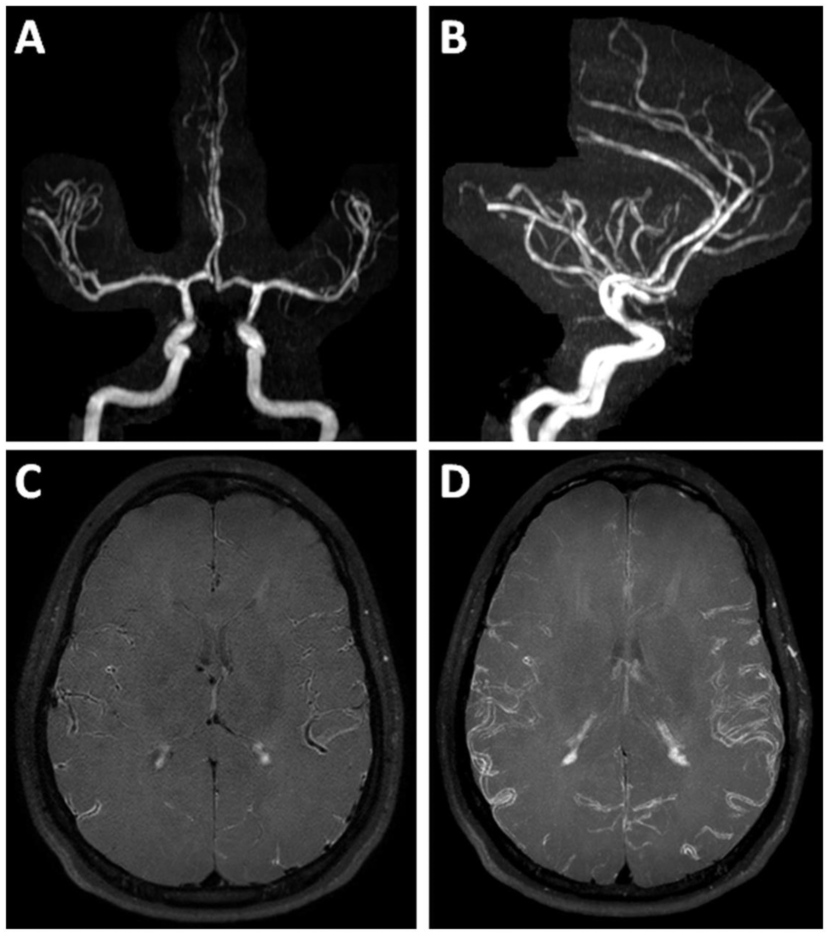
Intracranial vessel wall MR imaging (VWI) is increasingly being used as a valuable adjunct to conventional angiographic imaging techniques. This article will provide an updated review on intracranial VWI protocols and image interpretation. We review VWI technical considerations, describe common VWI imaging features of different intracranial vasculopathies and show illustrative cases. We review the role of VWI for differentiating among steno-occlusive vasculopathies, such as intracranial atherosclerotic plaque, dissections and Moyamoya disease. We also highlight how VWI may be used for the diagnostic work-up and surveillance of patients with vasculitis of the central nervous system and cerebral aneurysms.


Figures

Figure 1:
Figure 1:. Common VWI Protocols Reported in the Literature.
The most common VWI protocols reported in the literature are as follows: 3D T1-weighted (26%), 2D T1-weighted and 2D T2-weighted (16%); 2D T1-weighted, 2D T2-weighted, 2D PD-weighted (15%); 2D T1-weighted (12%); and 2D T2-weighted (5%). Other protocols include a combination of these pulse sequences and are further detailed in Song et al 2020.
Figure 2:
Figure 2:. VWI of Symptomatic ICAS with Vessel Wall Enhancement.
(A) A patient with a right middle cerebral artery (MCA) territory infarct showed (B) severe flow-limiting stenosis of the proximal right MCA on 3D TOF MRA. Postcontrast T1w VWI (C) coronal and (D) sagittal-oblique images show eccentric enhancement and is likely the causative culprit plaque.
Figure 3:
Figure 3:. VWI of Nonstenotic Basilar Artery Plaque.
(A) A 3D TOF MRA shows no significant stenosis of the basilar artery. (B) However, on precontrast T1w VWI, there is eccentric vessel wall thickening along the left side-wall revealing plaque. (C) Axial VWI image through the basilar artery confirms eccentric vessel wall thickening.
Figure 4:
Figure 4:. VWI of Symptomatic ICAS with T2 Hyperintensity and Enhancement
(A) A patient with acute infarction in the left centrum semiovale showed (B) severe flow-limiting stenosis in the left supraclinoid internal carotid artery on 3D TOF MRA (arrowhead). (C) Cerebral angiogram showed trace flow through this severely stenotic segment (arrowhead). (D) At the site of stenosis, sagittal T2w VWI showed eccentric T2 hyperintense signal (arrowhead). (E) Precontrast T1w VWI and (F) postcontrast T1w VWI reveals plaque enhancement (arrowhead).
Figure 5:
Figure 5:. VWI of Symptomatic ICAS with T1 Hyperintensity
(A) A 3D TOF MRA shows flow-limiting right M1 MCA stenosis (arrowhead). (B) Precontrast T1w VWI coronal and (C) sagittal planes through the MCA stenosis shows eccentric wall thickening (arrowhead) with intrinsic T1 hyperintense signal (arrowhead) of a symptomatic plaque.
Figure 6:
Figure 6:. VWI of Moyamoya Disease
(A) 3D TOF MRA shows severe stenosis of the left internal carotid artery terminus, severe irregularity of the left A1 anterior cerebral artery and nonvisualization of the left M1 middle cerebral artery (MCA) with lenticulostriate collaterals in a patient with moyamoya disease (MMD). (B) Sagittal-oblique precontrast T1w VWI of the right M1 MCA shows a thin vessel wall with preservation of the lumen diameter and appears normal. (C) The affected left M1 MCA shows small outer and inner diameters consistent with inward (negative) wall remodeling, a feature reported in MMD. (D) Axial T2w VWI shows lenticulostriate collaterals at the site of the left M1 MCA.
Figure 7:
Figure 7:. VWI of Primary Angiitis of the Central Nervous System
(A) A patient with a diagnosis of primary angiitis of the CNS shows flow-limiting stenosis of the right cavernous and supraclinoid internal carotid artery (ICA, bracket). Similar findings are seen in the contralateral left ICA. (B) Postcontrast T1w VWI shows avid concentric vessel wall thickening and enhancement of the stenotic ICA segments (right, arrowhead and left, arrow). (C) Coronal postcontrast T1w VWI confirms concentric vessel wall thickening and enhancement of the right supraclinoid ICA (arrowhead). By contrast, the right cavernous ICA (arrowhead) shows features of atherosclerotic plaque with eccentric wall thickening and a linear overlying enhancing rim favored to be a fibrous cap (arrow).
Figure 8:
Figure 8:. VWI of VZV vasculitis
(A) A patient with a diagnosis of varicella zoster vasculitis shows multiple segments of avid wall thickening and enhancement along the course of the bilateral anterior cerebral arteries (arrows) including the pericallosal artery (arrowhead) on postcontrast T1w VWI. (B) Coronal plane of the enhancing pericallosal artery segment shows both concentric wall enhancement as well as enhancement extending beyond the vessel wall borders, characteristic of perivascular enhancement (arrowhead). (C–D) After 2 months of treatment with acyclovir, there was improvement in the degree of vessel wall and perivascular enhancement.
Figure 9:
Figure 9:. Intracranial Vertebral Artery Dissection on VWI
(A) Coronal 3D surface rendered CTA image shows occlusion of the right intradural vertebral artery (arrowhead). (B) Coronal postcontrast T1w VWI shows abnormal signal in a long segment of the right intradural vertebral artery (arrowheads). (C) Coronal oblique precontrast T1w VWI shows mural hematoma (arrow) and intraluminal thrombus (asterisk). (D) The intraluminal thrombus enhances on coronal oblique postcontrast T1w VWI (asterisk). A thin intimal flap is detected (arrowheads) spiraling through the lumen and separating the intraluminal thrombus from the intramural hematoma.
Figure 10:
Figure 10:. Stable basilar tip aneurysm without enhancement
(A) A 3D TOF MRA shows a basilar tip aneurysm with a small daughter sac along the superolateral wall (arrowhead). (B–C) Axial postcontrast PDw VWI shows no aneurysm wall enhancement near the aneurysm daughter sac (B) nor near the aneurysm neck (C).
Figure 11:
Figure 11:. Stable fusiform pericallosal aneurysm with enhancement
(A) A fusiform pericallosal artery aneurysm seen on 3D TOF MRA was imaged with postcontrast T1w VWI. (B–C) A thin rim of aneurysm wall enhancement was present on postcontrast T1w VWI in this asymptomatic patient.
Figure 12:
Figure 12:. VWI of Reversible Cerebral Vasoconstriction Syndrome
(A–B) 3D TOF MRA maximum image projection images show diffuse irregularity of the cerebral vasculature. (C) Axial postcontrast T1w VWI and (D) a maximum image projection shows diffuse vessel wall enhancement. Clinical symptoms of severe headache resolved after starting a calcium channel blocker. Patient was diagnosed with RCVS after a negative laboratory work-up. A 1-month follow-up MRI showed complete resolution of the imaging findings (not shown).

References

    1. Mandell DM, Mossa-Basha M, Qiao Y, et al.Intracranial Vessel Wall MRI: Principles and Expert Consensus Recommendations of the American Society of Neuroradiology. AJNR Am J Neuroradiol 38:218–29, 2017. - PMC - PubMed
    1. Song JW, Obusez EC, Raymond SB, et al.Vessel Wall MRI Added to MR Angiography in the Evaluation of Suspected Vasculopathies. J Neuroimaging 29:454–7, 2019. - PubMed
    1. Song JW, Guiry SC, Shou H, et al.Qualitative Assessment and Reporting Quality of Intracranial Vessel Wall MR Imaging Studies: A Systematic Review. AJNR Am J Neuroradiol 40:2025–32, 2019. - PMC - PubMed
    1. Schaafsma JD, Rawal S, Coutinho JM, et al.Diagnostic Impact of Intracranial Vessel Wall MRI in 205 Patients with Ischemic Stroke or TIA. AJNR Am J Neuroradiol 40:1701–6, 2019. - PMC - PubMed
    1. Antiga L, Wasserman BA, Steinman DA On the overestimation of early wall thickening at the carotid bulb by black blood MRI, with implications for coronary and vulnerable plaque imaging. Magn Reson Med 60:1020–8, 2008. - PubMed

MeSH terms