. 2021 Oct;297(4):101202.
doi: 10.1016/j.jbc.2021.101202. Epub 2021 Sep 16.

Structures of synthetic nanobody-SARS-CoV-2 receptor-binding domain complexes reveal distinct sites of interaction

Affiliations

Structures of synthetic nanobody-SARS-CoV-2 receptor-binding domain complexes reveal distinct sites of interaction

Javeed Ahmad et al. J Biol Chem. 2021 Oct.

Abstract

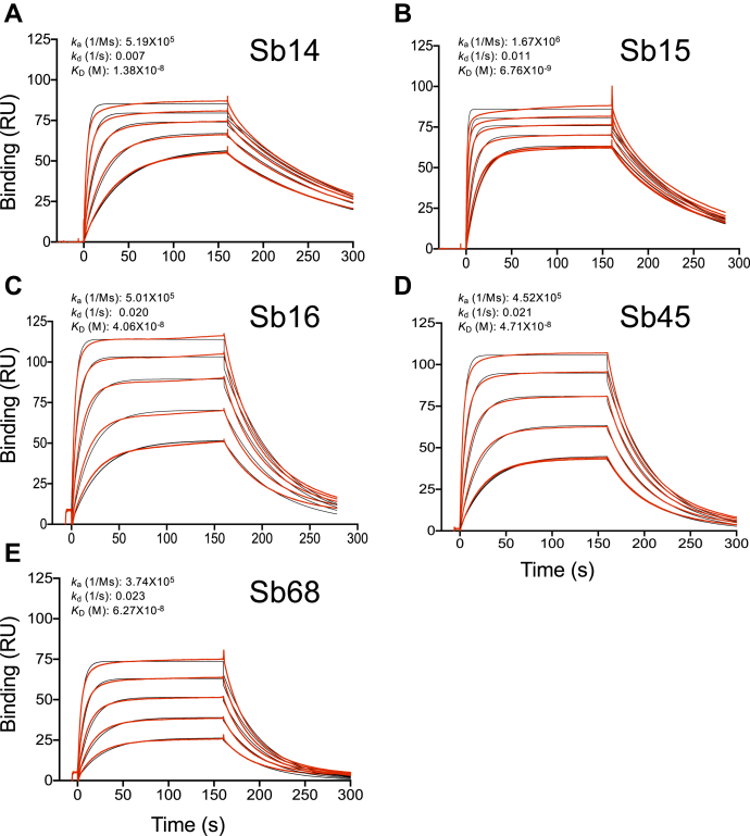
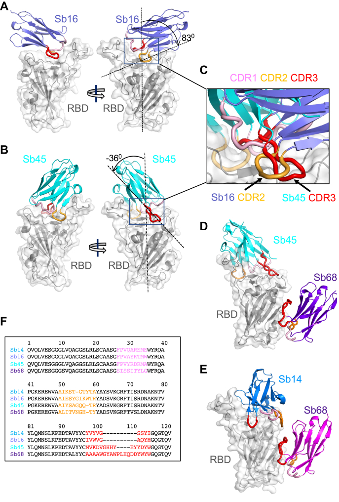
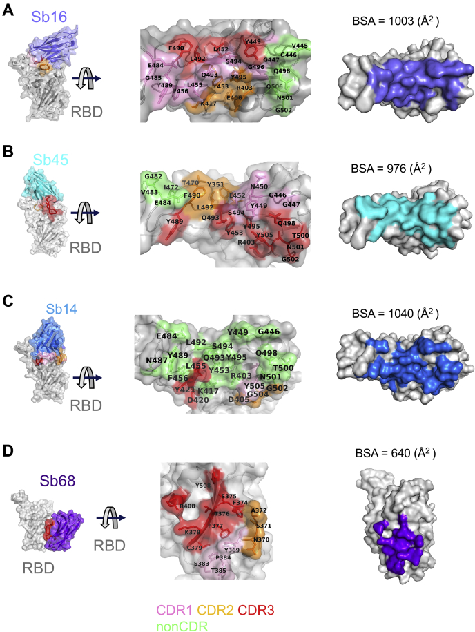
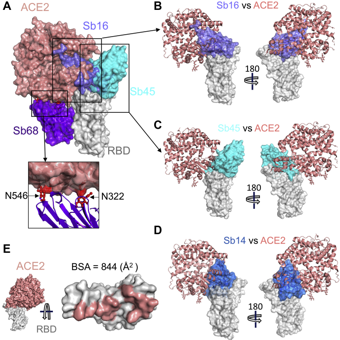
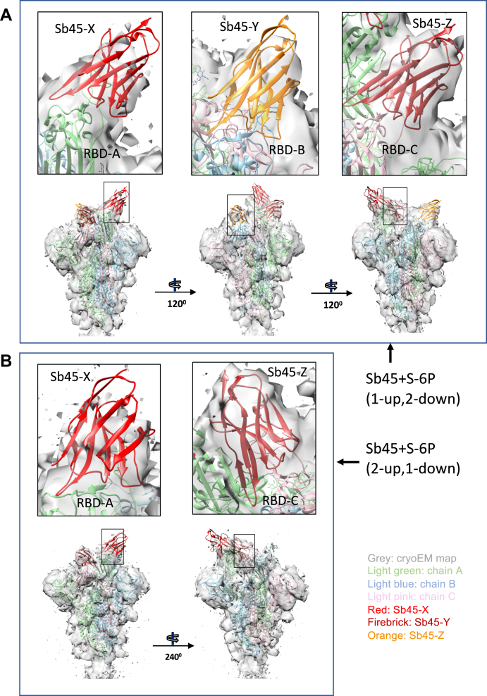
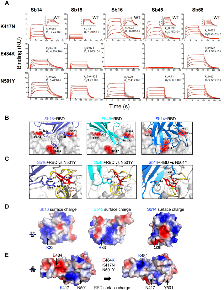
Combating the worldwide spread of severe acute respiratory syndrome coronavirus 2 (SARS-CoV-2) and the emergence of new variants demands understanding of the structural basis of the interaction of antibodies with the SARS-CoV-2 receptor-binding domain (RBD). Here, we report five X-ray crystal structures of sybodies (synthetic nanobodies) including those of binary and ternary complexes of Sb16-RBD, Sb45-RBD, Sb14-RBD-Sb68, and Sb45-RBD-Sb68, as well as unliganded Sb16. These structures reveal that Sb14, Sb16, and Sb45 bind the RBD at the angiotensin-converting enzyme 2 interface and that the Sb16 interaction is accompanied by a large conformational adjustment of complementarity-determining region 2. In contrast, Sb68 interacts at the periphery of the SARS-CoV-2 RBD-angiotensin-converting enzyme 2 interface. We also determined cryo-EM structures of Sb45 bound to the SARS-CoV-2 spike protein. Superposition of the X-ray structures of sybodies onto the trimeric spike protein cryo-EM map indicates that some sybodies may bind in both "up" and "down" configurations, but others may not. Differences in sybody recognition of several recently identified RBD variants are explained by these structures.

Keywords: SARS-CoV-2; cryo-EM; crystallography; single-domain antibody (sdAb, nanobody); surface plasmon resonance (SPR).


Conflict of interest statement

Conflict of interest The authors declare that they have no conflicts of interest with the contents of this article.

Figures

Figure 1
Figure 1
Sybodies bind the RBD with KDvalues in the nanomolar range. The RBD was coupled to a biosensor chip as described in Experimental procedures. Graded concentrations (31–500 nM) of each of the indicated sybodies were offered to the coupled surface (from t = 0 to t = 160 s) followed by buffer washout and measurement of net binding (in RU). Experimental curves (red) were fit by global analysis using Biacore T200 evaluation software 3.1 (Cytiva) (black). The curves shown are representative of at least two determinations. A, B, C, D, and E represent data and curve fits for Sb14, Sb15, Sb16, Sb45, and Sb68, respectively. RBD, receptor-binding domain; RU, resonance units.
Figure 2
Figure 2
Overall structures of Sb14, Sb16, Sb45, and Sb68 complexed with the SARS-CoV-2 RBD.Ribbon (sybodies) and ribbon plus surface (RBD) representations of the complex of (A) Sb16 (slate) with the RBD (gray) (7KGK); (B) Sb45 (cyan) with the RBD (7KGJ); (D) Sb45 and Sb68 (purple) with the RBD (7KLW), and (E) Sb14 (blue) and Sb68 (magenta) with the RBD (7MFU). Sb16–RBD and Sb45–RBD superimposed based on the RBD are shown in panel C, to highlight CDR loops, which are color-coded as CDR1 (pink), CDR2 (orange), and CDR3 (red). The CDR2 of Sb16 and CDR3 of Sb45 interact similarly with the RBD surface. Panel F shows a sequence alignment of the four sybodies. CDR1, complementarity-determining region 1; CDR2, complementarity-determining region 2; CDR3, complementarity-determining region 3; RBD, receptor-binding domain; SARS-CoV-2, severe acute respiratory syndrome coronavirus 2.
Figure 3
Figure 3
Interfaces and interactions of sybodies with the RBD.A, Sb16–RBD; (B) Sb45–RBD; (C) Sb14–RBD; and (D) Sb68–RBD. (Individual contacting residues are listed in Table S1). CDR1, CDR2, and CDR3 are painted pink, orange, and red, respectively. Additional non-CDR-contacting residues are colored lime. On the RBD surface, the epitopic residues that contact the sybodies are colored according to the sybody CDR. CDR1, complementarity-determining region 1; CDR2, complementarity-determining region 2; CDR3, complementarity-determining region 3; RBD, receptor-binding domain.
Figure 4
Figure 4
Sybodies clash with ACE2 in RBD complex structures.A, Sb16 (slate), Sb45 (cyan), Sb14 (blue), and Sb68 (purple)—RBD complexes were superposed on the ACE2–RBD structure (salmon) (6M0J) based on the RBD. Views of Sb16 (B), Sb45 (C), and Sb14 (D) are shown alone as well. Sb14 and Sb16 are buried inside ACE2, Sb45 is partially buried in ACE2, and Sb68 has major clashes with two N-glycan sites (N322 and N546) of ACE2 (inset). E, epitopic area (on the RBD) captured by ACE2 (salmon) is indicated along with its BSA. ACE2, angiotensin-converting enzyme 2; BSA, buried surface area; RBD, receptor-binding domain.
Figure 5
Figure 5
X-ray model of sybody superposed on cryo-EM structures of SB45–S-6P.A, model of Sb45+S-6P (1-up, 2-down) is fitted to the map with Sb45-X bound to RBD-A (up); Sb45-Y to RBD-B (down), and Sb45-Z to RBD-C (down). CCs (Sb45-X/Sb45-Y/Sb45-Z) are 0.52/0.49/0.57 respectively. B, model of Sb45+S-6P (2-up, 1-down) is fitted to the map with Sb45-X bound to RBD-A (up) and Sb45-Z bound to RBD-C (down), and CCs (Sb45-X/Sb45-Z) are 0.47/0.70, respectively. CCs, correlation coefficients; RBD, receptor-binding domain.
Figure 6
Figure 6
RBD mutations affect sybody binding.A, SPR binding of each of the indicated sybodies (across top) to each of the individual RBD mutants. Inset shows binding of sybodies to the WT RBD (from Fig. 1). Experimental tracings are shown in red, curve fits in black, and kd (s−1) and KD (M) values as determined from global fitting with BIAeval 2.0 are provided in each panel. B, location of contacts of Sb16, Sb45, and Sb14 is shown. E484, K417, and N501 of the RBD (WT) interact with K32, Y54, and R60 of Sb16, respectively; E484 and N501 of the RBD (WT) interact with R33 and H103 of Sb45, respectively; and E484, K417, and N501 of the RBD (WT) interact with Q39, E35, and Y60 of Sb14, respectively. C, comparison of complex structures with minimized models involving the N501Y mutation. In silico mutagenesis of N501Y was performed using 7KGK (Sb16+RBD), 7KGJ (Sb45+RBD), and 7MFU (Sb14+RBD+Sb68). After amino acid substitution in Coot, local energy minimization (within 15–20 Å of the mutant residue) was performed through three rounds in PHENIX. For the Sb16–RBD complex, when N501 is mutated to Y501, the loop (496–506, from yellow to wheat) extends about 2.4 Å, but R60 (revealing a double conformation) still forms hydrogen bonds with the Y501 loop; for the Sb45–RBD complex, when N501 is mutated to Y501, the loop (496–506, from yellow to wheat) extends about 1.0 Å, but H103 of Sb45 would still interact with Y501; for the Sb14–RBD complex, when N501 is mutated to Y501, the loop (496–506, from yellow to wheat) is extended about 2.0 Å, but T58 and K65 still form hydrogen bonds with Y501. D, the surface charge of Sb16; K32 forms a hydrogen bond with E484 of the RBD with the opposite charge; the surface charge of Sb45, R33 forms a hydrogen bond with E484 of the RBD with the opposite charge; the surface charge of Sb14, Q39 (a neutral residue) interacts with E484 of the RBD. E, surface charge of the WT RBD and surface charge of the RBD with the three mutations (E484, K417N, and N501Y). When E484 is mutated to K484, the surface charge is changed from negative to positive. Therefore, the hydrogen bonds are broken, pushing Sb16 and Sb45 out of contact, whereas because Q39 of Sb14 is not a charged residue, it still may interact with K484 of the mutated RBD. RBD, receptor-binding domain; SPR, surface plasmon resonance.

Update of

References

    1. Conti P., Caraffa A., Gallenga C.E., Kritas S.K., Frydas I., Younes A., Di Emidio P., Tete G., Pregliasco F., Ronconi G. The British variant of the new coronavirus-19 (Sars-Cov-2) should not create a vaccine problem. J. Biol. Regul. Homeost Agents. 2021;35:1–4. - PubMed
    1. Kirby T. New variant of SARS-CoV-2 in UK causes surge of COVID-19. Lancet Respir. Med. 2021;9:e20–e21. - PMC - PubMed
    1. Tang J.W., Tambyah P.A., Hui D.S.C. Emergence of a novel coronavirus causing respiratory illness from Wuhan, China. J. Infect. 2020;80:350–371. - PMC - PubMed
    1. Wibmer C.K., Ayres F., Hermanus T., Madzivhandila M., Kgagudi P., Oosthuysen B., Lambson B.E., de Oliveira T., Vermeulen M., van der Berg K., Rossouw T., Boswell M., Ueckermann V., Meiring S., von Gottberg A. SARS-CoV-2 501Y.V2 escapes neutralization by South African COVID-19 donor plasma. Nat. Med. 2021;27:622–625. - PubMed
    1. Wang Q., Zhang Y., Wu L., Niu S., Song C., Zhang Z., Lu G., Qiao C., Hu Y., Yuen K.Y., Wang Q., Zhou H., Yan J., Qi J. Structural and functional basis of SARS-CoV-2 entry by using human ACE2. Cell. 2020;181:894–904.e9. - PMC - PubMed

Publication types

MeSH terms

Substances