. 2021 Sep:103-104:1-21.
doi: 10.1016/j.matbio.2021.09.001. Epub 2021 Sep 17.

EphA2 signaling within integrin adhesions regulates fibrillar adhesion elongation and fibronectin deposition

Affiliations

EphA2 signaling within integrin adhesions regulates fibrillar adhesion elongation and fibronectin deposition

Alexandra C Finney et al. Matrix Biol. 2021 Sep.

Abstract

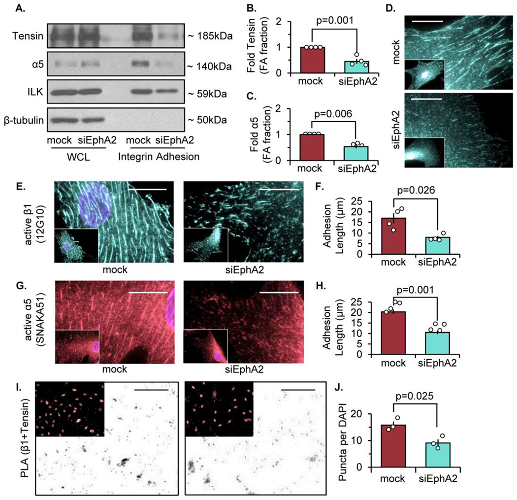
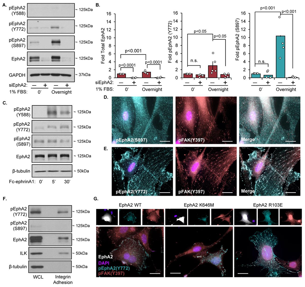
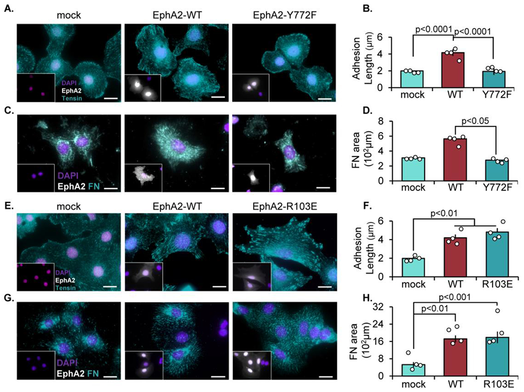
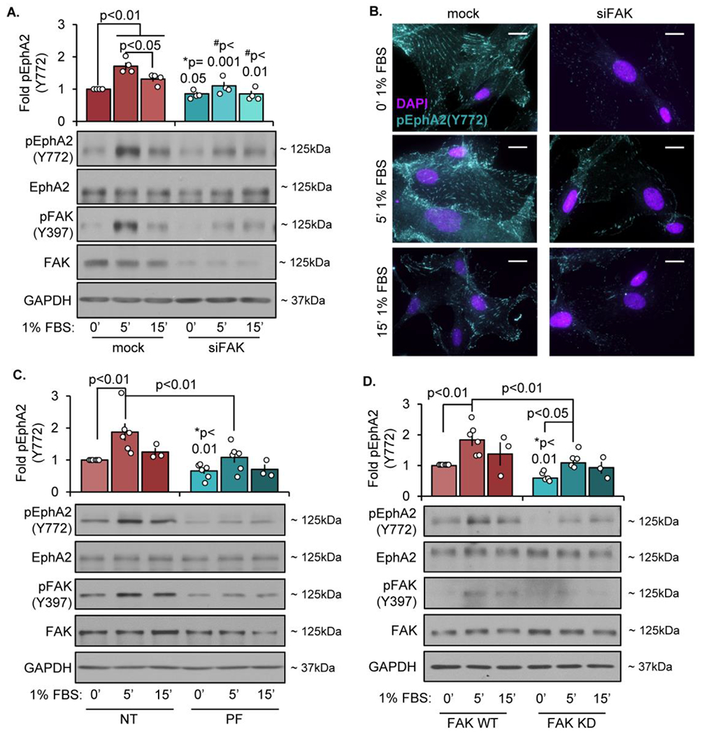
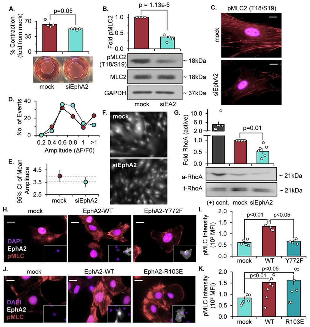
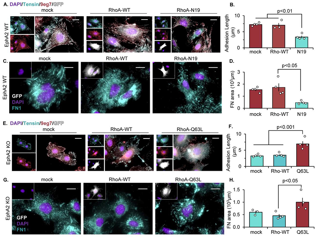
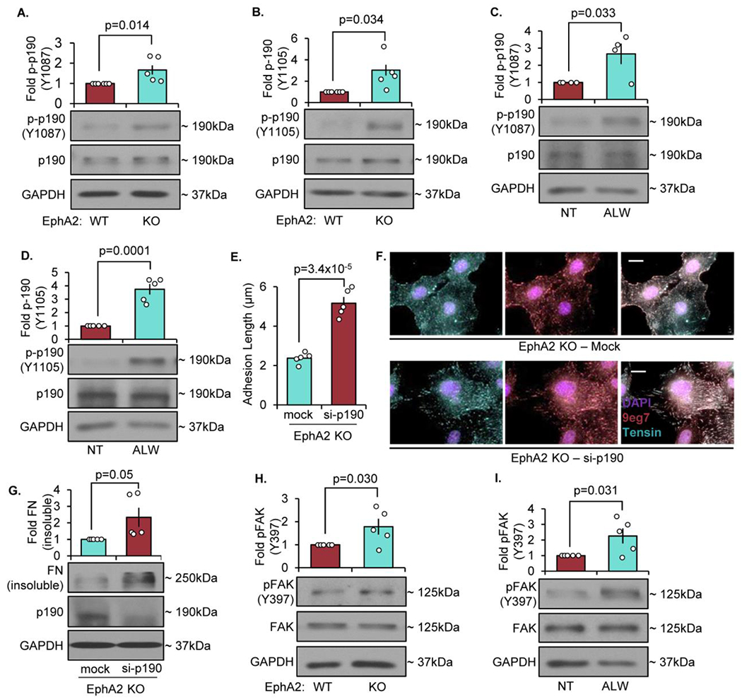
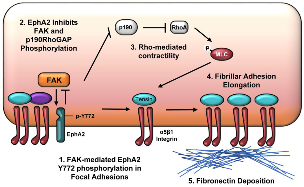
The multifunctional glycoprotein fibronectin influences several crucial cellular processes and contributes to multiple pathologies. While a link exists between fibronectin-associated pathologies and the receptor tyrosine kinase EphA2, the mechanism by which EphA2 promotes fibronectin matrix remodeling remains unknown. We previously demonstrated that EphA2 deletion reduces smooth muscle fibronectin deposition and blunts fibronectin deposition in atherosclerosis without influencing fibronectin expression. We now show that EphA2 expression is required for contractility-dependent elongation of tensin- and α5β1 integrin-rich fibrillar adhesions that drive fibronectin fibrillogenesis. Mechanistically, EphA2 localizes to integrin adhesions where focal adhesion kinase mediates ligand-independent Y772 phosphorylation, and mutation of this site significantly blunts fibrillar adhesion length. EphA2 deficiency decreases smooth muscle cell contractility by enhancing p190RhoGAP activation and reducing RhoA activity, whereas stimulating RhoA signaling in EphA2 deficient cells rescues fibrillar adhesion elongation. Together, these data identify EphA2 as a novel regulator of fibrillar adhesion elongation and provide the first data identifying a role for EphA2 signaling in integrin adhesions.

Keywords: EphA2 receptor tyrosine kinase; Fibrillar adhesions; Fibronectin deposition; Focal adhesion kinase; P190RhoGAP; RhoA Contractility.


Conflict of interest statement

Declaration of Interest: None.

Figures

Figure 1:
Figure 1:. EphA2 depletion reduces tensin localization and fibrillar adhesion length.
Human vascular smooth muscle cells (VSMCs) were transfected with either mock or siRNA targeted against EphA2 for 24 hours. A-C) Cells were plated onto Matrigel-coated glass slides overnight in 1% serum, and integrin adhesion isolation was performed. Protein expression was measured by immunoblot and normalized to integrin-linked kinase (ILK). β-tubulin from the whole cell lysate (WCL) fraction was shown for integrin adhesion isolation purity. B,C) Tensin and α5 integrin from the integrin adhesion fraction were quantified. D) Cells were stained for tensin. E,F) Cells were stained for active β1 integrin (12G10) and adhesion length was quantified in microns. G,H) Cells were stained for active α5 integrin (SNAKA51) and adhesion length was quantified in microns. (I) Proximity ligation assay (PLA) was performed for β1 and tensin interactions, and counterstained with DAPI (pink). Scale bar = 25μm. J) PLA puncta were quantified per high powered field and normalized to number of DAPI per high powered field. Scale bar = 25um. n=3-4. Data are expressed as mean ±SEM. Statistical comparisons were made using Student’s T-test (B,F,H,J,L). A p-value less than 0.05 is considered significant.
Figure 2:
Figure 2:. Serum treatment induces ligand-independent EphA2 phosphorylation.
Human VSMCs were transfected with either mock or siRNA targeted against EphA2 for 24 hours. Cells were plated onto Matrigel coated plates overnight in either serum-free or 1% serum. Protein was isolated and analyzed via immunoblotting (A) Representative immunoblots and (B) densitometry analysis of phospho-Y772, phospho-S897, and total EphA2. Total EphA2 was normalized to GAPDH, pEphA2 Y772 and pEphA2 S897 were normalized to total EphA2. Values expressed as fold change from baseline (mock 0’). C) Human VSMCs were serum-starved for four hours, treated with 1μg/mL Fc-EphrinA1 for 5 or 30 minutes, followed by immunoblotting for phosphorylated (Y588, Y772, and S897) and total EphA2. D-E) Human VSMCs were seeded onto Matrigel-coated glass coverslips overnight in 1% serum-containing media and stained for (D) S897-phosphorylated EphA2 (teal), (E) Y772-phosphorylated EphA2 (teal), (D,E) Y397-phosphorylated FAK (red), and DAPI (nuclei). F) Cells were plated onto Matrigel-coated glass slides overnight in 1% serum, followed by integrin adhesion isolation and protein expression was measured via immunoblot. G) EphA2 KO mouse aortic VSMCs cells were transfected with EphA2-WT, K646M, or R103E, plated overnight in 1% serum, and stained for phospho-EphA2 Y772 (teal), Y397-pFAK (pink), EphA2 (white) and DAPI (nuclei). n=4. Data are expressed as mean ±SEM. Statistical comparisons were made using 2-way ANOVA with Bonferroni post-test. A p-value less than 0.05 is considered significant.
Figure 3:
Figure 3:. EphA2-dependent fibrillar adhesion elongation requires Y772 phosphorylation but not EphA2-ligand interactions.
EphA2 KO mouse aortic VSMCs were transfected with either mock, EphA2-WT, (A-D) EphA2-Y772F, or (E-H) EphA2-R103E constructs for 24 hours, then plated onto Matrigel-coated coverslips overnight in 1% serum. Cells were stained for EphA2 (white), and EphA2-positive cells were quantified. A-B,E-F) Cells were stained fortensin (teal) and adhesion length was measured in microns. C-D,G-H) Cells were stained for fibronectin (teal) and quantified as fibronectin-positive area in microns. Scale bar = 25μm. n=4-5. Data are expressed as mean ±SEM. Statistical comparisons were made using One-way ANOVA with Bonferroni post-test. A p-value less than 0.05 is considered significant.
Figure 4:
Figure 4:. FAK is required for EphA2 phosphorylation within the focal adhesion.
Human VSMCs cells were transfected with either mock or siRNA targeting FAK for 24 hours. Cells were then plated onto Matrigel overnight, serum starved for 4 hours, and treated with 1% serum at the indicated timepoints. A) Phospho-EphA2 was analyzed by immunoblot and normalized to total EphA2 or (B) stained for Y772-phospho EphA2 (teal) and DAPI (nuclei). C) Cells were treated with PF-573228 (PF, 4μM) for 30 minutes, then treated with 1% serum at the indicated timepoints. Phospho-EphA2 was analyzed by immunoblot and normalized to total EphA2. D) FAK WT or FAK KD cells were serum-starved for 4 hours, then treated with 1% serum at the indicated timepoints. Phospho-EphA2 was measured by immunoblot and normalized to total EphA2. Fold changes are relative to mock/NT/WT 0’ timepoint. n=4. Data are expressed as mean ±SEM. Statistical comparisons were made using 2way ANOVA with Bonferroni post-test. #p-values compare between conditions for each treatment (mock vs. siFAK). *p-values compare baseline (0’ 1%FBS) between conditions using Student’s T-test. A p-value less than 0.05 is considered significant.
Figure 5:
Figure 5:. EphA2 promotes VSM cell contractility independent of calcium signaling.
Human VSMCs were transfected with either mock or siRNA targeted against EphA2 for 24 hours. A) Cells were embedded in 0.8% collagen isolated from rat tail and allowed to polymerize at 37°C. The cells and collagen gels were incubated in 1% serum overnight, and collagen gel diameter was measured and expressed as a percent contraction from the diameter of the well. B,C) Cells were plated onto Matrigel overnight in 1% serum and phospho-MLC was measured by B) Western blot and normalized to total MLC or C) staining (pink) with DAPI (nuclei). D-F) Cells were labeled with 1μM Fluo4-AM 45 minutes prior to treatment with 1% serum. Fluo4-AM fluorescence was visualized by microscopy every 15 seconds over a duration of 15 minutes. D) Fluorescence intensity (amplitude, ΔF/F0) per each cell was represented as a histogram. E) Mean amplitude of fluorescence intensity shown as 95% confidence interval, with dashed lines representing overlapping SEM. F) Representative image of Fluo4-AM-labeled cells. G) Cells were plated onto Matrigel overnight in 1% serum and RhoA activity was measured by Rho activation assay. Active RhoA was measured by Western blot and normalized to total RhoA. Non-hydrolyzable GTPγS was utilized as a positive control ((+) cont). N=6 (H-K) EphA2 KO mouse VSMCs transiently expressing EphA2-WT, EphA2-Y772F, and EphA2-R103E were seeded overnight on Matrigel in 1% serum, then (H,J) stained: phospho-MLC (pink), EphA2 (white), and DAPI (nuclei), and (l,K) quantified as mean fluorescence intensity (MFI). Scale bar = 25um. n=4-7. Data are expressed as mean ±SEM. Statistical comparisons were made using Student’s T-test (A,B,G) or Two-way ANOVA with Bonferroni post-test (I,K). A p-value less than 0.05 is considered significant.
Figure 6:
Figure 6:. EphA2 signals through RhoA to promote fibrillar adhesion elongation and fibronectin deposition.
A-D) EphA2 WT mouse VSMCs were transfected with either mock, RhoA-WT, or dominant-negative RhoA (RhoA-N19) for 24 hours. (E-H) EphA2 KO mouse VSMCs were transfected with mock, RhoA-WT, or constitutively active RhoA (RhoA-Q63L) for 24 hours. Cells were plated onto Matrigel overnight post-transfection, and GFP-positive cells (white) were quantified. A/B,E/F) Cells were stained for tensin (teal), active β1 integrin (9eg7, pink), and DAPI (purple) and fibrillar adhesion length was measured in microns. C/D,G/H) Cells were stained for fibronectin (teal) and DAPI (purple) and fibronectin area was measured in microns. Scale bar = 25μm. n=4. Data are expressed as mean ±SEM. Statistical comparisons were made using One-way ANOVA with Bonferroni post-test A p-value less than 0.05 is considered significant.
Figure 7:
Figure 7:. Deletion of EphA2 enhances p190Rho-GAP phosphorylation to inhibit fibronectin fibrillogenesis.
A/B) EphA2 WT or EphA2 KO mouse VSMCs were plated onto Matrigel overnight in 1% serum, and phospho-p190RhoGAP was quantified by Western blot and normalized to total p190RhoGAP. C/D) EphA2 WT mouse VSMCs were plated onto Matrigel overnight in 1% serum, then treated with ALW-II-41-27 (0.5μM) for 30 minutes. Phospho-p190RhoGAP was quantified by Western blot and normalized to total p190RhoGAP. E-G) EphA2 KO mouse VSMCs were transfected with either mock or siRNA against p190Rho-GAP for 24 hours, then plated onto Matrigel overnight. G,H) Cells were stained for tensin (teal), active β1 integrin (9eg7, pink), and DAPI (purple), and fibrillar adhesion length was measured in microns. Scale bar = 25μm. n=4-5. G) Cells were analyzed for fibronectin deposition with deoxycholate extraction. Deoxycholate-insoluble (deposited) fibronectin was normalized to deoxycholate-soluble GAPDH. H/I) EphA2 WT or EphA2 KO mouse VSMCs were plated onto Matrigel overnight in 1% serum, and phospho-FAK Y397 was quantified by Western blot and normalized to total FAK. Data are expressed as mean ±SEM. Statistical comparisons were made using Student’s T-test. A p-value less than 0.05 is considered significant.
Figure 8:
Figure 8:. Schematic diagram of proposed signaling pathway:
Schematic summary of the proposed mechanism of EphA2-regulated fibronectin deposition. 1) EphA2 within the focal adhesion undergoes ligand-independent phosphorylation on Y772 by focal adhesion kinase (FAK). EphA2 expression limits FAK activity, suggesting a novel reciprocal relationship between FAK and EphA2 signaling in the focal adhesion. 2) Elevated EphA2 signaling and reduced FAK signaling are associated with reduced phosphorylation and activation of p190RhoGAP. 3) Elevated RhoA activity due to inactive p190RhoGAP mediates myosin light chain phosphorylation and cellular contractility. 4) RhoA-driven contractility supports the recruitment of α5β1 integrins into tensin-rich fibrillar adhesions that drive 5) fibronectin deposition.

Similar articles

Cited by

References

    1. Lock JG, Wehrle-Haller B, Stromblad S, Cell-matrix adhesion complexes: master control machinery of cell migration, Seminars in cancer biology 18(1) (2008) 65–76. - PubMed
    1. Wight TN, Potter-Perigo S, The extracellular matrix: an active or passive player in fibrosis?, Am J Physiol Gastrointest Liver Physiol 301(6) (2011) G950–5. - PMC - PubMed
    1. Saunders JT, Schwarzbauer JE, Fibronectin matrix as a scaffold for procollagen proteinase binding and collagen processing, Mol Biol Cell 30(17) (2019) 2218–2226. - PMC - PubMed
    1. Valiente-Alandi I, Potter SJ, Salvador AM, Schafer AE, Schips T, Carrillo-Salinas F, Gibson AM, Nieman ML, Perkins C, Sargent MA, Huo J, Lorenz JN, DeFalco T, Molkentin JD, Alcaide P, Blaxall BC, Inhibiting Fibronectin Attenuates Fibrosis and Improves Cardiac Function in a Model of Heart Failure, Circulation 138(12) (2018) 1236–1252. - PMC - PubMed
    1. Wang WY, Twu CW, Liu YC, Lin HH, Chen CJ, Lin JC, Fibronectin promotes nasopharyngeal cancer cell motility and proliferation, Biomed Pharmacother 109 (2019) 1772–1784. - PubMed

Publication types