Review
. 2021 Dec;63(12):1957-1967.
doi: 10.1007/s00234-021-02813-9. Epub 2021 Sep 18.

Radiomics, machine learning, and artificial intelligence-what the neuroradiologist needs to know

Affiliations
Review

Radiomics, machine learning, and artificial intelligence-what the neuroradiologist needs to know

Matthias W Wagner et al. Neuroradiology. 2021 Dec.

Abstract

Purpose: Artificial intelligence (AI) is playing an ever-increasing role in Neuroradiology.

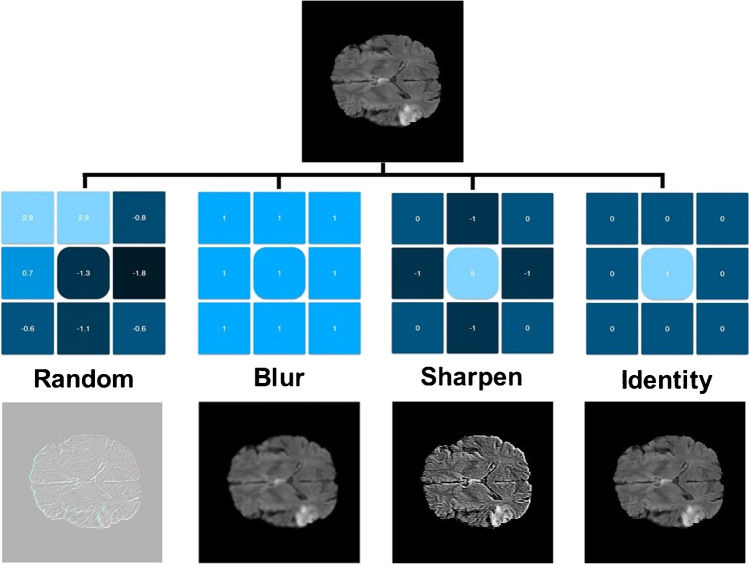
Methods: When designing AI-based research in neuroradiology and appreciating the literature, it is important to understand the fundamental principles of AI. Training, validation, and test datasets must be defined and set apart as priorities. External validation and testing datasets are preferable, when feasible. The specific type of learning process (supervised vs. unsupervised) and the machine learning model also require definition. Deep learning (DL) is an AI-based approach that is modelled on the structure of neurons of the brain; convolutional neural networks (CNN) are a commonly used example in neuroradiology.

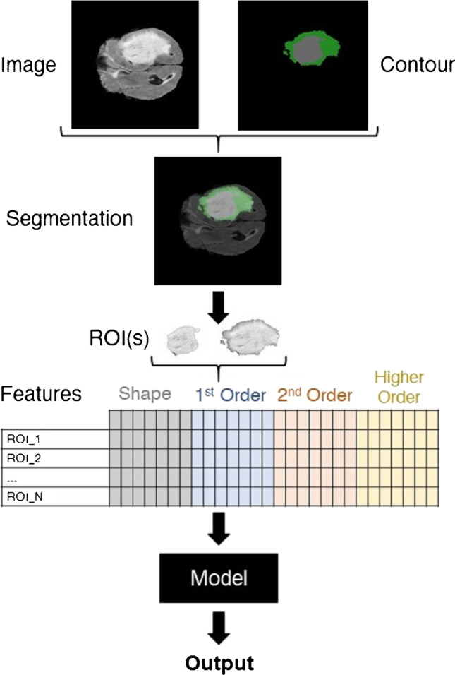
Results: Radiomics is a frequently used approach in which a multitude of imaging features are extracted from a region of interest and subsequently reduced and selected to convey diagnostic or prognostic information. Deep radiomics uses CNNs to directly extract features and obviate the need for predefined features.

Conclusion: Common limitations and pitfalls in AI-based research in neuroradiology are limited sample sizes ("small-n-large-p problem"), selection bias, as well as overfitting and underfitting.

Keywords: Artificial intelligence; Machine learning; Neuroradiology; Radiomics.


Conflict of interest statement

There is no conflict of interest for this review article for any of the authors.

Figures

Fig. 1
Fig. 1
Example of a typical radiomics pipeline. Regions of interests (ROI) are created based on the neuroradiological images and binary masks are created. The corresponding radiomics features are extracted through applying predefined formulae to ROI numerical representations. A model is used to infer the output based on the input radiomics. The task for which the pipeline is implemented determines type of output. Classification, risk score assessment (regression), and survival analysis are the most common purposes of radiomics-based pipelines
Fig. 2
Fig. 2
Schematic of a convolutional neural network. Convolutional neural networks consist of convolution layers and fully connected layers, also known as dense layers. The convolution layers serve as feature extractors, and the fully connected layers are classifiers. Output of the network depends on the target task. For an N-class classification scenario, the network has N nodes in its output layer. Each of these will generate the probability of the input image belonging to their corresponding class
Fig. 3
Fig. 3
Utilizing kernels to manipulate images. Kernels are the essence of convolutional neural networks. These are predefined matrices customized for specific tasks such as sharpening and blurring images. In convolutional neural networks, the idea is to learn multiple kernels and utilize them to extract informative features from the input images (images were created using https://github.com/generic-github-user/Image-Convolution-Playground)
Fig. 4
Fig. 4
Schematic for an optimum point in training, validation, and test cohorts in relation to the number of iterations

References

    1. Eltorai AEM, Bratt AK, Guo HH. Thoracic radiologists' versus computer scientists' perspectives on the future of artificial intelligence in radiology. J Thorac Imaging. 2020;35:255–259. doi: 10.1097/RTI.0000000000000453. - DOI - PubMed
    1. European Society of Radiology (ESR) Impact of artificial intelligence on radiology: a EuroAIM survey among members of the European Society of Radiology. Insights Imaging. 2019;10:105. doi: 10.1186/s13244-019-0798-3. - DOI - PMC - PubMed
    1. Weikert T, Cyriac J, Yang S, Nesic I, Parmar V, Stieltjes B. A practical guide to artificial intelligence-based image analysis in radiology. Invest Radiol. 2020;55:1–7. doi: 10.1097/RLI.0000000000000600. - DOI - PubMed
    1. Rizzo S, Botta F, Raimondi S, Origgi D, Fanciullo C, Morganti AG, Bellomi M. Radiomics: the facts and the challenges of image analysis. Eur Radiol Exp. 2018;2:36. doi: 10.1186/s41747-018-0068-z. - DOI - PMC - PubMed
    1. van Timmeren JE, Cester D, Tanadini-Lang S, Alkadhi H, Baessler B. Radiomics in medical imaging-"how-to" guide and critical reflection. Insights Imaging. 2020;11:91. doi: 10.1186/s13244-020-00887-2. - DOI - PMC - PubMed

LinkOut - more resources