Review
. 2022 Jan;480(1):177-189.
doi: 10.1007/s00428-021-03185-4. Epub 2021 Sep 18.

Apocrine lesions of the breast

Affiliations
Review

Apocrine lesions of the breast

Cecily M Quinn et al. Virchows Arch. 2022 Jan.

Abstract

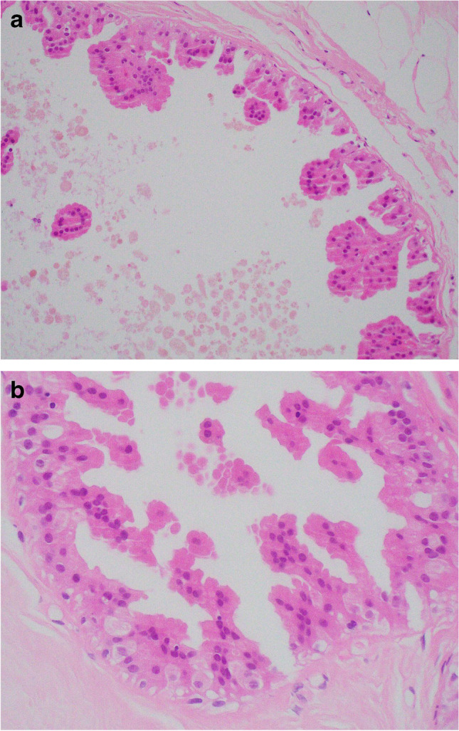
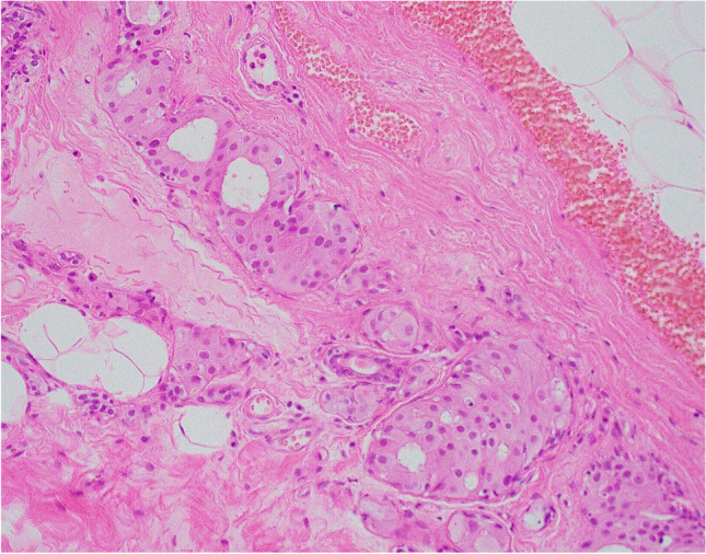
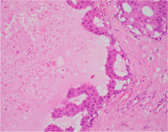
Apocrine change is recognised in benign, atypical and malignant lesions of the breast. Apocrine metaplasia, a frequent finding in the breast of women over the age of 25 years, is most commonly seen in benign cysts with a simple or papillary configuration. Apocrine change is also recognised in other benign lesions including sclerosing adenosis, now known as apocrine adenosis. Apocrine atypia usually refers to cytological atypia in which there is at least threefold variation in nuclear size but architectural atypia may also occur. The distinction between atypical apocrine hyperplasia and non-high-grade apocrine ductal carcinoma in situ may be difficult due to the relative rarity of these entities and the lack of validated diagnostic criteria. Lobular carcinoma in situ (LCIS) with apocrine change is considered to be a variant of pleomorphic LCIS. An apocrine variant of encapsulated papillary carcinoma is also recognised. Apocrine change is described in invasive carcinoma, including no special type, lobular, micropapillary and mucinous variants. The recent WHO 2019 update recognises 'carcinoma with apocrine differentiation' as a special type breast carcinoma based on the presence of apocrine morphology in at least 90% of the tumour. Tumours with apocrine morphology are usually but not always hormone receptor negative. Human epidermal growth factor receptor 2 (HER-2) status is variable. Molecular studies have identified breast tumours with apocrine features and high expression of androgen receptor mRNA including 'luminal androgen receptor tumours' and 'molecular apocrine tumours'. The term 'pure apocrine carcinoma' has been proposed to describe an invasive carcinoma with apocrine morphology that is oestrogen and progesterone receptor negative and androgen receptor positive. HER-2 status may be positive or negative. This article reviews the pathology of benign, atypical and malignant apocrine lesions of the breast, with emphasis on diagnostic criteria including an approach to evaluation of apocrine lesions on needle core biopsy, and recent advances in our understanding of invasive apocrine carcinoma.

Keywords: Apocrine; Atypia; Breast; Carcinoma with apocrine differentiation; In situ carcinoma; Molecular apocrine tumour.


Conflict of interest statement

The authors declare no competing interests.

Figures

Fig. 1
Fig. 1
Benign cyst lined by a single, flat, layer of apocrine epithelium
Fig. 2
Fig. 2
a Simple papillary apocrine change in which the apocrine cells are 3 or more cells thick focally and form mounds of cells, broader at the base than at the tip with no anastomoses. b Complex papillary apocrine change in which are taller and broader than those seen in simple papillary apocrine change with a tendency to anastomose within the lumen
Fig. 3
Fig. 3
Apocrine adenosis (sclerosing adenosis with apocrine change)
Fig. 4
Fig. 4
Atypical apocrine change in which there is at least threefold variation in nuclear size of the component apocrine cells
Fig. 5
Fig. 5
Atypical apocrine hyperplasia in which the atypia is mainly architectural due to a proliferation of apocrine cells with a solid and cribriform architecture
Fig. 6
Fig. 6
High-grade apocrine ductal carcinoma in situ (DCIS). The cells showed marked nuclear pleomorphism and abnormal mitoses. In this example, there is also dense peri-ductal chronic inflammation
Fig. 7
Fig. 7
Non-high-grade apocrine DCIS showing mild cytological atypia (at least threefold variation in nuclear size) and architectural complexity. In this example, there is early necrosis in the lumen which assists the diagnosis
Fig. 8
Fig. 8
Lobular carcinoma in situ (LCIS) with apocrine morphology a H&E and b negative e-cadherin immunohistochemistry
Fig. 9
Fig. 9
Encapsulated papillary carcinoma with apocrine change in the epithelial cell component (a, b)
Fig. 10
Fig. 10
Examples of invasive carcinoma with apocrine differentiation (a, b)
Fig. 11
Fig. 11
Invasive carcinoma with apocrine morphology composed of cells with foamy cytoplasm resembling histiocytes (histiocytoid carcinoma)
Fig. 12
Fig. 12
Gross cystic disease fluid protein-15 (GCDFP-15) expression, demonstrated on immunohistochemistry, in carcinoma with apocrine differentiation (a). GCDFP-15 expression in invasive lobular carcinoma with apocrine morphology (b)
Fig. 13
Fig. 13
Androgen receptor positivity, demonstrated on immunohistochemistry, in carcinoma with apocrine differentiation
Fig. 14
Fig. 14
Carcinoma with apocrine differentiation showing HER-2 positivity, demonstrated on immunohistochemistry

References

    1. Wells CA, El-Ayat GA. Non-operative breast pathology: apocrine lesions. J Clin Pathol. 2007;60(12):1313–1320. doi: 10.1136/jcp.2006.040626. - DOI - PMC - PubMed
    1. Selim AG, Wells CA. Immunohistochemical localisation of androgen receptor in apocrine metaplasia and apocrine adenosis of the breast: relation to oestrogen and progesterone receptors. J Clin Pathol. 1999;52(11):838–841. doi: 10.1136/jcp.52.11.838. - DOI - PMC - PubMed
    1. Mazoujian G, Haagensen DE. The immunopathology of gross cystic disease fluid proteins. Ann N Y Acad Sci. 1990;586:188–197. doi: 10.1111/j.1749-6632.1990.tb17806.x. - DOI - PubMed
    1. Sciarra A, Lopez G, Corti C, Runza L, Ercoli G, Bonometti A, et al. Columnar Cell Lesion and Apocrine Hyperplasia of the Breast: Is There a Common Origin? The Role of Prolactin-induced Protein. Appl Immunohistochem Mol Morphol. 2019;27(7):508–514. doi: 10.1097/PAI.0000000000000604. - DOI - PubMed
    1. Tsung JS, Wang TY, Wang SM, Yang PS. Cytological and biochemical studies of breast cyst fluid. Breast. 2005;14(1):37–41. doi: 10.1016/j.breast.2004.09.006. - DOI - PubMed