

The behavior and functions of embryonic microglia
- PMID: 34537900
- PMCID: PMC8732885
- DOI: 10.1007/s12565-021-00631-w
The behavior and functions of embryonic microglia
Abstract
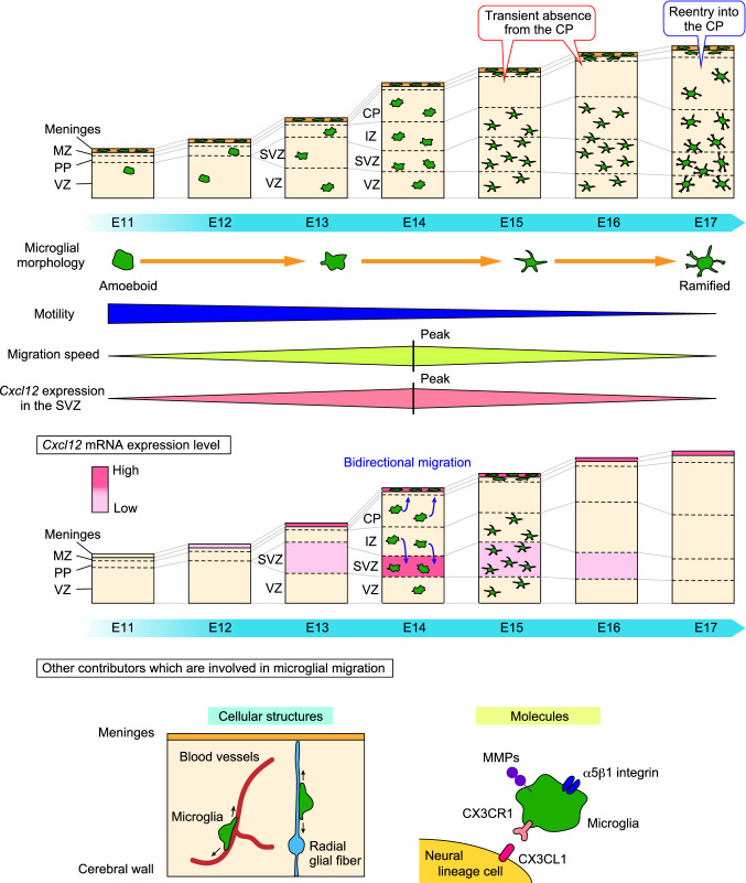
Microglia are the resident immune cells of the central nervous system. Microglial progenitors are generated in the yolk sac during the early embryonic stage. Once microglia enter the brain primordium, these cells colonize the structure through migration and proliferation during brain development. Microglia account for a minor population among the total cells that constitute the developing cortex, but they can associate with many surrounding neural lineage cells by extending their filopodia and through their broad migration capacity. Of note, microglia change their distribution in a stage-dependent manner in the developing brain: microglia are homogenously distributed in the pallium in the early and late embryonic stages, whereas these cells are transiently absent from the cortical plate (CP) from embryonic day (E) 15 to E16 and colonize the ventricular zone (VZ), subventricular zone (SVZ), and intermediate zone (IZ). Previous studies have reported that microglia positioned in the VZ/SVZ/IZ play multiple roles in neural lineage cells, such as regulating neurogenesis, cell survival and neuronal circuit formation. In addition to microglial functions in the zones in which microglia are replenished, these cells indirectly contribute to the proper maturation of post-migratory neurons by exiting the CP during the mid-embryonic stage. Overall, microglial time-dependent distributional changes are necessary to provide particular functions that are required in specific regions. This review summarizes recent advances in the understanding of microglial colonization and multifaceted functions in the developing brain, especially focusing on the embryonic stage, and discuss the molecular mechanisms underlying microglial behaviors.
Keywords: Cortex; Developing brain; Microglia; Neural progenitors; Neurogenesis.
© 2021. The Author(s).
Conflict of interest statement
The author declares no competing financial interests.
Figures

Similar articles
-
The multifaceted roles of embryonic microglia in the developing brain.Front Cell Neurosci. 2023 May 12;17:988952. doi: 10.3389/fncel.2023.988952. eCollection 2023. Front Cell Neurosci. 2023. PMID: 37252188 Free PMC article. Review.
-
The microglia-blood vessel interactions in the developing brain.Neurosci Res. 2023 Feb;187:58-66. doi: 10.1016/j.neures.2022.09.006. Epub 2022 Sep 24. Neurosci Res. 2023. PMID: 36167249 Review.
-
Microglia extensively survey the developing cortex via the CXCL12/CXCR4 system to help neural progenitors to acquire differentiated properties.Genes Cells. 2018 Oct;23(10):915-922. doi: 10.1111/gtc.12632. Epub 2018 Aug 24. Genes Cells. 2018. PMID: 30144249
-
Embryonic Pericytes Promote Microglial Homeostasis and Their Effects on Neural Progenitors in the Developing Cerebral Cortex.J Neurosci. 2022 Jan 19;42(3):362-376. doi: 10.1523/JNEUROSCI.1201-21.2021. Epub 2021 Nov 24. J Neurosci. 2022. PMID: 34819341 Free PMC article.
-
Mechanisms underlying microglial colonization of developing neural retina in zebrafish.Elife. 2021 Dec 7;10:e70550. doi: 10.7554/eLife.70550. Elife. 2021. PMID: 34872632 Free PMC article.
Cited by
-
Maternal SARS-CoV-2 impacts fetal placental macrophage programs and placenta-derived microglial models of neurodevelopment.J Neuroinflammation. 2024 Jun 25;21(1):163. doi: 10.1186/s12974-024-03157-w. J Neuroinflammation. 2024. PMID: 38918792 Free PMC article.
-
Microglia Colonization Associated with Angiogenesis and Neural Cell Development.Adv Neurobiol. 2024;37:163-178. doi: 10.1007/978-3-031-55529-9_10. Adv Neurobiol. 2024. PMID: 39207692 Review.
-
Circadian Regulation of the Neuroimmune Environment Across the Lifespan: From Brain Development to Aging.J Biol Rhythms. 2023 Oct;38(5):419-446. doi: 10.1177/07487304231178950. Epub 2023 Jun 26. J Biol Rhythms. 2023. PMID: 37357738 Free PMC article. Review.
-
Transplanting Microglia for Treating CNS Injuries and Neurological Diseases and Disorders, and Prospects for Generating Exogenic Microglia.Cell Transplant. 2023 Jan-Dec;32:9636897231171001. doi: 10.1177/09636897231171001. Cell Transplant. 2023. PMID: 37254858 Free PMC article. Review.
-
FlashTag-mediated Labeling for Intraventricular Macrophages in the Embryonic Brain.Bio Protoc. 2025 Jan 20;15(2):e5166. doi: 10.21769/BioProtoc.5166. eCollection 2025 Jan 20. Bio Protoc. 2025. PMID: 39872722 Free PMC article.
References
-
- Alliot F, Godin I, Pessac B. Microglia derive from progenitors, originating from the yolk sac, and which proliferate in the brain. Brain Res Dev Brain Res. 1999;117:145–152. - PubMed
-
- Antony JM, Paquin A, Nutt SL, Kaplan DR, Miller FD. Endogenous microglia regulate development of embryonic cortical precursor cells. J Neurosci Res. 2011;89:286–298. - PubMed
-
- Arlotta P, Molyneaux BJ, Chen J, Inoue J, Kominami R, Macklis JD. Neuronal subtype-specific genes that control corticospinal motor neuron development in vivo. Neuron. 2005;45:207–221. - PubMed
Publication types
MeSH terms
Grants and funding
LinkOut - more resources
Full Text Sources
Miscellaneous
