Review
. 2022 Jan 1;95(1129):20210699.
doi: 10.1259/bjr.20210699. Epub 2021 Sep 24.

Dual-energy CT in pulmonary vascular disease

Affiliations
Review

Dual-energy CT in pulmonary vascular disease

Ioannis Vlahos et al. Br J Radiol. .

Abstract

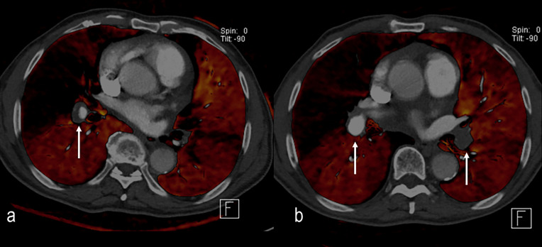
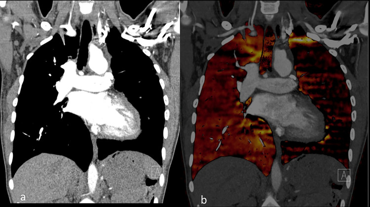
Dual-energy CT (DECT) imaging is a technique that extends the capabilities of CT beyond that of established densitometric evaluations. CT pulmonary angiography (CTPA) performed with dual-energy technique benefits from both the availability of low kVp CT data and also the concurrent ability to quantify iodine enhancement in the lung parenchyma. Parenchymal enhancement, presented as pulmonary perfused blood volume maps, may be considered as a surrogate of pulmonary perfusion. These distinct capabilities have led to new opportunities in the evaluation of pulmonary vascular diseases. Dual-energy CTPA offers the potential for improvements in pulmonary emboli detection, diagnostic confidence, and most notably severity stratification. Furthermore, the appreciated insights of pulmonary vascular physiology conferred by DECT have resulted in increased use for the assessment of pulmonary hypertension, with particular utility in the subset of patients with chronic thromboembolic pulmonary hypertension. With the increasing availability of dual energy-capable CT systems, dual energy CTPA is becoming a standard-of-care protocol for CTPA acquisition in acute PE. Furthermore, qualitative and quantitative pulmonary vascular DECT data heralds promise for the technique as a "one-stop shop" for diagnosis and surveillance assessment in patients with pulmonary hypertension. This review explores the current application, clinical value, and limitations of DECT imaging in acute and chronic pulmonary vascular conditions. It should be noted that certain manufacturers and investigators prefer alternative terms, such as spectral or multi-energy CT imaging. In this review, the term dual energy is utilised, although readers can consider these terms synonymous for purposes of the principles explained.


Conflict of interest statement

Conflicts of interest: RRL serves as a member of an advisory board for GE Healthcare. IV has had prior remote research support from Siemens Medical Systems and GE Healthcare (>5years previously). MCG has no disclosures. KS has no disclosures. MCJ has no disclosures.

Figures

Figure 1.
Figure 1.
(a). A subsegmental pulmonary embolus is suspected in the right lower lobe vasculature on weighted average images 100kVp/Sn140kVp. (b). Diagnostic confidence is increased by review of the simultaneous low energy100kVp images with greater contrast conspicuity. (c) Conversely 140kVp images are unhelpful with poorer iodine conspicuity but are essential for the reconstruction of dual-energy material-specific images.
Figure 2.
Figure 2.
(a) 1 mm 100 kVp image from dual-energy acquisition reconstructed by filtered back projection, (b) by iterative reconstruction SAFIRE level three and (c) by higher level iterative reconstruction, SAFIRE level 5. Increasing iterative reconstruction improves image quality without sacrificing ability to evaluate for small pulmonary emboli.
Figure 3.
Figure 3.
(a). 1.5 mm 100kVp/Sn140kVp perfused blood volume (PBV) map reconstructed with filtered back projection in a patient with PE (not shown) and right lung central defect. (b) image data reconstructed with SAFIRE three iterative reconstructions demonstrate improvement in image noise with reduced mottle in the central pulmonary arteries as well as in the PBV image.
Figure 4.
Figure 4.
(a). Weighted average 100 kVp/Sn140 kVp image. Single subsegmental right lower lobe pulmonary embolus suspected (arrow) but diagnosis is not certain. (b) Pulmonary perfused blood volume images demonstrate a corresponding single well-defined triangular defect (arrow) increasing radiological and clinical confidence in the diagnosis.
Figure 5.
Figure 5.
Occlusive pulmonary emboli in the right lower lobe segmental vasculature are associated with extensive perfusion defects. The right middle lobe demonstrates extensive perfusion reduction due to central embolus more cephalad (not shown). Patchy perfusion in the left lung is due to non-occlusive PE centrally.
Figure 6.
Figure 6.
(a–c) Multifocal pulmonary perfused blood volume defects in a patient with multifocal pulmonary emboli. Defects of this extent are more often associated with right ventricular dysfunction, poor oxygenation and worse prognosis.
Figure 7.
Figure 7.
Differences in image reconstruction appearance. Same patient imaged initially with dual-source CT pulmonary angiography (a), with study interrupted due to patient-related factors. Repeated 4 h later with single-source rapid kVp switching scanner (b). Dual-source imaging thresholds out the central vasculature and mediastinum for perfused blood volume analysis, evaluating only the smaller vessels and parenchyma. The central vessels are depicted as per conventional grayscale angiography. In single-source technique, the entire image, including central vessels and mediastinum, is evaluated and colour-coded for calculated enhancement. Note that the difference in range of enhancement assessed also results in differences in the scale depicted and the conspicuity of a perfusion defect in the right upper lobe secondary to embolus.
Figure 8.
Figure 8.
(a). Initial diagnosis of chronic thromboembolic pulmonary hypertension (CTEPH) with eccentric thrombus in the right interlobar artery and extensive peripheral defects in perfusion on perfused blood volume (PBV) imaging. (b) 4 months following anticoagulation and vasodilator therapy there is improvement of the right sided central thrombus but the large central defect in perfusion persists as well as several smaller peripheral defects bilaterally. Thrombus persists in the left-sided central vessels. The detection of smaller peripheral perfusion defects in CTEPH appears to be an advantage of dual-energy CT over conventional CT pulmonary angiography.
Figure 9.
Figure 9.
(a) Initial evaluation with dual energy CT demonstrates a right lower lobe embolus (arrow) with peripheral perfusion defect. (b) Follow-up study 14 months later demonstrates near complete resolution of the embolus but a residual linear defect in the vessel. The resolution of the PBV defect aids in discriminating that this reflects chronic thrombus rather than chronic thromboembolic pulmonary hypertension.
Figure 10.
Figure 10.
Dual time point dual-energy CT images in a patient with pulmonary hypertension. (a) Pulmonary arterial phase imaging demonstrates peripheral perfusion reduction defects. (b) Imaging performed 7 s later in the systemic arterial phase demonstrates global increase in pulmonary parenchymal perfusion and in-filling of pulmonary peripheral defects. This delayed increase in pulmonary parenchymal enhancement is a feature of pulmonary hypertension and correlates with pulmonary vascular resistance.
Figure 11.
Figure 11.
Congenital abnormality evaluated by dual-energy CT pulmonary angiography in a patient with Fallot’s tetralogy (a) coronal image demonstrates marked reduction in size of the left-sided central pulmonary arterial vasculature. (b) corresponding perfused blood volume image highlights the marked reduction in perfusion of the left lung.
Figure 12.
Figure 12.
25-year-old male presented with symptoms of acute PE and large central filling defect on CT pulmonary angiography. Central linear high attenuation was concerning for possible enhancement within the filling defect. (b) Dual-energy calculated perfused blood volume images confirm reduction in perfusion of the right lung. (c) Iodine calculation performed of the central vessels and soft tissues demonstrates intermediate iodine uptake in the filling defect, above background uptake in the paraspinal musculature. The appearances were concerning for an enhancing intravascular tumour (d). Confirmation of corresponding metabolic activity in the same location at 18-fluorodeoxyglucose PET-CT imaging. Lesion was histologically confirmed as a leiomyosarcoma at resection.

References

    1. Hounsfield GN. Computerized transverse axial scanning (tomography). 1. description of system. Br J Radiol 1973; 46: 1016–22. doi: 10.1259/0007-1285-46-552-1016 - DOI - PubMed
    1. Alvarez RE, Macovski A. Energy-Selective reconstructions in X-ray computerized tomography. Phys Med Biol 1976; 21: 733–44. doi: 10.1088/0031-9155/21/5/002 - DOI - PubMed
    1. Flohr TG, McCollough CH, Bruder H, Petersilka M, Gruber K, Süss C, et al. . First performance evaluation of a dual-source CT (DSCT) system. Eur Radiol 2006; 16: 256–68. doi: 10.1007/s00330-005-2919-2 - DOI - PubMed
    1. McCollough CH, Boedeker K, Cody D, Duan X, Flohr T, Halliburton SS, et al. . Principles and applications of multienergy CT: report of AAPM task group 291. Med Phys 2020; 47: e881–912. doi: 10.1002/mp.14157 - DOI - PubMed
    1. Zhang D, Li X, Liu B. Objective characterization of Ge discovery CT750 HD scanner: gemstone spectral imaging mode. Med Phys 2011; 38: 1178–88. doi: 10.1118/1.3551999 - DOI - PubMed

MeSH terms