. 2021 Dec 1;90(11):756-765.
doi: 10.1016/j.biopsych.2021.07.018. Epub 2021 Jul 26.

Hyperexcitable Phenotypes in Induced Pluripotent Stem Cell-Derived Neurons From Patients With 15q11-q13 Duplication Syndrome, a Genetic Form of Autism

Affiliations

Hyperexcitable Phenotypes in Induced Pluripotent Stem Cell-Derived Neurons From Patients With 15q11-q13 Duplication Syndrome, a Genetic Form of Autism

James J Fink et al. Biol Psychiatry. .

Abstract

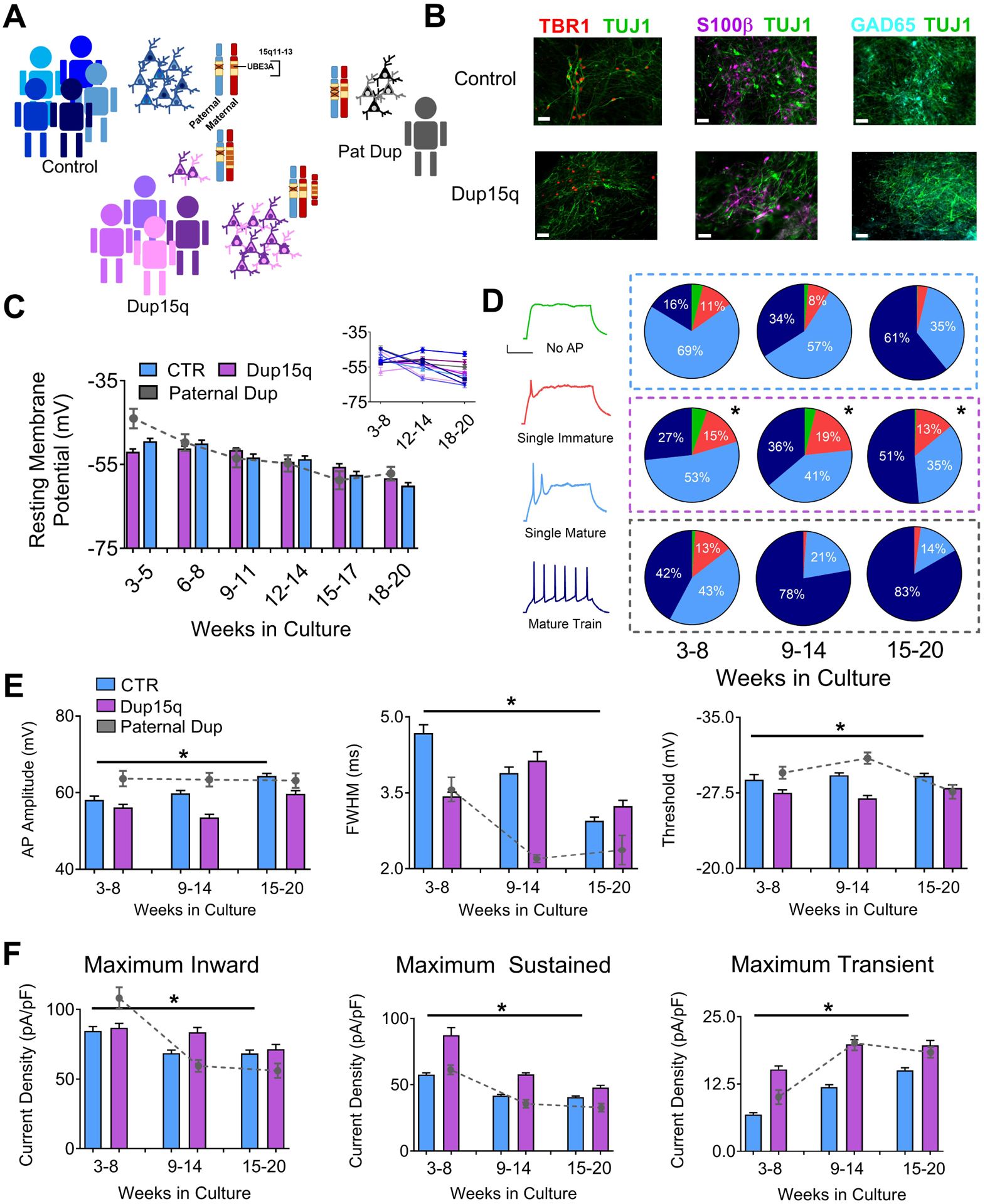
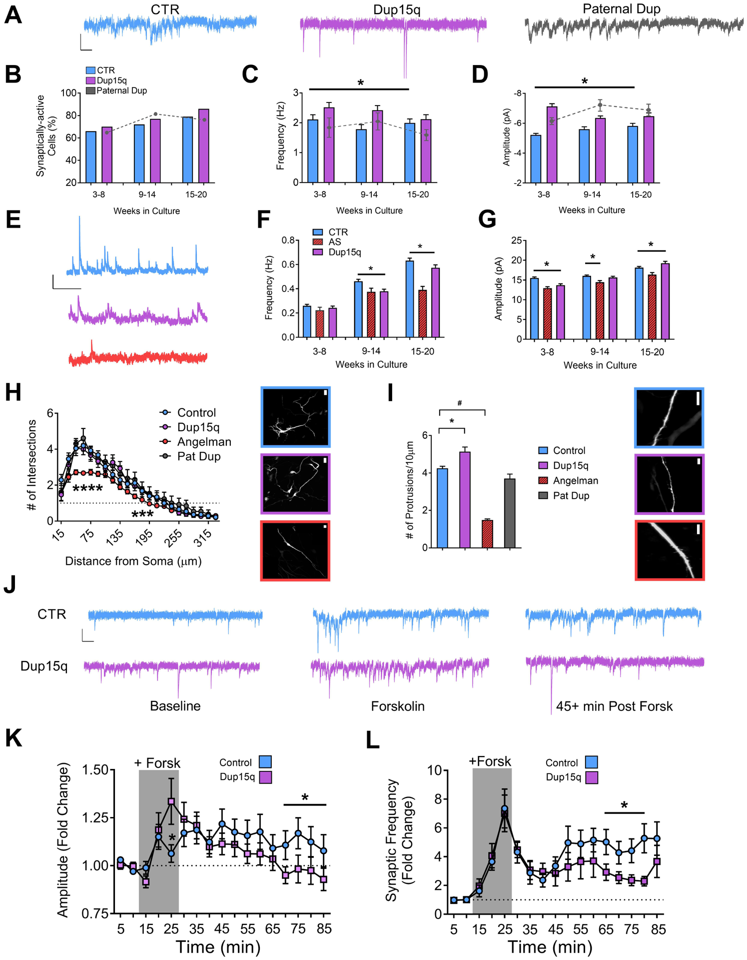
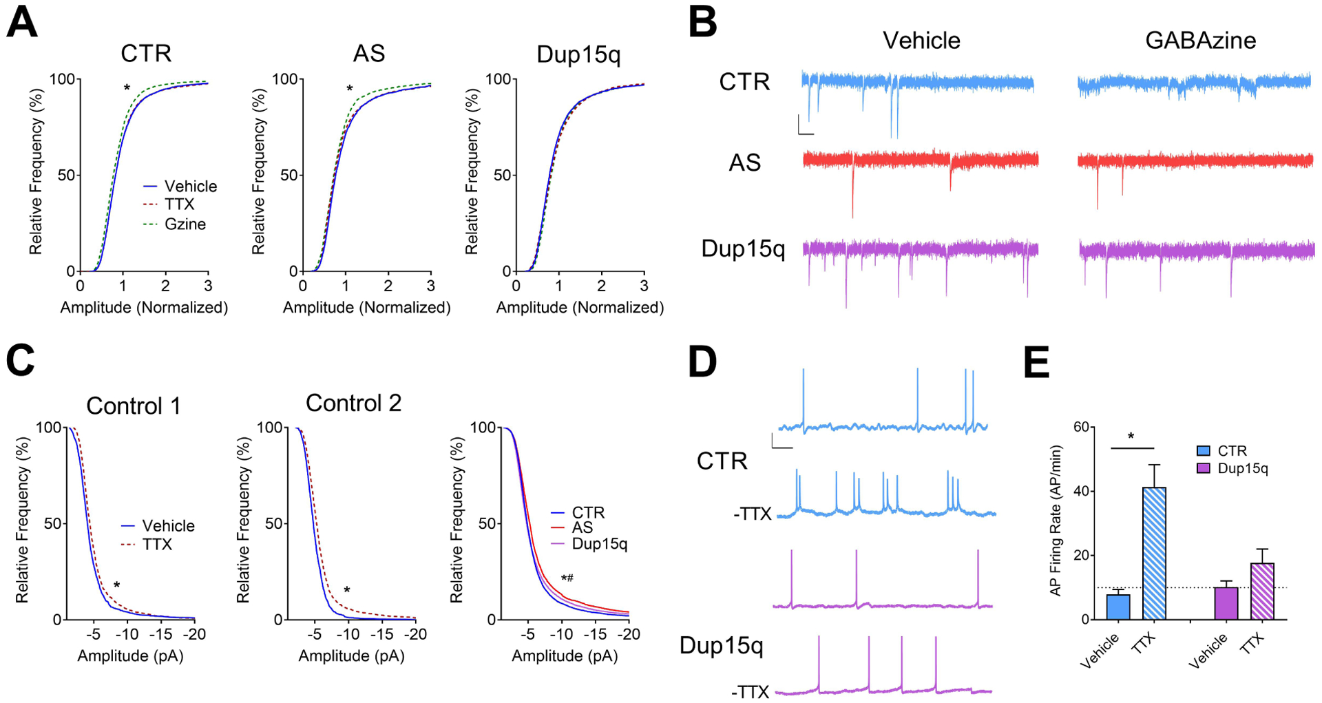
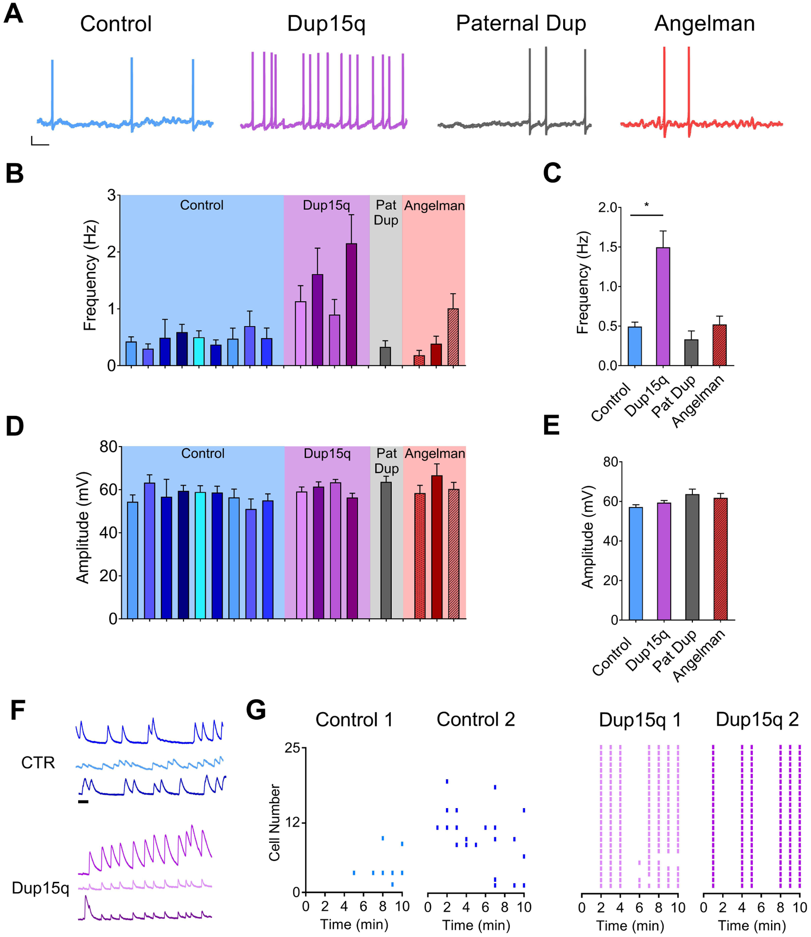
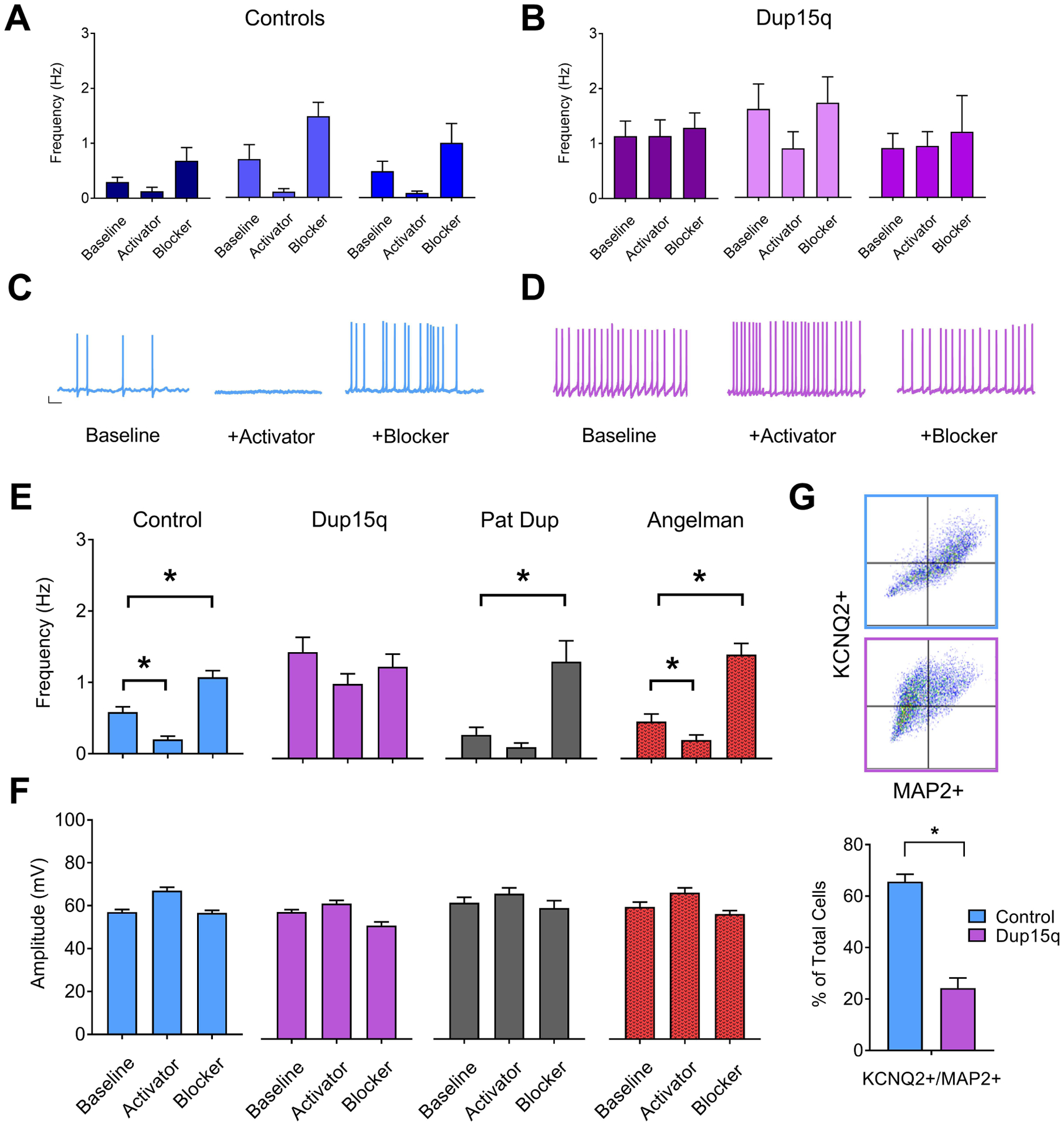
Background: Chromosome 15q11-q13 duplication syndrome (Dup15q) is a neurogenetic disorder caused by duplications of the maternal copy of this region. In addition to hypotonia, motor deficits, and language impairments, patients with Dup15q commonly meet the criteria for autism spectrum disorder and have a high prevalence of seizures. It is known from mouse models that synaptic impairments are a strong component of Dup15q pathophysiology; however, cellular phenotypes that relate to seizures are less clear. The development of patient-derived induced pluripotent stem cells provides a unique opportunity to study human neurons with the exact genetic disruptions that cause Dup15q.

Methods: Here, we explored electrophysiological phenotypes in induced pluripotent stem cell-derived neurons from 4 patients with Dup15q compared with 6 unaffected control subjects, 1 patient with a 15q11-q13 paternal duplication, and 3 patients with Angelman syndrome.

Results: We identified several properties of Dup15q neurons that could contribute to neuronal hyperexcitability and seizure susceptibility. Compared with control neurons, Dup15q neurons had increased excitatory synaptic event frequency and amplitude, increased density of dendritic protrusions, increased action potential firing, and decreased inhibitory synaptic transmission. Dup15q neurons also showed impairments in activity-dependent synaptic plasticity and homeostatic synaptic scaling. Finally, Dup15q neurons showed an increased frequency of spontaneous action potential firing compared with control neurons, in part due to disruption of KCNQ2 potassium channels.

Conclusions: Together, these data point to multiple electrophysiological mechanisms of hyperexcitability that may provide new targets for the treatment of seizures and other phenotypes associated with Dup15q.

Keywords: Autism; Autism spectrum disorder; Chromosome 15; Dup15q syndrome; Electrophysiology; Hyperexcitability; Neurodevelopment; iPSC.


Conflict of interest statement

Conflict of Interest

The authors report no biomedical financial interests or potential conflicts of interest.

Figures

Figure 1.
Figure 1.. Electrophysiological profile of iPSC-derived neurons from control and Dup15q subjects during in vitro development.
(A) Schematic of patient line genetics of the chromosome 15q11–13 genomic locus. (B) Immunocytochemical staining for TBR1/TUJ1, S100β/TUJI, and GAD65/TUJ1, in control and Dup15q-derived cultures between 20 and 25 weeks in culture. Scale bar, 50 μm (C) Group data for resting membrane potential (RMP) of control (CTR; 5 subjects; n>200 at each time point), Dup15q (4 subjects; n>175 at each time point) and a subject with a paternal duplication of chromosome 15q11-q13 (Paternal Dup; 1 subject; n=45 at each time point) during development. Inset: RMP for all individual lines. For each time bin, n≥30 for all lines. (D) Left: Example traces representing four AP firing patterns used for characterization. Scale bar, 20 mV, 200 ms. Right: Distribution of AP firing patterns for control (blue box; 5 subjects; n>430 at each time point), Dup15q (purple box; 4 subjects; n>430 at each time point), and a 15q11-q13 paternal duplication (grey box; 1 subject; n>60 at each time point) neurons at three developmental time bins. *P<0.0001 for differences between control and Dup15q; χ2 test. #P<0.0001 for differences between control and paternal duplication; χ2 test. (E) AP amplitude (left), full width at half-maximum amplitude (FWHM; middle), and AP threshold (right), for control, Dup15q, and paternal duplication cultures at three time points (n>350 for both control and dup15q at all time points; n>80_for paternal duplication at all time points). *P<0.001 for significant differences between control and Dup15q, #P<0.001 for significant differences between control and paternal duplication (two-way ANOVA). (F) Group data for maximum inward current density (left), maximum sustained outward current density (middle), and maximum transient outward current density (right), for control, Dup15q, and paternal duplication cultures at three time points (n>350 for both control and dup15q at all time points; n>80_for paternal duplication at all time points). *P<0.001 for significant differences between control and Dup15q, #P<0.001 for significant differences between control and paternal duplication (two-way ANOVA).
Figure 2.
Figure 2.. Development of synaptic activity and expression of activity-dependent synaptic plasticity.
(A) Example traces of spontaneous excitatory synaptic currents from control (CTR), Dup15q, and 15q11–13 paternal duplication (Paternal Dup) neurons at 15–20 weeks in culture. Scale bar: 10 pA, 100 ms. (B) Percent of synaptically-active neurons (event frequency at >0.2 Hz) derived from CTR (5 subjects; n>415 at every time point), Dup15q (4 subjects; n>350 at every time point), and paternal duplication (1 subject; n>80 at every time point). *P<0.05 for significant differences between control and Dup15q (χ2 test). (C) Mean frequency of spontaneous synaptic events for active neurons derived from CTR (5 subjects, n>250 cells at every time point), Dup15q (4 subjects, n>315 cells at every time point) and 15q11–13 paternal duplication (1 line; n>50 cells at every time point). (D) Mean amplitude of spontaneous synaptic events for neurons plotted in (c). *P<0.05 for differences between CTR and AS (two-way ANOVA). (E) Example traces of spontaneous inhibitory synaptic currents from control (CTR; blue), Dup15q (purple), and Angelman syndrome (AS; red) neurons at 15–20 weeks in culture. Scale bar: 20 pA, 1000 ms. (F) Mean frequency of spontaneous inhibitory synaptic events from CTR (6 subjects, n>600 cells at every time point), Dup15q (4 subjects, n>300 cells at every time point) and AS (1 line; n>100 cells at every time point). (G) Mean amplitude of spontaneous inhibitory synaptic events for neurons plotted in (F). *P<0.05 for differences between CTR, AS, and Dup15q as indicated by the bracket above the bars in the graph (Student’s t-test). (H) Left: Grouped Sholl analysis for neurons (28–30 weeks) from control- (n>20), Dup15q- (n=11), AS- (n>40), and 15q11-q13 paternal duplication-derived (n=5) cultures. *P<0.05 indicated significant differences between control and AS (Student’s t-test). Right: example images of transfected neurons used for analysis from control (top; blue), Dup15q (purple; middle), and AS (red; bottom). Scale bar: 25μm. (I) Left: Grouped analysis for number of dendritic protrusions for control (n>45), Dup15q (n=20), AS (n>40), and 15q11–13 paternal duplication cultures (n=9). Data from weeks 15–20 and 28–30 were collapsed into a single bin. *P<0.05 indicates significant differences between control and Dup15q. #P<0.05 indicates significant differences between control and AS (Student’s t-test). Right: example images of transfected neurons used for analysis from control (top; blue), Dup15q (purple; middle), and AS (red; bottom). Scale bar: 10μm. (J) Example traces of spontaneous excitatory synaptic currents from control (CTR; top) and Dup15q (bottom) neurons at (left to right) baseline, during forskolin/rolipram/0 Mg (Forsk), and 45+ min post-Forsk (see Methods for details). Scale bar, 10 pA, 100 ms. (L,K) Group data for CTR (n>60) and Dup15q (n>45) neurons showing (L) mean frequency and (K) mean amplitude of spontaneous synaptic currents during baseline, plasticity induction (indicated by grey box; see Methods for details) and post induction. *P<0.05, indicates differences for CTR (blue) vs Dup15q (purple) (Student’s t-test).
Figure 3.
Figure 3.. Impaired synaptic scaling in Dup15q-derived neurons.
(A) Cumulative frequency histograms of amplitudes (normalized) of miniature spontaneous excitatory synaptic currents for control (CTR; left; 6 subjects; n>140 neurons per treatment), Angelman (AS; middle; 4 subjects; n>80 neurons per treatment), and Dup15q (right; 4 subjects; n>100 neurons per treatment) cultures. *P<0.0001 indicated significant differences between control and GABAzine treatments (Student’s t-test). (B) Example traces of miniature spontaneous excitatory synaptic currents from CTR, AS, and Dup15q neurons following treatment with either vehicle (left) or GABAzine (right). Scale bar: 10 pA, 100ms. (C) Cumulative frequency histograms of raw amplitudes of miniature spontaneous excitatory synaptic currents for 2 control subjects treated with TTX (Control 1; left. Control 2; middle), with baseline synaptic frequencies >3 Hz. Right: Cumulative frequency histograms of raw baseline (vehicle-treated) amplitudes for control, AS, and Dup15q cultures. *P<0.0001 indicates significant differences between control and Dup15q, #P<0.0001 indicates significant differences between control and AS (Kolmogorov-Smirnov test). (D) Example traces of spontaneous action potential firing from control (CTR) and Dup15q neurons from data presented in (E). Scale bar: 20 mV, 1s. (E) Spontaneous action potential (AP) firing rate of vehicle- or TTX-treated neuronal cultures from control and Dup15q subjects following washout of TTX. *P<0.05 indicates significant differences between vehicle- and TTX-treated cultures (Student’s t-test).
Figure 4.
Figure 4.. Spontaneous action potential firing.
(A) Example traces of spontaneous action potential firing from control (CTR; blue), Dup15q (purple), 15q11–13 paternal duplication (Paternal Dup; grey) and AS (red) neurons at 25+ weeks in culture. Scale bar: 10 mV, 1s. (B,D) Spontaneous action potential frequency (B) and amplitude (D) of neurons derived from individual control lines (blue; 9 subjects; n>15 neurons per bar), Dup15q lines (purple; 4 subjects; n>15 neurons per bar), 15q11-q13 paternal duplication (grey; 1 subject; n>25), and AS (red; 3 subjects; n>25 per bar). (C,E) Grouped data of data presented in (B,D) (n=224, n=110, n=32, n=89 for control, Dup15q, Pat Dup, and AS, respectively). *P<.05 indicates significant differences between control and Dup15q (Student’s t-test). (F) Example traces of spontaneous baseline calcium transients from 3 neurons each from CTR (top; blue) and Dup15q (bottom; purple). Scale bar, 1 min. (G) Raster plots of frequency of calcium transients from cultures derived from control (left; 2 subjects; 25 neurons each) and Dup15q (right; 2 subjects; 25 neurons each).
Figure 5.
Figure 5.. Impaired KCNQ2 channels in Dup15q-derived neurons.
(A,B) Frequency of spontaneous action potential firing for 3 individual control (A) and Dup15q (B) lines at baseline or in the presence of either pharmacological blocker or activator of KCNQ2 channels (n>15 neurons for each bar). (C) Example traces of spontaneous action potentials recorded from control neurons. Scale bar: 10mV, 1s (D) Example traces of spontaneous action potentials recorded from Dup15q neurons (E) Group data for frequency of spontaneous action potential firing for control (left; blue; 9 lines; n=251, 160, 204 neurons for baseline, activator, and blocker, respectively), Dup15q (middle left; purple; 4 lines; n=110, 108, 93 neurons for baseline, activator, and blocker, respectively), 15q11–13 paternal duplication (middle right; grey; 1 line; n=30 neurons for all bars), and Angelman (right; red; 3 lines; n>89 neurons for all bars), recorded at baseline or in the presence of either pharmacological blockers or activators of KCNQ2 channels. *P<0.05 indicates significant differences (student’s t-test). (F) Group data for amplitude of spontaneous action potential firing for data presented in (E). (G) Top: Examples of flow cytometry plots for cultures stained with KCNQ2 and MAP2. Bottom: Flow cytometry quantification of percent of cells positive for both MAP2 and KCNQ2 for control (2 subjects/8 coverslips) and Dup15q (2 subjects/8 coverslips).*P<0.05 indicates significant differences (Student’s t-test).

Comment in

References

    1. Chamberlain SJ, Chen P-F, Ng KY, Bourgois-Rocha F, Lemtiri-Chlieh F, Levine ES, Lalande M (2010): Induced pluripotent stem cell models of the genomic imprinting disorders Angelman and Prader-Willi syndromes. Proc Natl Acad Sci USA 107: 17668–17673. - PMC - PubMed
    1. Judson MC, Sosa-Pagan JO, Del Cid WA, Han JE, Philpot BD (2013): Allelic specificity of Ube3a expression in the mouse brain during postnatal development. J Comp Neurol 522: 1874–1896. - PMC - PubMed
    1. Chamberlain SJ, Lalande M (2010): Neurodevelopmental disorders involving genomic imprinting at human chromosome 15q11-q13. Neurobiology of Disease 39: 13–20. - PubMed
    1. Cassidy SB, Driscoll DJ (2008): Prader–Willi syndrome. European Journal of Human Genetics 17: 3. - PMC - PubMed
    1. Chamberlain SJ, Lalande M (2010): Angelman syndrome, a genomic imprinting disorder of the brain. J Neurosci 30: 9958–9963. - PMC - PubMed

Publication types